BREAKTHROUGH DISCOVERIES IN NEPHROLOGY
←
→
Page content transcription
If your browser does not render page correctly, please read the page content below
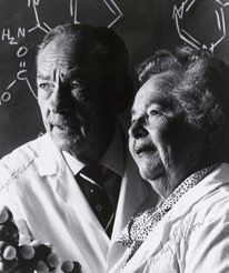
table of contents   Breakthrough Discoveries in Nephrology

         BREAKTHROUGH
         DISCOVERIES IN
         NEPHROLOGY

Page 1                                            Advancing kidney health worldwide. Together.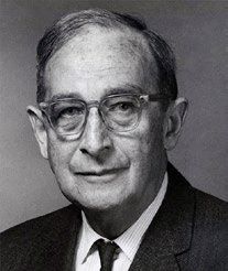
Breakthrough Discoveries in Nephrology

   In celebration of the 60th Anniversary of the ISN, the ISN Research Working Group
   (RWG), under the leadership of Chair, Adeera Levin, and Deputy Chair, Masaomi
   Nangaku, published a monthly series, “Breakthrough Discoveries.”

   The series highlighted 60 + 1 historical discoveries of significant impact to
   the nephrology community. So that the selection of discoveries was globally
   representative, the leadership of the ten ISN Regional Boards were asked to provide
   references from their respective regions. Subsequently, Leon Fine, Pierre Ronco,
   and John Feehally were advisors, and the ISN RWG leadership and the ISN Executive
   Committee reviewed and voted for the final selection.

   Every month in 2020, the ISN RWG, supported by the ISN Young Nephrologists
   Committee (YNC), highlighted and discussed five breakthrough discoveries. This
   series frames the year of celebratory activities and highlights ISN’s commitment to
   research, collaboration, and global community, as well as the achievements ISN has
   made through its members and supporters.

   We want to thank the ISN Breakthrough Discoveries Narratives Contributors:

     • Abduzhappar Gaipov                       • Lani Shochet
     • Caner Alperslan                          • Lili Zhou
     • Daisuke Nakano                           • Marco van Londen
     • Eishin Yaoita                            • Mirna Aleckovic-Halilovic
     • Enisa Mesic                              • Mirha Pjanic
     • Fabian Braun                             • Muzamil Hassan
     • Fumiaki Ando                             • Nikolay Bulanov
     • Georgina Irish                           • Rhys Evans
     • Hirotaka Komaba                          • Rolando Claure-Del Granado
     • Huang-Yu Yang                            • Sabine Karam
     • HuiYi Shan                               • Shankar Yadav
     • Kate Robson                              • Yosuke Hirakawa

   The ISN would like to particularly thank Masaomi Nangaku, ISN RWG Deputy Chair,
   on leading the publication of the monthly series, “Breakthrough Discoveries”,
   in collaboration with Yosuke Hirakawa, YNC member, to celebrate the 60th
   Anniversary of the ISN in 2020.

Page 2                                               Advancing kidney health worldwide. Together.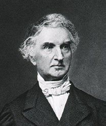
Breakthrough Discoveries in Nephrology

                                        TABLE OF CONTENTS
   JANUARY............................................................................................ 6
         First microscopic description of the ‘glomeruli’....................................................7
         Identification of kidney disease.............................................................................8
         Description of the nephron...................................................................................9
         Description of the urinary sediment as a tool for the diagnosis
         of nephropathies.................................................................................................10
         Routine measurement of urea and creatinine in clinical practice......................11

   FEBRUARY........................................................................................ 12
         Discovery of the Nephron Loop...........................................................................13
         Micropuncture Technique...................................................................................14
         Demonstration of Renal Physiology....................................................................15
         Description of Acute Renal Failure with Crush Syndrome..................................16
         Laboratory Methods for Clinical Application to treat Kidney Disease................18

   MARCH............................................................................................. 19
         Volhard & Fahr’s book.........................................................................................20
         Percutaenous kidney biopsy................................................................................21
         Application of immune fluorescence technique to kidney biopsy –
         Mellors 1950s......................................................................................................22
         Application of electron microscopy to analyze ultrastructural
         changes of the kidney..........................................................................................23
         Structure-function correlation in the glomerulus...............................................24

   APRIL................................................................................................ 25
         Understanding of acid excretion in kidney disease.............................................26
         Perfusion of isolated tubules...............................................................................27
         Understanding of nephron membrane transport systems by means
         of isolated membranes and cells.........................................................................28
         Countercurrent multiplication system without active transport........................29
         Identification of molecules..................................................................................30

Page 3                                                                      Advancing kidney health worldwide. Together.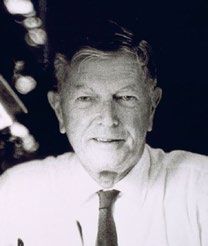
Breakthrough Discoveries in Nephrology

   MAY.................................................................................................. 31
         Description of Minimal Change Nephrotic Syndrome, “Lipoid Nephrosis”........32
         Culture of Glomerular Cells.................................................................................33
         Identification of Antigen of Membranous Nephropathy....................................34
         First Description of IgA Glomerulonephritis (Berger’s Disease).........................35
         IgA Hinge Glycosylation in IgA Nephropathy and IgA Vasculitis..........................36

   JUNE................................................................................................. 37
         Discovery of the link between kidney disease and hypertension.......................38
         Experimental Hypertension.................................................................................39
         Macula densa.......................................................................................................40
         Expression of glomerular receptor for angiotensin II.........................................41
         Discovery of Renin...............................................................................................42

   JULY.................................................................................................. 43
         Description of familial kidney disease.................................................................44
         Description of Alport syndrome..........................................................................45
         APOL1 and kidney disease...................................................................................46
         Cloning of Nephrin...............................................................................................47
         Cloning of PKD1 and PKD2 and Treatment of Autosomal Dominant
         Polycystic Kidney Disease....................................................................................48

   AUGUST............................................................................................ 50
         Kidney Transplantation........................................................................................51
         Knights of Hemodialysis.......................................................................................52
         Vascular Access....................................................................................................54
         Peritoneal Dialysis................................................................................................55
         First Continuous Renal Replacement Treatment (CRRT).....................................56

   SEPTEMBER...................................................................................... 57
         Development of the Animal Model of Nephrotoxic Serum Nephritis................58
         Immune Tolerance...............................................................................................59
         Introduction of Azathioprine and Cyclosporin....................................................60
         Rituximab Therapy for Renal Diseases................................................................61
         Immune Complex-mediated Glomerulonephritis...............................................62

Page 4                                                                      Advancing kidney health worldwide. Together.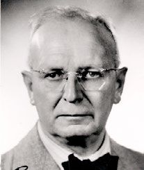
Breakthrough Discoveries in Nephrology

   OCTOBER.......................................................................................... 63
         Historic Breakthroughs in Preeclampsia.............................................................64
         Antineutrophil Cytoplasmic Antibodies..............................................................65
         Plasma Exchange for TTP and Anti-GBM Nephritis.............................................66
         Diabetic Kidney Disease.......................................................................................67
         Aristolochic Acid Nephropathy............................................................................68

   NOVEMBER...................................................................................... 69
         eGFR.....................................................................................................................70
         CKD Classification and Staging.............................................................................72
         Purification of Erythropoietin..............................................................................73
         Renal Osteodystrophy and CKD-MBD.................................................................75
         Cardio-renal Association......................................................................................77

   DECEMBER....................................................................................... 78
         Corticosteroids as effective (and therefore lifesaving) in nephrotic
         syndrome in children...........................................................................................79
         Hyperfiltration Theory.........................................................................................80
         Kidney protection by RAS inhibition....................................................................81
         HIF and hypoxia of the kidney.............................................................................83
         Identification of SGLT2 and Clinical Application of its Inhibitor..........................85

   SIXTY PLUS ONE:
         An overview of recent discoveries as potential breakthroughs
         to future knowledge............................................................................................87

Page 5                                                                          Advancing kidney health worldwide. Together.
Breakthrough Discoveries in Nephrology
JANUARY
Page 6 Advancing kidney health worldwide. Together.table of contents Breakthrough Discoveries in Nephrology
FIRST MICROSCOPIC DESCRIPTION
OF THE ‘GLOMERULI’
By Sabine Karam MD
Saint George Hospital University Medical Center, Beirut, Lebanon
The Italian anatomist Marcello Malpighi (1628–1694),
often referred to as the founder of microscopical
anatomy, is credited for the first microscopic description
of the glomeruli. He described them as dark, vascular
structures resembling fruit suspended from a branch (…
quae sanguineis vasis atro liquore turgidis in speciolae
arbori formam productis, velut poma appenduntur). He
demonstrated their continuity with the renal vasculature
in ‘De renibus,’ a section of ‘De Viscerum Structura Exercitatio Anatomica’, originally
published in Bologna, Italy, in 1666 and then in London, in 16691.
In 1782, Alexander Schlumlanski (1758-1795) described ‘de Structura renum’ in his
dissertation as a connection between the circulation and the uriniferous tubules,
deduced by experimenting on pig kidneys2. However, it was the surgeon and
anatomist William Bowman (1816-1892) who elucidated the capillary architecture
of the glomerulus and the continuity between its surrounding capsule and the
proximal tubule in detail (see Discovery #3 by Lili Zhou). He presented his findings
in the paper “On the Structure and Use of the Malpighian Bodies of the Kidney”3.
Nonetheless, the term glomerulus would come into usage only a few years later in
the mid-nineteenth century. It seems to be derived from the Latin word ‘glomus’,
which means ‘ball of thread’4.
1 M. M. De Viscerum Structura Exercitatio Anatomica. Londini: Typis T.R. Impensis Jo.Martyn;(London). MDCLXIX1669. p. 83–4.
2 A. S. Dissertatio inauguralis anatomica De Structura Renum MDCCLXXXII. : Argentorati (Strasbourg): Typis Lorenzii & Schuleri;
1782.
3 Todd Bentley R BW. The physiological anatomy and physiology of man. West Strand,London:John W Parker and sons. 1859;2:482-
507.
4 Merriam Webster Dictionary. Glomerulus.
Page 7 Advancing kidney health worldwide. Together.table of contents Breakthrough Discoveries in Nephrology
IDENTIFICATION OF KIDNEY DISEASE
By Yosuke Hirakawa
Division of Nephrology and Endocrinology, the University of Tokyo Hospital, Japan
The symptoms of kidney disease are non-specific;
therefore, diagnosis of kidney disease without laboratory
testing is difficult. This was especially the case in the early
19th century. Surprisingly, the first identification of kidney
disease was made by Richard Bright in 1827 before the
identification of creatinine and routine measurement of
urea. He extensively examined patients with proteinuria,
anasarca, and uremia by checking urinary albumin and
renal morbid anatomy in his work as a physician at Guy’s
Hospital in London1,2. Today, some of his cases would be diagnosed as having
nephrotic syndrome caused by glomerulonephritis3. Identification of what became
known as “Bright’s disease” made a huge contribution to medicine. The most
important aspect is the distinction Bright made between patients with kidney
diseases and patients with cardiac diseases. After this differentiation, the
characteristics of kidney disease patients started to be eagerly examined, and it was
followed by increased urea and creatinine levels being identified shortly afterwards
as major hallmarks of kidney disease.
1 Boss J. Richard Bright’s Reports of Medical Cases (1827): A sesquicentennial note. Bristol Med Chir J. 1978;93:5-6, 18.
2 Cameron JS. Bright’s Disease Today: The Pathogenesis and Treatment of Glomerulonephritis – I. Br Med J. 1972;4:87-90.
3 Weller RO, Nester B. Histological reassessment of Three Kidneys Originally Described by Richard Bright in 1827-36. Br Med J. 1972
Jun 24;2:761-3.
Page 8 Advancing kidney health worldwide. Together.table of contents Breakthrough Discoveries in Nephrology
DESCRIPTION OF THE NEPHRON
By Lili Zhou
Division of Nephrology, Nanfang Hospital, Southern Medical University, China
The kidney is the most important organ in the human
body to excrete urine and maintain the balance of water
and electrolytes. These processes take place through
the subtle mechanisms of the nephron, a tiny unit of
workforce within the kidney. In 1842, after two years of
exploration, Dr. William Bowman, a famous English surgeon
and anatomist, discovered the nephron’s true nature.
Through repeated injections from the arteries, tubes,
and veins in multiple species’ kidneys, he found the real
structure of the Malpighian bodies, as well as their connecting tubes and circulation.
The Malpighian bodies, called glomerulus today, originate and gradually subdivide
from the afferent artery terminal twigs to become two rounded capillary vessels
tufts. These vessels ultimately converge to become one efferent channel (smaller
in size than the afferent) to enter the capillary plexus surrounding the uriniferous
tubes (proximal convoluted tubules, loop of Henle, and distal convoluted tubules).
The interconnected capillary plexus surrounding the tubes serves as the portal
system in contact with the tubes’ basement membrane to renal veins. The tubes
are the extension of Malpighian bodies’ capsules (Bowman’s capsule) and expand
tortuously near the Malpighian bodies but straighten when proceeding toward the
excretory channel (collecting duct). All these features retard blood flow and delay
the excretion of nutrients into urine to maintain the balance of water, sugar, and
electrolyte assimilation and excretion. This revolutionary discovery opened a new
era in physiological and pathological research in kidneys1.
1 W. Bowman. On the Structure and Use of the Malpighian Bodies of the Kidney, with Observations on the Circulation through That
Gland. Philosophical Transactions of the Royal Society of London Vol. 132 (1842), pp. 57-80.
Page 9 Advancing kidney health worldwide. Together.table of contents Breakthrough Discoveries in Nephrology
DESCRIPTION OF THE URINARY SEDIMENT AS A
TOOL FOR THE DIAGNOSIS OF NEPHROPATHIES
By Rolando Claure
Universidad Mayor de San Simon School of Medicine, Cochabamba, Bolivia
Urinary sediment has been used as a diagnostic tool since
the 17th century1and Pierre Rayer occupies a special
place in the history of nephrology for his attempt to
classify various diseases using this important diagnostic
tool. Alongside his intern, Eugene Napoleon Vigla, Rayer
revolutionized the study of kidney diseases by using
microscopy to analyze urinary sediments, describing
crystals, cells, casts, and yeasts2,3.
At the Hôpital de la Charité, a microscope became available in 1835 and Rayer
promptly set up a program to investigate urinary sediment findings in various forms
of kidney diseases. His proposed classification was based on clinical findings, urinary
microscopy, and gross specimens whenever possible. Renal diseases were divided
into acute nephritis with many red blood cells and too much albumin in the urine,
and chronic albuminuric nephritis corresponding to what is now known as nephrotic
syndrome. There was also mention of suppurative forms of nephritis, with pus cells
in the urine, the result of either blood-borne or ascending infection of the kidneys4.
After this first description, routine chemical analysis of urine and microscopic
examination of the sediment were introduced during the first half of the 19th
century. After a first wave of interest, the use of urinary sediment has progressively
decreased5; urinary microscopy analysis is now performed mostly in central
laboratories and is infrequently performed by nephrologists who have lost the
expertise to identify some types of casts and/or cells in order to perform clinical
correlations6. Nephrologists should reclaim this noninvasive test, since combining
it with a comprehensive clinical evaluation and new biomarkers would provide new
insights into renal diseases7.
1 Armstrong JA. Urinalysis in Western culture: a brief history. Kidney Int. 2007;71(5):384-7.
2 Fogazzi GB, Cameron JS. The introduction of urine microscopy into clinical practice. Nephrol Dial Transplant. 1995;10(3):410-3.
3 Fogazzi GB, Cameron JS. Urinary microscopy from the seventeenth century to the present day. Kidney Int. 1996;50(3):1058-68.
4 Richet G. From Bright’s disease to modern nephrology: Pierre Rayer’s innovative method of clinical investigation. Kidney Int.
1991;39(4):787-92.
5 Eknoyan G. Looking at the urine: the renaissance of an unbroken tradition. Am J Kidney Dis. 2007;49(6):865-72.
6 Fogazzi GB, Garigali G. The clinical art and science of urine microscopy. Curr Opin Nephrol Hypertens. 2003;12(6):625-32.
7 Claure-Del Granado R, Macedo E, Mehta RL. Urine microscopy in acute kidney injury: time for a change. Am J Kidney Dis.
2011;57(5):657-60.
Page 10 Advancing kidney health worldwide. Together.table of contents Breakthrough Discoveries in Nephrology
ROUTINE MEASUREMENT OF UREA AND
CREATININE IN CLINICAL PRACTICE
By Yosuke Hirakawa
Division of Nephrology and Endocrinology, the University of Tokyo Hospital, Japan
Routine measurement of urea and creatinine, sensitive
indicators of renal function, forms the present-day basis
of clinical nephrology. Around 1850, two important events
related to measurement of urea and creatinine occurred.
The word “creatinine” was probably first used by Justus
von Liebig in 18471,2. He found the ingredient creatinine
in beef tea. Beef tea was a traditional English remedy,
made using only beef and salt, not tea leaves. It had
previously been known that creatine, the precursor of creatinine, was abundant
in animal muscle. Liebig, who established the Justus von Liebig’s Extract of Meat
Company, found that the addition of hydrochloric acid to creatine resulted in the
production of creatinine. Around this time, creatinine was not used as an indicator
of renal function; researchers focused on urea as an indicator of renal function, with
Joseph Picard having established the reproducible and sensitive method of urea
measurement in 1856. He later found that urea concentration in the renal vein fell
from that in the renal artery3. Around the same time, toxic mechanism came to be
accepted as the etiology of uremic syndrome; therefore, the establishment of the
urea measurement technique and the discovery of the urea fall in the renal vein led
to the concept that urea was the causative substance of uremic syndrome. However,
a brave study of urea loading in human patients performed in 1972 revealed that
urea itself is not a uremic toxin4.
1 Pierre Delanaye (2012). “Serum Creatinine: An Old and Modern Marker of Renal Function” in Pierre Delanaye (ed.) Nephrology and
Clinical Chemistry: The Essential Link pp9-20.
2 Kramer H, Rosas SE, Matsushita K. Beef Tea, Vitality, Creatinine, and the Estimated GFR. Am J Kidney Dis. 2016;67:169-72.
3 Gabriel Richet. Early history of uremia. Kidney Int 1988;33:1013-5
4 Johnson WJ, Hagge WW, Wagoner RD, Dinapoli RP, Rosevear JW. Effects of urea loading in patients with far-advanced renal failure.
Mayo Clin Proc 1972;47:21-29
Page 11 Advancing kidney health worldwide. Together.Breakthrough Discoveries in Nephrology
FEBRUARY
Page 12 Advancing kidney health worldwide. Together.table of contents Breakthrough Discoveries in Nephrology
DISCOVERY OF THE NEPHRON LOOP
By Yosuke Hirakawa
Division of Nephrology and Endocrinology, the University of Tokyo Hospital, Japan
In 1862, anatomist Jacob Henle presented the existence of
nephron loops, widely known today as the loop of Henle1.
He provided a morphological description; the importance
of electrolyte reabsorption had not yet been determined
and the existence of tubular reabsorption was only proved
in the 1920s by Alfred Newton Richards. Henle is famous
for his description of epithelial tissue: large sheets of cells
free from blood vessels, blood components, and nerve
endings. Henle described first epithelial tissue in the digestive tract followed by
glandular and tubular organs, including the kidney2. In renal medulla, Henle found
two tubular subtypes: one type was a papillary collecting duct with a diameter
of 0.05-0.06 mm; the other type had a much smaller diameter of approximately
0.02-0.03 mm, running parallel to the collecting ducts but returning in a narrow
hairpin curve toward the surface. The latter is the well-known loop of Henle. As an
anatomist, Henle examined other epithelial tissues also: Henle’s gland in the eyelids
and Henle’s layer in the hair follicle3.
1 Morel F. The loop of Henle, a turning-point in the history of kidney physiology. Nephrol Dial Transplant. 1999;14:2510-5
2 Kinne-Saffran E, Kinne RK. Jacob Henle: the kidney and beyond. Am J Nephrol. 1994;14:355-60.
3 Weyers W. Jacob Henle–a pioneer of dermatopathology. Am J Dermatopathol. 2009;31:6-12
Page 13 Advancing kidney health worldwide. Together.table of contents Breakthrough Discoveries in Nephrology
MICROPUNCTURE TECHNIQUE
By Yosuke Hirakawa
Division of Nephrology and Endocrinology, the University of Tokyo Hospital, Japan
It is now common knowledge that urine is produced by
glomerular filtrate and tubular reabsorption of substances
such as electrolytes and glucose. This phenomenon was
initially understood by Alfred Newton Richards and his
colleagues in the 1920s. At that time, many researchers
had tried to observe glomerular circulation but did not
have the capacity or methods to do so. Richards was the
pioneer who decided to observe frog kidneys, which are
thin and flat1. He investigated the effect of adrenaline on
glomerular circulation with a micropipette introduced into glomerular space with
the help of a micromanipulator. In the process, he obtained enough glomerular
fluid for quantitative tests. He found that glomerular filtrate contained both
chloride and sugar, detectable in blood but undetectable in bladder urine, leading
to the conclusion that there must be 2 different processes: glomerular filtration
and tubular reabsorption respectively. This description is thought to be one of the
most important contributions in our understanding of renal physiology2 on which
subsequent understandings of glomerular filtration rate (GFR) and solute transport
have been built. Richards’ achievements are widely known and the ISN ensures that
his outstanding and fundamental contribution to basic research is honored through
the Alfred Newton Richards Award for basic science.
1 Schmidt CF. Alfred Newton Richards 1876-1966. Ann Intern Med. 1969: Suppl 8:15-27.
2 Sands JM. Micropuncture: unlocking the secrets of renal function. Am J Physiol Renal Physiol. 2004; 287:F866-7.
Page 14 Advancing kidney health worldwide. Together.table of contents Breakthrough Discoveries in Nephrology
DEMONSTRATION OF RENAL PHYSIOLOGY
By Sabine Karam MD
Saint George Hospital University Medical Center, Beirut, Lebanon
Homer W. Smith (1895-1962), physiologist and medical
writer, was a pioneer in renal physiology. He recognized
the clinical importance of renal clearance methods, a
concept initially introduced by Donald Van Slyke in 19281.
He introduced them as a tool for the precise measurement
of renal function in medical practice and elaborated on
the concepts of glomerular filtration, effective renal
plasma flow, and intrarenal resistance2. Smith also played
an essential role in elucidating tubular transport capacity,
the reabsorption and secretion of various substances such as urea and creatinine,
as well as providing novel insights into the mechanisms of the excretion of water
and electrolytes3. Finally, he was instrumental in setting the perfect example of
collaboration between basic scientists and clinicians, a model which has since been
followed worldwide2. Dr. Smith’s studies of the kidney culminated in 1951 with the
authoritative summary, “The Kidney, Structure and Function in Health and Disease“.
He also had a remarkable career in philosophy and literature, as illustrated by the
reflective essay, “From Fish to Philosopher,” describing the evolutionary role of the
kidney in enabling survival in both water and on land4. Today, his legacy endures
through the Homer W. Smith annual award in renal physiology established by the
American Society of Nephrology in 1964.
1 SE B. Clearance concept in renal physiology. In: GOTrSCHALK CW BR, GIEBISCH GH, editor. Renal Physiology, People and Ideas.
Bethesda: American Physiological society; 1987.
2 Baldwin DS, Neugarten J. Homer Smith: his contribution to the practice of nephrology. J Am Soc Nephrol. 1995;5(12):1993-9.
3 Giebisch G. Homer W. Smith’s contribution to renal physiology. J Nephrol. 2004;17(1):159-65.
4 Fishman AP. Homer W. SMITH (1895-1962). Circulation. 1962;26:984-5.
Page 15 Advancing kidney health worldwide. Together.table of contents Breakthrough Discoveries in Nephrology
DESCRIPTION OF ACUTE RENAL FAILURE WITH
CRUSH SYNDROME
By Mirna Aleckovic-Halilovic and Enisa Mesic
Clinic for Internal Diseases, Department of Nephrology, Dialysis and Renal Transplantation,
University Clinical Center Tuzla, Tuzla, Bosnia and Herzegovina
In 1941, during the London Blitz, E.G. Bywaters and D. Beall
described four victims of trauma-related crush syndrome,
with limb edema and shock, who showed oliguria and
brownish urine. All four patients died in about a week with
nitrogen retention, and necropsy revealed pigment casts,
polymorphonuclear invasion, and acute tubular necrosis
in the kidney1.
In 1943, using animal models, E.G. Bywaters & J.K. Stead
identified myoglobin as the offending agent and formulated the first treatment plan2.
It was later understood that crush syndrome is a systemic response to traumatic
rhabdomyolysis and that acute kidney injury is one of its severest complications.
It was only after 25 000 people died in an Armenian Earthquake in 1988 that
this entity got the attention it deserved through the establishment of ISN’s Renal
Disaster Relief Task Force and its recommendations for the management of crush
victims in mass disasters3. Furthermore, this was the origin of the term ‘disaster
nephrology’4,5.
This condition, first recognized as a single broad pathophysiologic entity in 1941,
remains pertinent in the current era: The number of victims from natural and man-
made disasters is growing, and crush syndrome is the second most common cause of
death, after asphyxia, caused by these disasters6. Moreover, despite being rescued
alive from the rubble. many crush victims die afterward due to the lack of access to
dialysis.
1 Bywaters EG and Beall D. Crush Injuries with Impairment of Renal Function. Br Med J. 1941 Mar 22;1(4185):427-32.
2 Bywaters EGL, Stead JK, The production of renal failure following injection of solutions containing myohaemoglobin. QJ Exp Physiol
1944;33:53
3 Sever MS, Vanholder R and the Workgroup. Recommendations for the management of crush victims in mass disasters. Nephrol
Dial Transplant 2012; 27(Suppl 1): i1–67
4 Vanholder R, Sever MS, Erek E et al. Acute renal failure related to the crush syndrome: towards an era of seismo-nephrology?
Nephrol Dial Transplant 2000; 15: 1517–1521
5 Gibney RT, Sever MS, Vanholder RC. Disaster nephrology: crush injury and beyond. Kidney Int 2014; 85: 1049–1057)
6 Ukai T. The Great Hanshin-Awaji Earthquake and the problems with emergency medical care. Ren Fail 1997; 19:633–645
Page 16 Advancing kidney health worldwide. Together.table of contents Breakthrough Discoveries in Nephrology
Since randomized control trials during mass disasters are unfeasible, the treatment
recommendations in the guidelines1 are based on observational data accrued from
case reports and case series written by physicians in the field using physiological
principles to treat these victims.
In honor of the achievements of Bywaters, the ISN established the Bywaters Award
in 1991 to recognize outstanding contributions to the understanding and treatment
of Acute Kidney Injury.
1 Sever MS, Vanholder R and the Workgroup. Recommendations for the management of crush victims in mass disasters. Nephrol
Dial Transplant 2012; 27(Suppl 1): i1–67
Page 17 Advancing kidney health worldwide. Together.table of contents Breakthrough Discoveries in Nephrology
LABORATORY METHODS FOR CLINICAL
APPLICATION TO TREAT KIDNEY DISEASE
By Nikolay Bulanov
Sechenov First Moscow State Medical University, Russia
In 1886, Max Jaffe (1841 – 1911), a German physician and
chemist, observed that creatinine produced an intensive
red color in alkaline picrate solution and detected needle-
formed crystals under the microscope which he reported
in his landmark paper1.
However, the quantitative analytical method used to
assess creatinine concentration was developed in the
first decade of the twentieth century by an outstanding
Swedish-born American biochemist Otto Folin (1867 – 1934), who called it the
“Jaffe method”2. Even over a century after its introduction into clinical practice,
this procedure is still widely used to measure creatinine levels due to its simplicity
and low-cost. However, several organic compounds called pseudochromogens (e.g.
acetone, glucose) that were first recognized by Jaffe can also react with alkaline
picrate and lead to an analytical bias.
In 1957, Alfred Free (1913 – 2000) and his co-authors working at the Ames
Corporation published a paper describing a new colorimetric test for urinary protein3.
The first dipstick was a yellow paper strip, impregnated with a citrate buffer and
tetrabromphenol blue, which turns green in the presence of protein. Free et al tested
their new method obtaining approximately 5000 turbid urine samples from patients
and healthy subjects and demonstrated its adequate sensitivity and specificity.
Today, urinary dipstick test is one of the most common screening techniques for
early detection of kidney diseases.
In 1945, Bowling Barnes, David Richardson, John Berry, and Robert Hood introduced
flame photometer to measure the low concentration of sodium and potassium in a
solution4. Flame photometer measures the intensity of emitted light when a metal
is introduced into the flame, giving information about the amount of the element
present in the sample. This technology allows for cheap and simple measurements
of electrolytes in serum and urine.
1 Jaffe M. Ueber den Niederschlag welchen Pikrinsäure in normalen Harn erzeugt und über eine neue reaction des Kreatinins. Z
Physiol Chem. 1886;10:391–400.
2 Shaffer PA. Otto Folin 1867-1934. Washington, DC: National academy of sciences;1952:47–82
3 Free AH, Rupe CO, Metzler I. Studies with a new colorimetric test for proteinuria. Clin Chem. 1957;3:716–727.
4 Barnes RB, Richardson D, Berry JW, Hood RL. Flame photometry; A rapid analytical procedure. Ind Eng Chem Anal Ed. 1945;17:605-
11.
Page 18 Advancing kidney health worldwide. Together.Breakthrough Discoveries in Nephrology
MARCH
Page 19 Advancing kidney health worldwide. Together.table of contents Breakthrough Discoveries in Nephrology
VOLHARD & FAHR’S BOOK
By Yosuke Hirakawa
Division of Nephrology and Endocrinology, the University of Tokyo Hospital, Japan
In 1914, Franz Volhard, a clinician, and Theodor
Fahr, a pathologist, published the textbook, Die
Brightsche Nierenkrankheit. Klinik, Pathologie
und Atlas (Bright’s disease. Clinical aspects,
Pathology and illustrations)1,2. This is one of
the most important textbooks because for the
first time several pathological entities were
described. ‘Bright’s disease’ was categorized
into nephrosis (nephrotic syndrome),
nephritis, and arteriosclerotic renal disease; now recognized as focal segmental
glomerulonephritis, crescentic glomerulonephritis, and membranoproliferative
glomerulonephritis3. The original classification described by Volhard and Fahr
formed the basis of current renal pathology constructs. This specific textbook is also
notable as one of the last to be hand-illustrated in color. The beautiful drawings can
be seen in the Nephrol Dial Transplant article from 1998.
1 Fogazzi GB, Ritz E. Novel classification of glomerulonephritis in the monograph of Franz Volhard and Theodor Fahr. Nephrol Dial
Transplant. 1998;13:2965-7.
2 Luft FC, Dietz R. Franz Volhard in historical perspective. Hypertension. 1993;22:253-6.
3 J Stewart Cameron. “Glomerular Disease – Before 1950” in John Feehally, Christopher McIntyre, J. Stewart Cameron (ed.). Landmark
Papers in Nephrology (English Edition) 1st Edition. pp102-3.
Page 20 Advancing kidney health worldwide. Together.table of contents Breakthrough Discoveries in Nephrology
PERCUTAENOUS KIDNEY BIOPSY
By Caner Alparslan
Diyarbakır Gazi Yaşargil Training and Research Hospital, Turkey
The use of percutaneous kidney biopsy technique has
become one of the most important tools in nephrology
practice. In conjunction with the introduction of
immunofluorescence and electron microscopy, the
technique of percutaneous kidney biopsy has contributed
to an improved understanding of kidney diseases. Nill
Alwall performed the first percutaneous kidney biopsy
in 1944. One year later, he presented preliminary results
in Lund, Sweden. However, he stopped performing
percutaneous kidney biopsies after one patient developed a hemorrhagic
complication1,2. Nonetheless, Antonio Perez-ara in Cuba (in 1948), and Paul Iverson
and Claus Brun in Copenhagen (in 1949), unaware of each other’s work3,4, began to
perform percutaneous kidney biopsies. A liver biopsy aspiration needle was used
by Iverson and Brun, whereas Perez-ara used a Vim-Silverman needle to perform
the procedure. In following years, Robert M. Kark and Robert C. Muehrcke further
developed the prone position percutaneous kidney biopsy technique which was
subsequently adopted by many countries5. In children, the first documented
attempts were made in 1955 in Cuba and 1957 in Europe1.
Localizing the kidney is an important component of obtaining a sample(s) and, in
part, reducing biopsy related complications. Until 1961, fluoroscopy and intravenous
pyelography were used to localize the kidney. G.M. Berlyne suggested the use
of ultrasound to localize kidneys, which subsequently became the standard in
percutaneous kidney biopsy technique1.
Today, with advances in technology and tools, disposable automatic biopsy
needles and higher resolution in imaging aid in the performance of safe and useful
percutaneous kidney biopsies to yield tissue samples. This tissue remains the ultimate
tool to aid in diagnosis, management, and identification of new therapeutic targets
in both pediatric and adult nephrology practice.
1 Cameron JS, Hicks J. The introduction of renal biopsy into nephrology from 1901 to 1961: a paradigm of the forming of nephrology
by technology. Am J Nephrol 1997;17:347-358.
2 Alwall N. Aspiration biopsy of the kidney, including report of a case of amyloidosis diagnosed in 1944 and investigated autopsy.
Acta Med Scand 1952;143:430-435.
3 Iversen P, Brun C. Aspiration biopsy of the kidney. Am J Med 1951;11:324-330.
4 Perez-Ara A. La biopsia-punctural del rinon no megalico-consideraciones generales y aportacion de un nuevo metodo. Bol Liga
Cubana Contra Cancer 1950;25:121-147.
5 Muehrcke RC, Kark RM, Pirani CL. Biopsy of the kidney in the diagnosis and management of renal disease. NEJM 1955;253:537-
546.
Page 21 Advancing kidney health worldwide. Together.table of contents Breakthrough Discoveries in Nephrology
APPLICATION OF IMMUNE FLUORESCENCE
TECHNIQUE TO KIDNEY BIOPSY – MELLORS 1950s
By Shankar Prasad Yadav
Department Of Pediatrics, B.P.Koirala Institute Of Health Sciences, Dharan, Nepal
The use of immunofluorescence techniques to detect
specific tissue antigens using fluorescein-labeled antibody
was first described in the 1940s by Albert Hewett Coons
and colleagues. The antibody, coupled with fluorescein
(immunochemical reagent), reacts with tissue containing
antigen and produces a light emission visible through
a fluorescence microscope. Coons used fluorescein
to detect pneumococcal antigen in Aschoff nodules, a
pathognomonic marker of rheumatic fever1,2.
It was not until the 1950s, however, that the use of this principle in kidney biopsy was
demonstrated by R.C. Mellors. The technique was modified to localize the antibody
in kidney tissue. In this landmark study3, fifteen healthy rabbits were injected with
bovine gamma globulin, while four rabbits were used as a control group: this led
to the description of different patterns of acute glomerulonephritis among twelve
of the experimental animals. In the second part of the experiment, antibody was
prepared from the globulin fraction of chicken anti-serum and rat immunoglobulin,
which was coupled with fluorescence thiocyanate to generate fluorochrome
reagent. The application of this reagent to kidney tissue, with subsequent fluorescent
microscopy, demonstrated that there was increased intensity of immunofluorescence
in the glomerulus of affected rabbits in comparison to tubules, or unaffected, or
control group. These findings helped to conceptualize the antigen-antibody reaction
as central in the pathogenesis of human glomerulonephritis.
Current use of immunofluorescence in diagnosing glomerulonephritis including
IgA nephropathy, C3 glomerulonephritis, lupus nephritis, or detection of C4d in
humoral anti-graft reactions are based on the fundamental principles elucidated by
Mellors more than 60 years ago.
1 Coons AH, Creech H J & Jones R N. Immunological properties of an antibody containing a fluorescent group. Soc. Expt. Biol.
Med.47, 200–202 (1941).
2 Coons AH., Creech HJ., Jones RN. & Berliner E. The demonstration of pneumococcal antigen in tissues by the use of fluorescent
antibody. Immunol.45, 159–170 (1942).
3 Mellors RC, Arias-Stella J, Siegei M, & Pressman D. Analytic Pathology II. Histopathologic demonstration of glomerular·localizing
antibodies in experimental glomerulonephritis. Am. J. Path., In Press, 1955.
Page 22 Advancing kidney health worldwide. Together.table of contents Breakthrough Discoveries in Nephrology
APPLICATION OF ELECTRON MICROSCOPY TO
ANALYZE ULTRASTRUCTURAL CHANGES
OF THE KIDNEY
By Nikolay Bulanov
Sechenov First Moscow State Medical University, Russia
In 1957, American scientists Marilyn Farquhar, Robert
Vernier, and Robert Good published the first paper
describing the implications of a new technique of electron
microscopy to study glomerular pathology1. They explored
ultrastructural changes in the glomeruli of sixteen patients
with ‘nephrosis’, seven patients with glomerulonephritis,
and three patients with systemic lupus erythematosus.
Electron microscopy revealed the effacement of podocyte
foot processes in ‘nephrosis,’ and the glomerular basement membrane thickening
in glomerulonephritis and lupus nephritis. Since then, electron microscopy is widely
employed in clinical practice and has contributed to the discovery of several renal
diseases, e.g. fibrillary glomerulonephritis. Today, electron microscopy is considered
essential for definite diagnosis of glomerular diseases associated with mutations in
type IV collagen genes, minimal change disease, and renal lesions associated with
monoclonal gammopathy, etc. This technique reveals changes in cell structure,
glomerular basement membrane, and localization of immune deposits that can’t be
visualized by light microscopy or immunofluorescence microscopy. However, electron
microscopy requires special processing of tissue samples and is therefore relatively
expensive, time-consuming, and not universally available in some countries.
1 Farquhar MG, Vernier RL, Good RA. An electron microscope study of the glomerulus in nephrosis, glomerulonephritis, and lupus
erythematosus. J Exp Med. 1957;106(5):649–660.
Page 23 Advancing kidney health worldwide. Together.table of contents Breakthrough Discoveries in Nephrology
STRUCTURE-FUNCTION CORRELATION
IN THE GLOMERULUS
By Lili Zhou
Division of Nephrology, Nanfang Hospital, Southern Medical University, China
Glomerular capillary tufts are responsible for filtration.
However, the structure and function of the different cell
types within the glomerular capillary tufts were not well
described until 1987. Described endothelial cells (ECs),
as highly attenuated and fenestrated cells, could not
be demonstrated to modulate blood flow. In 1987, Dr.
Kriz Wilhelm discovered that mesangial cells (MCs) are
important components that influence filtration dynamics.
Filled with microtubules and intermediate filaments, these
stellate-like MCs could contract and easily regulate the area size of mesangium.
They also protrude tongue-like cell processes that extend to the mesangial angle
(i.e. the sites where glomerular basement membrane (GBM) deviates from its
pericapillary course and covers the mesangium). These processes can change the
width of the GBM channel to permit the constriction and relaxation of capillaries
and then modulate the intraglomerular blood flow and filtration. In addition to
mesangial cells, he described podocytes, which sit outside the GBM. In 1995,
Wilhelm summarized the podocyte structure-function relationships and those of
other cell types. Through his work, a variety of cell types (MC, EC, podocytes) were
described, and their structural relationship to the slit diaphragms and GBM with
functional implications in the perfusion and filtration functions of the kidneys were
subsequently elucidated1,2,3,4.
1 Sakai T, Kriz W. The structural relationship between mesangial cells and basement membrane of the renal glomerulus. Anat
Embryol (Berl) 176 (3), 373-86. 1987.
2 Mundel P, Kriz W. Structure and function of podocytes: an update. Anat Embryol (Berl) 192 (5), 385-97. Nov 1995.
3 Kriz W. Maintenance and breakdown of glomerular tuft architecture. J Am Soc Nephrol 29 (4), 1075-1077. Apr 2018.
4 Kriz W. The inability of podocytes to proliferate: cause consequences and origin. Anat Rec (Hoboken) 2019 Oct 12
Page 24 Advancing kidney health worldwide. Together.Breakthrough Discoveries in Nephrology
APRIL
Page 25 Advancing kidney health worldwide. Together.table of contents Breakthrough Discoveries in Nephrology
UNDERSTANDING OF ACID EXCRETION
IN KIDNEY DISEASE
By Mirna Aleckovic-Halilovic and Enisa Mesic
Clinic for Internal Diseases, Department of Nephrology, Dialysis and Renal Transplantation,
University Clinical Center Tuzla, Tuzla, Bosnia and Herzegovina
Fully understanding renal aspects of acid-base regulation
has always been a challenge. A landmark paper that
paved the way to current knowledge on acid excretion in
different renal diseases and became a citation classic in
1979 was written by Oliver Wrong and H.E.F. Davies and
published in 19591.
At the time, knowledge on acid excretion variation in
different renal diseases was limited and there was a clear
need for the development of a clinically useful test to
detect impairment of kidney acid elimination. The authors devised a test, still widely
used today, using ammonium chloride as an external acidification stimulus. They
tested 10 subjects with normal renal function and 58 patients with different renal
conditions. The cut-off point of pH 5.3 became the accepted criterion for defining a
defect in renal acid excretion.
Contrary to accepted belief, they found that renal ability to acidify urine was not
impaired in subjects with renal failure, and that systemic acidosis was, in fact, the
result of greatly reduced excretion of ammonium and, to a lesser extent, reduced
excretion of buffer and therefore reduced excretion of titratable acid, all due to
reduced renal mass and nephron number.
On the other hand, they found that renal ability to acidify urine in renal tubular
acidosis (RTA) was greatly impaired, and although buffer excretion was reduced,
reflecting the reduced hydrogen ion secretion, urinary ammonium excretion was
relatively well-preserved; this gave an explanation as to why many patients with RTA
were not acidotic and had what the authors named “incomplete RTA”.
The authors further recognized that the form of RTA associated with features of
renal Fanconi syndrome was different from the classical form, known today as distal
RTA, and suggested an abnormality of proximal nephron function2.
This first major work by Oliver Wrong3, as well as his very last paper4, was on RTA, a
clinical disorder he returned to throughout his life. He was rightly named a ‘salt and
water’ physician and a prize for innovative research in nephrology at the University
College of London is named in his honor.
1 Wrong O, Davies HE. The excretion of acid in renal disease. Q J Med. 1959;28(110):259-313.
2 Unwin, RJ (2012). “Back to the future: renal tubular acidosis then and now”. QJM. 105 (9): 915–916. doi:10.1093/qjmed/hcs134.
PMID 22855286.
3 Wrong O, Davies HE. The excretion of acid in renal disease. Q J Med. 1959;28(110):259-313.
4 Khositseth S, Bruce LJ, Walsh SB, Bawazir WM, Ogle GD, Unwin RJ, et al. Tropical distal renal tubular acidosis: clinical and
epidemiological studies in 78 patients. Q J Med 2012;105:861–77.
Page 26 Advancing kidney health worldwide. Together.table of contents Breakthrough Discoveries in Nephrology
PERFUSION OF ISOLATED TUBULES
By Nikolay Bulanov
Sechenov First Moscow State Medical University, Russia
Investigation of renal physiology is impossible without
studying the function of different parts of the nephron.
The first experiments on isolated tubules were described
in 1924 by Wearn et al. who performed renal tubule
micropuncture in vivo1. However, this technique was
complicated and only able to assess surface tubule
segments. In the 1950s, Maurice B. Burg began to
consider the possibility of perfusing single renal tubules in
vitro. After several years of hard work in the Laboratory of
Kidney and Electrolyte Metabolism, Burg et al. published a paper describing
the dissection of different tubule segments in single rabbit nephrons and their
electrolyte and water composition2. They demonstrated that proximal tubules
maintained transcellular gradients for sodium, potassium, and chloride ions. To
assess transcellular transport, the authors measured the volume and composition
of the effluent perfusion fluid. Decades later, Maurice Burg recalled that this
experiment required considerable time, collaboration, and effort, including the
development of special concentric perfusing micropipettes, and the application of
a wide range of microdissection and analytical techniques3. This study contributed
to a better understanding of cellular structure and function of both normal and
diseased kidneys.
1 Wearn JT, Richards AN. Observations on the composition of glomerular urine with particular reference to the problem of
reabsorption in the renal tubules. Am J Physiol. 1924; 71:209–227.
2 Burg M, Grantham J, Abramow M, Orloff J. Preparation and study of fragments of single rabbit nephrons. Am J Physiol. 1966;
210:1293–1298.
3 Burg M. Introduction: Background and development of microperfusion technique. Kidney Int. 1982; 22:417–424.
Page 27 Advancing kidney health worldwide. Together.table of contents Breakthrough Discoveries in Nephrology
UNDERSTANDING OF NEPHRON MEMBRANE
TRANSPORT SYSTEMS BY MEANS OF ISOLATED
MEMBRANES AND CELLS
By Sabine Karam MD
Saint George Hospital University Medical Center, Beirut, Lebanon
Rolf Kinne from the Max Planck Institute of Molecular
Physiology and Heini Murer from the University of Zurich
made significant contributions to the understanding of
transport mechanisms in human epithelial cells and,
most notably, in the proximal tubular cells. They used the
membrane-molecular approach1,2 to isolate intestinal and
renal brush-border-membrane vesicles in order to study
their transport properties in vitro. Intestinal and renal
brush-border membranes were found to contain an Na/H antiport system that
catalyzes an electroneutral exchange of Na+ against protons and can subsequently
produce a proton gradient in the presence of a concentration difference for Na+.
They concluded that there was an active proton secretion in the small intestine
and the proximal tubule of the kidney3. This technique allowed to localize transport
elements situated in the two opposite sides of the cell (luminal and basolateral); and
to characterize the driving forces, molecular properties, and regulatory influence
of these transport elements. They summarized their findings in a seminal paper
published in 19804.
1 Kinne R, Schwartz IL. Isolated membrane vesicles in the evaluation of the nature, localization, and regulation of renal transport
processes. Kidney Int. 1978;14(6):547-56. Epub 1978/12/01. doi: 10.1038/ki.1978.163. PubMed PMID: 219287.
2 Kinne R. Membrane-Cellular aspects of tubular transport. In: K.Thureau, editor. MTP International Review of Sciences Kidney and
Urinary Tract Physiology, Vol11. London: Butterworths, Baltimore: University Park Press; 1976. p. 169-210.
3 Murer H, Hopfer U, Kinne R. Sodium/proton antiport in brush-border-membrane vesicles isolated from rat small intestine and
kidney. Biochem J. 1976;154(3):597-604. Epub 1976/03/15. PubMed PMID: 942389; PubMed Central PMCID: PMCPMC1172760.
4 Murer H, Kinne R. The use of isolated membrane vesicles to study epithelial transport processes. J Membr Biol. 1980;55(2):81-95.
Epub 1980/07/15. doi: 10.1007/bf01871151. PubMed PMID: 6997489.
Page 28 Advancing kidney health worldwide. Together.table of contents Breakthrough Discoveries in Nephrology
COUNTERCURRENT MULTIPLICATION SYSTEM
WITHOUT ACTIVE TRANSPORT
By Rolando Claure-Del Granado
Universidad Mayor de San Simon School of Medicine, Cochabamba, Bolivia
Total body water content is largely determined by the total
amount of salt in the body, and the kidneys ultimately
control the salt and water concentration. Despite wide
fluctuations in the intake of salt and water, the renal
mechanisms maintain the serum sodium chloride
concentration within a very narrow range. The kidneys can
perform this critically important regulatory role by virtue
of being the target organ of various stimuli regulating salt
and water homeostasis. Our understanding of the mechanisms by which the kidney
can generate both dilute and concentrated urine was made possible by a description
of how the operation of the countercurrent multiplication system works.
Wirtz et al. initially developed the general architecture of the renal countercurrent
multiplication system in 19511. Since that initial description, several alternate models
of countercurrent multiplication systems were proposed; however, most of these
models were advanced by theoretic arguments without experimental basis.
In 1972, Kokko and Rector2 proposed a completely new model of the countercurrent
multiplication system. The fundamental difference between this and previous models
was that the new model removed the necessity of postulating active transport
processes in the thin ascending limb. This model was therefore consistent with
experimental results and satisfied the mathematical formulations simultaneously
developed by Stephenson3. The model developed by Kokko and Rector stressed
the importance of urea recirculation and allowed for an understanding of the
pathophysiology behind many of the clinical states associated with a deranged
balance of sodium and water.
1 Kokko JP. The role of the renal concentrating mechanisms in the regulation of serum sodium concentration. Am J Med.
1977;62(2):165-9.
2 Kokko JP, Rector FC, Jr. Countercurrent multiplication system without active transport in inner medulla. Kidney Int. 1972;2(4):214-
23.
3 Stephenson JL. Concentration of urine in a central core model of the renal counterflow system. Kidney Int. 1972;2(2):85-94.
Page 29 Advancing kidney health worldwide. Together.table of contents Breakthrough Discoveries in Nephrology
IDENTIFICATION OF MOLECULES
By Fumiaki Ando
Tokyo Medical and Dental University, Tokyo, Japan
Water and sodium homeostasis are closely interrelated
and precisely regulated by the kidneys. The disruption of
homeostatic balance is a common problem encountered
in clinical practice. Water channels and amiloride-
sensitive epithelial Na+ channel (ENaC) are representative
molecules to determine body fluid-electrolyte parameters
in blood and urine.
The first water channel, aquaporin-1 (AQP1), was
identified as a 28-kDa membrane protein (CHIP28) in erythrocytes by Peter Agre and
coworkers1. Agre was awarded the 2003 Nobel Prize in Chemistry for this discovery.
AQP1 is a constitutively open water channel present at the luminal membrane of
the proximal tubule cells and the descending thin limbs of the loop of Henle in
the kidney. AQP2 is another important aquaporin localized in the renal collecting
ducts (CD) that is critical in regulating urine volume2. In contrast to AQP1, AQP2 is
dynamically regulated and is translocated from intracellular vesicles to the apical
plasma membrane in response to dehydration leading to water reabsorption from
urine via the luminal AQP2. Loss-of-function mutations in the AQP2 cause congenital
nephrogenic diabetes insipidus.
ENaC is a plasma membrane protein localized primarily in the renal CD that plays a
fundamental role in sodium reabsorption and regulates body sodium content and
blood pressure. Canessa et al. found the first ENaC subunit (α), cloned from the
colon of salt-deprived rats, in 19933. Two other subunits (β and γ) were identified by
functional complementation of the α subunit4. Liddle syndrome is caused by gain-
of-function mutations in the ENaC that induce impairment of its degradation by the
ubiquitin-proteasome system and a subsequent increase in ENaC expression.
1 Preston GM, Carroll TP, Guggino WB, Agre P. Appearance of water channels in Xenopus oocytes expressing red cell CHIP28 protein.
Science. 1992;256(5055):385-7.
2 Fushimi K, Uchida S, Hara Y, Hirata Y, Marumo F, Sasaki S. Cloning and expression of apical membrane water channel of rat kidney
collecting tubule. Nature. 1993;361(6412):549-52.
3 Canessa CM, Horisberger JD, Rossier BC. Epithelial sodium channel related to proteins involved in neurodegeneration. Nature.
1993;361(6411):467-70.
4 Canessa CM, Schild L, Buell G, Thorens B, Gautschi I, Horisberger JD, Rossier BC. Amiloride-sensitive epithelial Na+ channel is made
of three homologous subunits. Nature. 1994;367(6462):463-7.
Page 30 Advancing kidney health worldwide. Together.You can also read