A TECHNOLOGISTS' GUIDE - European Association of ...
←
→
Page content transcription
If your browser does not render page correctly, please read the page content below
ADVANCES IN
PET/CT IMAGING
A TECHNOLOGISTS’ GUIDE
Produced with the kind support of
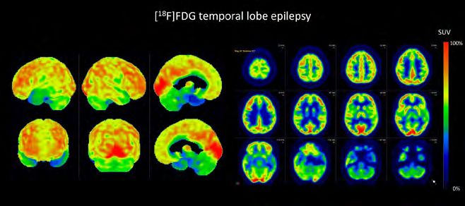
Table of contents
Foreword 4
Andrea Santos
Introduction 6
David Bogović, Luca Camoni, Christelle Terwinghe, Agata Pietrzak
Chapter 1 Hardware updates 8
Igor Iskra
Chapter 2 Updates in reconstruction algorithms 22
Dimitris Visvikis, Michel Koole, Ian Armstrong, Christian Vanhove
Chapter 3 Diagnostic CT in the oncological PET applications and protocols 32
Ivana Zagar
Chapter 4 PET/CT artefacts and pitfalls 46
Tyler Middlebrooks
Chapter 5 Advanced radiopharmaceuticals 58
Guy Bormans, Frederik Cleeren
Chapter 6 Advances in neuroimaging 76
Donatienne Van Weehaeghe
Chapter 7 Novelties in cardiac imaging 88
Elisabetta Cerudelli
Chapter 8 New solutions in the oncology 104
Andrej Doma
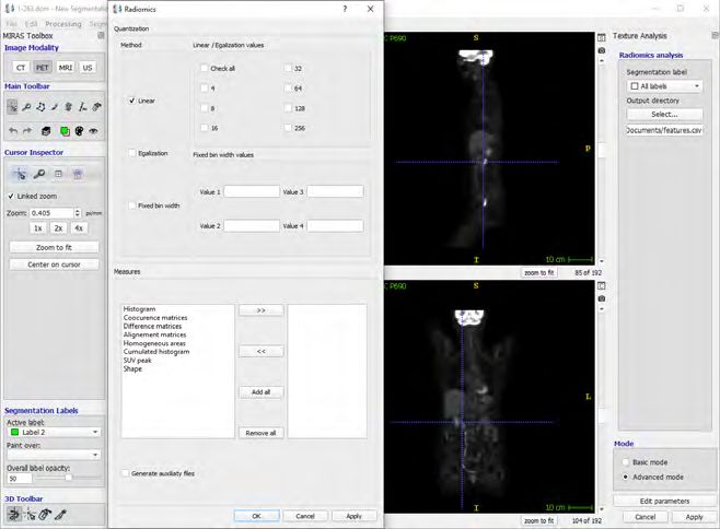
Chapter 9 Radiomics 122
Florent Tixier
Imprint 131
EANM TECHNOLOGISTS’ GUIDE 3
ADVANCES IN PET/CT IMAGING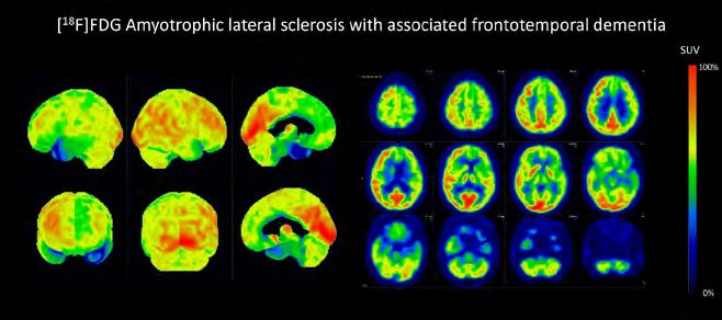
FOREWORD                                                                                                                                                         FOREWORD

    Foreword
    Positron emission tomography (PET) is one of the most promising areas
    of current medicine, considering the constant evolution of the technical
    equipment and the ongoing research into new and promising radiotracers
    for the study of human physiology and pathology. Since the advent of the
    hybrid imaging era, with computed tomography (CT) incorporated into
    the PET scanner, PET imaging has undergone a great evolution in terms of
    improved image quality and, consequently, better clinical output.

    Nuclear medicine technologists are              of the Technologists’ Guide, which           this project. A special thank you goes       to Angela Parker for her editing, reviewing
    the professionals when it comes to the          addressed the topic of hybrid imaging        to the Physics Committee of the EANM,        and support. Last but not least, I thank the
    imaging process.                                in conventional nuclear medicine, the        who have supported us throughout             EANM Board and Executive Office for their
      Acting as the link between the sciences       European Association of Nuclear Medicine’s   and made a valuable contribution to          tireless efforts, which have made this series
    that are the backbone of PET/CT imaging         Technologist Committee (EANM-TC) has         this Guide. I also gratefully acknowledge    of publications possible. Advances in PET/
    and the patient, we combine the necessary       kept this year’s focus on hybrid imaging.    the valuable content provided by our         CT Imaging was only possible thanks to
    theoretical background with practical skills    This time, however, we have embarked         overseas colleagues from the Society of      the contributions of everyone mentioned
    under the principle of best practice. To this   on the topic of PET-CT imaging, inviting     Nuclear Medicine and Molecular Imaging       above. My most sincere thanks to you all!
    end, an updated publication documenting         specialists in each specific area to write   – Technologist Section (SNMMI-TS). A
    the advances in this imaging technique is       about the advances in their field.           special word of appreciation goes to the
    of major importance in order to ensure            I should like to express my tremendous     editorial team who have dedicated their
    that everyone is working at the same level.     gratitude to all the authors who             time to this publication: Agata, Luca,         Andrea Santos
      Following on from last year’s edition         contributed their time and expertise to      Christelle and David – thank you! and also     Chair, EANM Technologists Committee

4       EANM TECHNOLOGISTS’ GUIDE                                                                                                                    EANM TECHNOLOGISTS’ GUIDE                5
        ADVANCES IN PET/CT IMAGING                                                                                                                 ADVANCES IN PET/CT IMAGING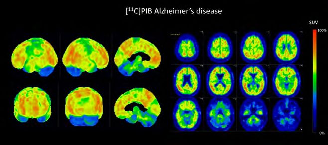
INTRODUCTION                                                                                                                                                   INTRODUCTION

    Introduction
    Every year, the Technologists Committee publishes a Technologists’
    Guide to develop and improve the nuclear medicine specialists’
    knowledge and personal skills in various aspects of nuclear medicine.
    This year, together with an interdisciplinary group of specialists,
    we decided to address the topic of “Advances in PET/CT Imaging”, outlining
    recent advancements in radioisotope imaging with a special emphasis on
    PET/CT techniques.

       PET/CT study allows us to evaluate the    the latest solutions that are improving         advantages, utilities and applications,         success of the process. We hope that this
    aetiology of various diseases, and has       technical and clinical study outcomes.          not forgetting the method’s limitations         latest edition of the Technologists’ Guide
    therefore become an essential element          This extensive guide encompassing             and ways of avoiding their impact on the        will be of benefit in helping our readers
    of diagnostic management. The rapidly        ten chapters provides the reader with           eventual PET/CT dataset.                        update their personal skillsets.
    evolving technology of medical imaging       a wide range of information: from                 We believe that hybrid imaging is not
    is permitting the constant improvement       hardware and software updates, PET/             only a set of techniques and properties,
    of existing solutions while simultaneously   CT artefacts and pitfalls through               but also and especially the interdisciplinary
    eliminating the method’s limitations.        radiopharmaceutical advancements to             and qualified medical team, who have
    Advances in PET/CT Imaging aims to           the latest solutions in oncology and the        a significant influence on diagnostic
    provide a technical and clinical overview    definition and applications of radiomics.       management. The complexity of PET/CT              David Bogović, Luca Camoni, Christelle
    of PET/CT principles and applications,       The contents guide readers through the          study means that the technician’s extensive     Terwinghe & Agata Pietrzak
    highlighting recent innovations and          long list of specific PET/CT characteristics,   knowledge and experience is vital to the          Editorial team

6      EANM TECHNOLOGISTS’ GUIDE                                                                                                                       EANM TECHNOLOGISTS’ GUIDE              7
       ADVANCES IN PET/CT IMAGING                                                                                                                    ADVANCES IN PET/CT IMAGING
H ARDWARE
U PDATES

by Igor Iskra

                1
CHAPTER 1                                                                                                                                                           CHAPTER 1

                       Positron emission tomography (PET) enables us to visualise molecular                         a transmission image was obtained using        array of crystal elements was coupled
                       processes in the human body. Certain positron-emitting radionuclides                         an external source of 68Ga and enabling        with four photomultiplier tubes (PMTs).

                                                                                                                                                                                                                    H AR D WAR E UPDATES
H AR D WAR E UPDATES

                       interact with specific molecular pathways, thus giving us the opportunity                    attenuation correction. In 1974, to reduce     The light produced in every scintillation
                       of imaging physiological or pathophysiological occurrences. PET scanners                     unwanted scattering effect, a hexagonal        event, caused by the interaction between
                       use the principle of coincidence detection of annihilation photons, a                        array of sodium iodide scintillation           the annihilation photon and the crystal
                       method in which two photons emerging from the same annihilation                              detectors was designed. This system            element, was shared among all four
                       process are detected on opposite sides of a detector system. Of course, like                 was heavily shielded with the assembly         photomultiplier tubes. The information
                       every diagnostic method, PET imaging has its own limitations and pitfalls,                   rotating around a transaxial plane, hence      from the detector thus contained the
                       scattering and attenuation of photons by the tissue being the two most                       being known as positron emission               coordinates of the scintillating element,
                       prominent ones. 1 2                                                                          transaxial tomography (PETT). This led         determined from the light distribution,
                                                                                                                    to a commercial version of this design,        and the energy of the incoming photon,
                          Coupling a PET scanner with a CT             patient’s exposure to radiation. The idea    known as the ECAT I (ECAT = emission           determined from the total amount of
                       scanner is an efficient way to correct          of using coincidence detection of positron   computerised axial tomograph).4 5              produced light. 1 4
                       most of the imperfections of PET imaging.       capture was born in the early 1950s. In         The next step towards the modern               Another problem in need of solving
                       “Low-dose” CT can greatly improve the           1953 a first paper was published on the      PET-CT scanner was a configuration with        was the small acceptance angle for
                       ability to obtain an exact anatomical           application of coincidence detection for     multi-ring geometry. This new design           coincidence photon detection due to
                       localisation    of      pathophysiological      localising brain tumours, describing a       allowed better usage of scanners for PET       the presence of the septa (2D mode).
                       processes in the patient. Furthermore, the      pair of sodium iodide detectors placed       quantification, but it also created some       Retracting or totally removing the septa
                       correlation between the intensity of focal      on opposite sides of the head. Then, in      new problems, mainly by reducing the           established a 3D mode of acquisition,
                       accumulation of radionuclide determined         1961, a ring of 32 sodium iodide-based       spatial resolution. First, there was the       improving sensitivity and enabling true
                       by the PET scanner and the degree of            coincidence detectors was developed          problem of size and spacing between the        3D reconstructions. The problem with the
                       morphological changes ascertained by            and several other designs for coincidence    detector elements. Another disadvantage        3D mode was an increased scatter fraction
                       the CT scanner is important in evaluating       detectors emerged in the subsequent          was the expensive and physically limiting      (35% or even more), but several scatter
                       the pathological potential of certain           years. 4                                     “one-to-one” coupling between the              correction models were subsequently
                       focal changes. Lastly, “low-dose” CT is            The first developmental breakthrough      scintillation crystal and the photodetector.   developed to compensate for this. 1
                       an irreplaceable tool for correcting the        came in the early 1970s, with the first      Lastly, the original designs included lead        The last and most important step
                       attenuation of annihilation photons by the      positron camera. Its unique design           or tungsten shields between the detector       towards modern PET-CT scanners was
                       tissue. 3                                       enabled smaller sodium iodide crystals       rings, which severely reduced the overall      the introduction of the CT scanner itself.
                                                                       to be coupled with fewer and larger          sensitivity. Nevertheless, the solutions       In the early days, a PET scanner was often
                                                                       photomultipliers, which resulted in          to these problems yielded new designs          equipped with some additional source of
                       HISTORICAL DEVELOPMENT OF                       reduced costs and improved spatial           which laid the foundations for modern          transmission scanning for the purpose of
                       PET-CT SCANNERS                                 resolution. Shortly after that, the idea     PET-CT scanners. 1 4                           attenuation correction, as well as for regular
                         Since the advent of the first PET scanners,   of a rotating positron camera emerged,          The problem of ineffective coupling         detector checks. 68Ge and 137Cs were the
                       the main developmental goals have been          producing multiple plain images              between the crystal and detector elements      main radionuclides used. As mentioned
                       to improve the sensitivity and spatio-          which were later back-projected onto         was overcome by introducing the block          before, the introduction of CT scanners in
                       temporal resolution and to reduce the           tomographic images. In every projection      detector concept. In this concept, an          PET acquisition not only enabled better

10                         EANM TECHNOLOGISTS’ GUIDE                                                                                                                      EANM TECHNOLOGISTS’ GUIDE                 11
                           ADVANCES IN PET/CT IMAGING                                                                                                                   ADVANCES IN PET/CT IMAGING
CHAPTER 1                                                                                                                                                                   CHAPTER 1

                       attenuation correction, but also paved           stopping power for 511 keV photons,              the real energy of the photons. Higher            orthosilicate (LYSO), a derivate of LSO with
                       the way for other advanced features. A           decay constant, light output and energy          energy resolution means that the energy           a small percentage of yttrium, is currently

                                                                                                                                                                                                                           H AR D WAR E UPDATES
H AR D WAR E UPDATES

                       main pitfall of the early CT scanners used       resolution.                                      variance is smaller, therefore photons            being used in the newest TOF-PET
                       in PET-CT acquisition was high radiation            Stopping power is described as the            whose energy is different from that of the        scanners since it has slightly better light
                       exposure, which was later remedied by the        inverse of the mean distance travelled           incident photons, i.e. Compton-scattered          output and energy resolution compared
                       advent of modern iterative reconstruction        by photons before they deposit energy            photons, will be less often detected. 1 7 8       to LSO.
                       techniques. 4 5 6                                in the crystal, and it is proportional to           Sodium iodide (NaI) was the first                 Instead     of    using     LSO,      some
                                                                        the density and effective atomic number          scintillator used in PET scanners. It is cheap    manufacturers opted to replace BSO
                                                                        of the material. Higher stopping power           and has the highest light output, but it has      with gadolinium orthosilicate (GSO),
                       MODERN PET-CT SCANNERS                           means that the electron will travel a            very low effective density and a very long        the main advantage of which is better
                          Although there are many specific              shorter distance in a material because           decay constant, so is no longer used today.       energy resolution. Other materials such
                       scanner designs, every PET scanner               it will interact more often with atoms in           Bismuth germinate (BGO) was the                as lanthanum (III) bromide (LaBr3) or LuAP
                       basically consists of three main parts:          the material, therefore indirectly enabling      main scintillator used in PET scanners            (lutetium aluminium perovskite, LuAlO₃:Ce)
                       scintillation detectors, photodetectors and      more effective detection of incident             throughout the 1980s and 1990s. It has            are currently being evaluated as possible
                       a computer. PET detectors are based on a         photons.                                         the highest known effective density of all        new scintillators in PET scanners.1 4 7 9
                       process called scintillation in which high-         The decay constant is determined by           scintillators, but its low light output and
                       energy photons interact with scintillating       the duration of the scintillation flash in the   long decay constant became a problem              Block detectors
                       crystals, creating a new electron either         crystal. A shorter decay constant means          with the widespread adoption of 3D                   As mentioned before, a block detector
                       by Compton scatter or by photoelectric           that the scintillation material will be able     acquisition mode. This resulted in a quest        setup was one of the major breakthroughs
                       absorption. This new electron loses its          to produce more individual scintillation         to find new scintillators with a shorter          in the development of modern PET
                       energy by passing through scintillation          flashes in a certain period of time, thus        decay constant, better light output and           scanners. Instead of using “one-to-one”
                       material and exciting new electrons. 7           allowing more incident photons to be             improved energy resolution.                       coupling between scintillators and
                          The excited electrons subsequently            counted.                                            Then came lutetium oxyorthosilicate            photodetectors, this concept proposed
                       return to their original energy state by            Light output can simply be described          (LSO), a component originally used for            using a block of scintillation crystals
                       releasing electromagnetic radiation in           as the yield of scintillation photons            nuclear well logging. It has a high light         divided into an array of single-functioning
                       the form of light. This light is registered by   produced by the incident photon. Higher          output and high efficient density, but            smaller elements, a few millimetres in
                       photodetectors and transformed into an           light output means that the incident             its main advantage is a very short decay          size. This was achieved by mechanical
                       electrical signal with the specific energy       photon will trigger the creation of more         constant. LSO’s superior time resolution          incision of channels in the primary crystal
                       and coordinates of the incident photon.          scintillation photons, thus increasing           has given PET scanners the ability to             block, with channels being filled with
                       All the signals are then processed by the        spatial and energy resolution.                   measure the time difference between               opaque material. This opaque material
                       computer, creating a final image.1 7                Finally, energy resolution is the ability     the arrivals of the two annihilation              prevents so-called “cross-talk”, i.e. a
                                                                        to accurately determine the energy of the        photons. This is called “time of flight” (TOF),   scatter of light between the elements. A
                       Scintillation crystals                           interacting photons. It depends on energy        and it provides otherwise unavailable             block is then usually coupled with four
                         Scintillation crystals are the most vital      variance, which is the ratio between             positioning information which enables             photomultiplier tubes. The sharing of
                       part of any detector. Every scintillation        the range of possible values of photon           us to determine an annihilation point to          light between the PMTs is converted to
                       material has four main characteristics:          energy determined by the detector and            within a few centimetres. Lutetium yttrium        their relative signal output, which is then

12                         EANM TECHNOLOGISTS’ GUIDE                                                                                                                              EANM TECHNOLOGISTS’ GUIDE                13
                           ADVANCES IN PET/CT IMAGING                                                                                                                           ADVANCES IN PET/CT IMAGING
CHAPTER 1                                                                                                                                                                  CHAPTER 1

                       used to determine the crystal element              analogue versions called avalanche                 exceptional light collection capability,      PMTs.12in terms of physical performance
                       involved in interaction with the photon.           photodiodes         (APDs)    and        silicon   which is due to their compact structure.      and technical features. Particular attention

                                                                                                                                                                                                                          H AR D WAR E UPDATES
H AR D WAR E UPDATES

                       There are also improved versions of block          photomultipliers (SiPMs). APDs, working in         Unlike the circular shape of PMTs, which      has been given to evaluate the whole-
                       detectors constructed from two different           Geiger mode, are generally not considered          was responsible for the significant gaps      body performance by sensitivity, spatial
                       scintillation materials with different decay       an effective choice for TOF-PET scanners           between them, SiMPs are square in shape,      resolution, dead time, noise equivalent
                       constants (photoswitch), enabling better           due to their significantly slower response         which allows them to be packed more           counting rate (NECR
                       determination of the depth of photon               time. Analogue SiPMs use single photon             tightly together. SiPMs mainly use “one-to-      An alternative type of scanner geometry
                       interaction with the detector material. 4 7        avalanche diode (SPAD) arrays to detect            one” coupling to crystals, which provides     is the “partial-ring” setup, in which two
                                                                          single scintillation photons, subsequently         a much higher count rate capability and       opposing matrices of crystal blocks
                       Photodetectors                                     turning all the generated pulses into one          improved spatial resolution.9                 rotate around the patient. They are not
                          Photomultiplier tubes were the most             analogue output signal. Like APDs, SiPMs                                                         perfectly opposed, thus achieving a
                       commonly used photodetectors in PET                initially didn’t have TOF capability.10 11         PET-CT scanner geometry                       better transversal field of view during
                       scanners. PMTs are glass-enveloped                    In recent years a new group of solid-              Most of the modern PET scanners have       the rotation, but their general sensitivity
                       vacuum tubes with three main parts: a              state detectors has emerged, called digital        “full-ring” geometry, with their detectors    is lower compared to “full-ring” scanners.
                       photocathode, dynodes and an anode.                photon counters (DPCs). In this version            covering a full 360° around the patient.      12
                                                                                                                                                                             in terms of physical performance and
                       Their functioning is based on the                  of detectors, every detector module                This setup provides optimum sensitivity,      technical features. Particular attention
                       photoelectric effect, in which an emission         consists of a 4 x 5 array of SiPM detector         fewer image artefacts due to tracer, organ    has been given to evaluate the whole-
                       of electrons is caused by interaction              tiles, additionally divided into a 4 × 4           or patient motion, and excludes the need      body performance by sensitivity, spatial
                       of light with photoelectric material. In           matrix of silicon sensor chips. Each chip          for additional calibrations since there are   resolution, dead time, noise equivalent
                       PMTs, the photoelectric component is               is a 2 × 2 matrix of digital photon counter        no moving components.5                        counting rate (NECR
                       called the photocathode. When electrons            detectors called silicon pixels. In every             Additionally, there are three different       Axially, PET scanners today consist of 2
                       are emitted from the photocathode,                 pixel there are 3200 SPADs or microcells,          types of detector surface structure. The      to 5 rings of detector elements, compared
                       induced by scintillation photons, they are         with every microcell functioning as an             first type is the common block detector       to several dozen in the earlier days. These
                       pulled towards the anode by the electric           individual detector. The total count of all        concept, in which there is a cylindrical      rings can be separated by thin annular
                       potential generated. On their way to the           photon–microcell interactions detected             assembly of crystal blocks in a ring          rings or septa made of photon-absorbing
                       anode, electrons collide with the dynodes,         in a certain interval of time, initiated by        structure several blocks deep. The second     material like tungsten and used for
                       creating secondary electron emissions              the arrival of the very first scintillation        option is a curved crystal structure, in      collimation. The presence or absence of
                       from the collision. The resulting electrical       photon, is translated into a digital number        which 6 bigger blocks of NaI crystals,        septa determines the type of acquisition.
                       current is proportional to the number of           proportional to the energy of the photon           coupled with 48 PMTs each, are placed side    If septa are present, then so-called “2D
                       initial scintillation photons, and is translated   that induced the scintillation event. PET          by side to achieve ring geometry. Finally,    acquisition” is performed, since the septa
                       into an electrical signal. PMTs are therefore      scanners equipped with DPCs show                   there is the PIXELAR module, consisting       shield “out of plane” coincidence photons.
                       considered as analogue photodetectors,             better image quality, improved lesion              of a curved matrix made up of 628 (22 x       Since the septa block a fairly large number
                       their main advantage being their lower             detectability, and are reported to perform         29) GSO crystals attached to a continuous     of true coincidences from ever reaching
                       sensitivity to temperature variations. 1 10        faster acquisitions with lower radiation           light guide, in which ring geometry is        the detector, this type of acquisition is
                          The development of modern solid-                exposure.11                                        achieved by placing 28 of these modules       characterised by reduced scattered and
                       state detectors started in the 1990s with             The main advantage of SiPMs is their            side by side and coupling them with 420       random coincidences, but it also has lower

14                         EANM TECHNOLOGISTS’ GUIDE                                                                                                                              EANM TECHNOLOGISTS’ GUIDE               15
                           ADVANCES IN PET/CT IMAGING                                                                                                                           ADVANCES IN PET/CT IMAGING
CHAPTER 1                                                                                                                                                              CHAPTER 1

                       sensitivity. Another type of acquisition is   on the vendor, although the basic                - by measuring the changes in air             because each gate can mix several tissue
                       so-called “full 3D acquisition”, performed    elements of design are largely similar. PET   temperature during respiration, either with      positions. Researchers have suggested

                                                                                                                                                                                                                        H AR D WAR E UPDATES
H AR D WAR E UPDATES

                       with or without retracted septa. In this      scanners have “full-ring” geometry and are    a sensor placed inside an oxygenation            either selecting PET events from gated
                       situation coincidences are accepted           integrated with CT scanners in a single       mask or with a sensor placed close to the        acquisitions or performing several PET
                       from all angles, thus providing higher        device. The patient is positioned only        patient’s nostrils: this mode of respiratory     acquisitions (corresponding to a breath-
                       sensitivity. However, that also implies an    once and the scans are acquired in close      gating uses the fact that the air is warmer      hold CT position
                       increase in random coincidences, higher       sequence, enabling improved registration      during exhalation;                                  Cardiac PET-CT scanners also use
                       scatter fraction and loss of events due to    accuracy and a single integrated scan. 4 5       - by using a spirometer placed close to       an external ECG device for gating and
                       an increased count rate, and subsequently                                                   the patient’s nostrils or mouth: this mode       detecting certain phases of the cardiac
                       increased dead time. It is worth mentioning   Additional hardware for PET-CT studies        of respiratory gating has not yet been used      cycle, in the same way as in gSPECT.
                       that today’s commercial PET scanners are         FDG PET-CT has an important role           in clinical practice.
                       not equipped with septa and thus acquire      in evaluation of pulmonary lesions               It is worth mentioning that nowadays,
                       data in this second acquisition mode. 14      and detection of occult intrathoracic         in view of the significant practical             CT SCANNER IMPROVEMENTS
                       12
                         in terms of physical performance and        metastases. In contrast to CT, which          limitations of hardware-based respiratory           Over the years, the two main goals of CT
                       technical features. Particular attention      covers only a part of one breathing           gating, the emphasis is on software              scanner development have been better
                       has been given to evaluate the whole-         cycle, PET acquisition lasts substantially    gating, Bayesian penalised likelihood            image resolution and lower radiation
                       body performance by sensitivity, spatial      longer through multiple breathing cycles.     (BPL) PET reconstruction, and texture            exposure to the patient. Alongside
                       resolution, dead time, noise equivalent       Respiratory motion can thus cause a lot       analysis. 13 14follow-up, and treatment          better software solutions and image
                       counting rate (NECR).                         of motion and attenuation correction          planning. For cancers located in the thorax      reconstruction algorithms, upgrades in
                                                                     artefacts on PET images. To eliminate         or abdomen, the patient’s breathing              hardware have been the main drivers of CT
                       PET-CT coupling                               this negative effect, respiratory gating is   causes artifacts and errors in PET and CT        scanner improvement. Nowadays, PET-CT
                          The first PET-CT scanner prototype         performed. In this process, data collected    images. Many different approaches for            systems are equipped with up to 128-slice
                       emerged in 1998. The PET scanner was          in each respiratory cycle is divided into     artifact avoidance or correction have            CT scanners, compared to the 16-slice
                       placed on the rear end of the CT scanner,     “bins”, either by detecting a certain         been developed; most are based on                scanners in use as little as 15 years ago. 4 5 15
                       creating a single integrated assembly. The    respiratory amplitude (amplitude-based        gated acquisition and synchronization               One other important area of
                       scanners rotated together at 30 rpm and       gating) or a phase of the respiratory cycle   between the respiratory signal and PET           improvement is the development of new
                       the patient bed could be moved axially        (phase-based gating).                         acquisition. The respiratory signal is usually   acquisition modes. Conventional PET-CT
                       from the CT to the PET scanner so the            Gating is usually performed using          produced by an external sensor that tracks       studies are performed in so-called “step-
                       corresponding images were accurately          external sensors which track respiratory      a physiological characteristic related to the    and-shoot” mode, in which the patient bed
                       registered. However, the acquisition,         motion. This can be accomplished in           patient’s breathing. Respiratory gating is       is moved between every step of image
                       reconstruction and operating systems          several ways:                                 a compensation technique in which time           acquisition outside the initial axial FOV
                       were separate and image fusion was               - by monitoring the displacement of the    or amplitude binning is used to exclude          range. An alternative mode is “continuous
                       performed by software. 5                      abdominal wall using an elastic chest belt;   the motion in reconstructed PET images.          table motion” mode, in which the patient
                          Nowadays, PET and CT scanners are             - by monitoring the displacement of        Although this technique is performed             table moves continuously throughout the
                       coupled through hardware fusion. The          two infrared markers positioned on the        in routine clinical practice, it fails to        whole scanning process. Data is acquired
                       characteristics of scanners vary depending    patient’s thorax;                             adequately correct for respiratory motion        uninterruptedly while the patient is being

16                         EANM TECHNOLOGISTS’ GUIDE                                                                                                                       EANM TECHNOLOGISTS’ GUIDE                    17
                           ADVANCES IN PET/CT IMAGING                                                                                                                    ADVANCES IN PET/CT IMAGING
CHAPTER 1                                                                                                                                                                                 CHAPTER 1

                       moved through the gantry, resulting in          effectively for this type of detection. 9        components and ambiguities regarding                        CONCLUSION
                       increased uniformity and sensitivity of            Monolithic       detector.    One      thin   the indications for whole-body scans,                         Modern PET-CT scanners are currently

                                                                                                                                                                                                                                   H AR D WAR E UPDATES
H AR D WAR E UPDATES

                       acquisition. 16                                 block of crystal coupled with multiple           which are still unclear. 17 18                              undergoing rapid development. New
                                                                       photodetectors can improve light                                                                             designs and software improvements
                                                                       transduction because of the larger crystal                                                                   are aimed at ensuring better image
                       FUTURE INNOVATIONS                              volume and absence of inter-crystal                                                                          resolution with lower radiation exposure
                          Improved ratio of deposited energy           boundaries. 9                                                                                                to the patient. New detector technologies
                       to light output of scintillators. Current                                                                                                                    using digital photon counters are set
                       Lu-based scintillation crystals have a                                                                                                                       to significantly improve the acquisition
                       somewhat non-linear light output in             EXPLORER – A WHOLE-BODY                                                                                      process, enabling faster and more reliable
                       response to the energy deposited by the         PET-CT SCANNER                                                                                               diagnostics.
                       interacting photon. In particular, this light      Current PET-CT scanners only capture a
                       yield ratio decreases with decreasing           small portion of the patient’s body inside
                       energy of photons. LuAP (lutetium               the field of view (FOV). What is more, only                                                  VEREOS           BIOGRAPH VI-
                                                                                                                                                                    (PHILLIPS        SION 600                      DISCOVERY MI
                       aluminium perovskite, LuAlO3) and LuYAP         3–5% of the available signal is detected by      PET-CT SCANNER         EXPLORER
                                                                                                                                                                    HEALTH-          (SIEMENS                    (GE HEALTHCARE)
                       (lutetium-yttrium aluminium perovskite)         scanners. These are the two main reasons                                                     CARE)            HEALTHINEERS)

                       scintillator crystals have a superior light     for the poor sensitivity of current whole-
                       yield ratio and hence superior energy           body PET-CT scanners.
                                                                                                                        crystals               LYSO                 LYSO             LSO                   LYSO
                       resolution. 9                                      EXPLORER is a PET-CT scanner prototype
                          Semiconductor        detectors.     These    which can perform a whole-body PET scan
                       non-scintillating detectors use dense           up to 40 times better than the currently                                2.76 x 2.76 x 18.1   3.86 × 3.86 ×
                                                                                                                        crystal size                                                 3.2 x 3.2 x 20 mm3    3.95 × 5.3 × 25 mm3
                       crystals which are capable of directly          available scanners. It has 40 rings and is                              mm3                  19 mm3
                       absorbing the interacting photon energy         able to scan the whole body within 30
                       and converting it into electrical charge        seconds. Its 564,480 LYSO crystals, 2.76 x       photon detectors       SiPM                 SiPM             SiPM                  SiPM
                       detected by previously placed electrodes.       2.76 x 18.1 mm each, are arranged in 7 x
                       The materials used in these detectors are       6 arrays and coupled with SiPM. This PET
                       CdZnTe (CZT) and TlBr. 9                        scanner has a diameter of 78.6 cm and an         number of crystals     564 480              23 040           60 800                19 584
                          Using Cherenkov light to improve time        axial length of 194 cm. EXPLORER is also
                       resolution. Cherenkov light is produced         equipped with an 80-row, 160-slice CT
                       instantaneously when a 511 keV photon           scanner.                                         number of detectors    53 760               23 040           304                   9 792
                       interacts with detector material, emitting         This setup allows EXPLORER to
                       a small number of transient, high-energy,       significantly increase acquisition sensitivity
                                                                       in whole-body scans and to produce more          CT slices              160                  128              128                   128
                       faster-than-light electrons. Lead-based
                       crystals, such as PbF2, show an excellent       reliable images along with rapid scanning
                       time resolution of 70–90 ps. Also, it seems     and lower radiation exposure. However,
                       that BGO scintillators could be used            its main disadvantages are expensive                Table 1. Comparison of some of the currently available PET-CT scanners 11 17 19 20 21

18                         EANM TECHNOLOGISTS’ GUIDE                                                                                                                                          EANM TECHNOLOGISTS’ GUIDE            19
                           ADVANCES IN PET/CT IMAGING                                                                                                                                       ADVANCES IN PET/CT IMAGING
CHAPTER 1
REFERENCES 2018;91.
1. Schmitz RE, Alessio AM, Kinahan PE. 1 The 14. Pépin A, Daouk J, Bailly P, Hapdey S, Meyer
ME. Management of respiratory motion in PET/
H AR D WAR E UPDATES
Physics of PET/CT Scanners. PET PET/CT. Published
online 2019:1-16. doi:10.1055/b-0039-166611 computed tomography: The state of the art. Nucl
2. Shukla AK, Kumat U. Positron emission Med Commun. 2014;35(2):113-122. doi:10.1097/
tomography: An overview. J Med Phys. 2006;31(1):13- MNM.0000000000000048
21. 15. Mccollough CH, Chen GH, Kalender W, et al.
3. Vaquero JJ, Kinahan P. Positron Emission Achieving routine submillisievert CT Scanning : Report
Tomography: Current Challenges and Opportunities from the Summit on Management of Radiation Dose.
for Technological Advances in Clinical and Preclinical Radiology. 2012;264(2):567-580.
Imaging Systems. Annu Rev Biomed Eng. 2015;17:385- 16. Rausch I, Cal-gonzález J, Dapra D, et al.
414. Performance evaluation of the Biograph mCT Flow
4. Jones T, Townsend D. History and future PET / CT system according to the NEMA NU2-2012
technical innovation in positron emission tomography. standard. EJNMMI Phys. 2015;2(26). doi:10.1186/
J Med Imaging. 2017;4(1):011013. doi:10.1117/1. s40658-015-0132-1
jmi.4.1.011013 17. Cherry SR, Jones T, Karp JS, Qi J, Moses WW,
5. Townsend DW, Carney JPJ, Yap JT, Hall NC. Badawi RD. Total-Body PET: Maximizing Sensitivity to
PET / CT Today and Tomorrow. 2019;45(1):4-15. Create New Opportunities for Clinical Research and
6. Alessio AM, Kinahan PE, Cheng PM, Vesselle Patient Care. J Nucl Med. 2018;59(1):3-12. doi:10.2967/
H, Karp JS. PET/CT scanner instrumentation, challenges, jnumed.116.184028
and solutions. Radiol Clin North Am. 2004;42(6):1017- 18. EXPLORER PET-CT scanner: a total
1032. doi:10.1016/j.rcl.2004.08.001 body experience. Accessed March 5, 2021. https://
7. Authors G of. PET/CT- Klinička Primjena. openmedscience.com/explorer-pet-ct-scanner-a-
Markulin d.o.o.; 2015. total-body-experience/#:~:text=EXPLORER PET%2FCT
8. Kapoor V, McCook BM, Torok FS. An scanner design and features,-Design&text=The
Introduction to PET-CT Imaging. RadioGraphics. EXPLORER combines positron emission,images with
2004;24:523-543. high temporal resolution.&text=40 Rings are present
9. Berg E, Cherry SR. Innovations in in,inclu
instrumentation for positron emission tomography. 19. Oddstig J, Leide Svegborn S, Almquist H, et
Semin Nucl Med. 2018;48(4):311-331. al. Comparison of conventional and Si-photomultiplier-
10. Spanoudaki VC, Levin CS. Photo-Detectors based PET systems for image quality and diagnostic
for Time of Flight Positron Emission Tomography (ToF- performance. BMC Med Imaging. 2019;19(1):1-9.
PET). Sensors. 2010;10:10484-10505. doi:10.1186/s12880-019-0377-6
11. Zhang J, Maniawski P, Knopp M V. 20. No Title. Accessed March 10, 2021. https://
Performance evaluation of the next generation solid- www.philips.co.uk/c-dam/b2bhc/gb/resource -
state digital photon counting PET/CT system. EJNMMI catalog/landing/brightontender/vereos-petct-
Res. 2018;8(97). product-specification-lr.pdf
12. Tarantola G, Zito F, Gerundini P. PET 21. No Title. Accessed March 10, 2021. https://
Instrumentation and Reconstruction Algorithms in www.siemens-healthineers.com/en-us/molecular-
Whole-Body Applications Continuing Education. J imaging/pet-ct/biograph-vision
Nucl Med. 2003;44(5):756-769. http://www.snm.org/
education/ce_online.html
13. Frood R, McDermott G, Scarsbrook A.
Respiratory-gated PET/CT for pulmonary lesion
characterisation—promises and problems. Br J Radiol.
20 EANM TECHNOLOGISTS’ GUIDE
ADVANCES IN PET/CT IMAGINGUPDATES IN
RECONSTRUCTION
ALGORITHMS
by Dimitris Visvikis
Michel Koole
Ian Armstrong
Christian Vanhove
2CHAPTER 2 CHAPTER 2
Image reconstruction generates a volume of image data that reflects the used to reconstruct the 3D distribution of LOR (see Figure 1a), while in TOF-PET it
distribution and quantities of the injected radiopharmaceutical within the PET tracer in the body of the patient. depends on the time difference between
UPDAT ES IN R ECO N S TR UC TIO N ALGO R ITH MS
UPDATES IN R ECO N S TR UC TIO N ALGO R ITH MS
the patient. A reconstructed image typically consists of a 3-dimensional the arrival of two coincidence photons,
array of discrete elements referred to as voxels. When all appropriate as well as detector characteristics, and
corrections have been applied, the value for each voxel typically indicates TIME OF FLIGHT (TOF) it is applied as a Gaussian distribution
the radioactive concentration of the radiopharmaceutical within this voxel In TOF-PET the difference in time on the LOR (see Figure 1b). The centre
volume in units of Bq/ml. Since reconstruction starts from the raw or between the coincidence photons’ arrival of this Gaussian distribution along the
acquired PET data, a short overview of the process for PET data acquisition is measured and stored. For list-mode LOR is the length equivalent of the time
is given below. acquisition the time difference (Δt) is difference between the arrival of the two
stored event by event, while in sinogram coincidence photons, computed as s = c ·
mode the sinogram is divided into several Δt, with c representing the speed of light.
PET DATA ACQUISITION of a transaxial section through the patient. TOF-bins, each corresponding to a time The full width at half maximum (FWHM) of
For PET imaging, positron-emitting This matrix is called a sinogram, since all difference. In conventional PET image the Gaussian distribution is related to the
radioisotopes are required. When the LORs passing through a point at different reconstruction, the probability of an time resolution of the detector.
positron is emitted, it travels for a few angles correspond to a sinusoid curve. This annihilation event is constant along the
millimetres through the body until it also explains the term sinogram. However,
interacts with a free electron, resulting in before the detected events are stored in a
an annihilation event which produces two sinogram, coincidence events can also be
back-to-back gamma photons with energy stored in a list as individual events in the
of 511 keV each. These photons are emitted order in which they are detected (1). This
in almost opposite directions, defining a is called list mode, but it should be noted
line along which the annihilation event that list-mode acquisition is not always
must have occurred. Since PET is based available on the PET system, or it needs to
on the coincident detection of these two be deliberately selected as the acquisition
opposite gamma photons along this line mode. List-mode acquisition provides
of response (LOR), a pair of detectors is more flexibility, however, as additional
needed. Clinical PET systems use a full information such as photon energy,
ring of detectors surrounding the patient, detection timing and detection location
which also explains the superior sensitivity can be stored as well (2). Based on this
of PET. If the two back-to-back gamma information, list-mode data can then be
photons are detected within a certain short used to generate sinograms. In addition,
time window (typically 4 ns depending on list-mode data also allow for retrospective
the PET system settings), it is assumed that rebinning of dynamic raw PET data into
they originate from the same annihilation sinograms corresponding to different time
event. These coincidence events are stored frames, where the frame duration can be
in a matrix which represents the number chosen after data acquisition is completed.
of detected events along different angles Once generated, sinogram data are Figure 1: Probability distributions during (a). conventional image reconstruction, (b). TOF-PET image
reconstruction
24 EANM TECHNOLOGISTS’ GUIDE EANM TECHNOLOGISTS’ GUIDE 25
ADVANCES IN PET/CT IMAGING ADVANCES IN PET/CT IMAGINGCHAPTER 2 CHAPTER 2
RECONSTRUCTION takes into account the Poisson statistics An accelerated variant of MLEM, ordered convert a measured count rate to activity
After acquisition of PET data, the next of radioactive decay and the statistical subsets expectation maximisation (OSEM) concentration, and the attenuation and
UPDAT ES IN R ECO N S TR UC TIO N ALGO R ITH MS
UPDATES IN R ECO N S TR UC TIO N ALGO R ITH MS
step is to reconstruct an estimate of the in nature of the measurement process. Since (5), is currently the most commonly used time-of-flight differences of the two 511
vivo tracer distribution. There are different this approach is iterative, it starts from an iterative reconstruction algorithm in PET keV gamma rays. The exceptions are
approaches to reconstruct the PET data. initial estimate and updates the previous clinical imaging. OSEM accelerates the corrections for scattered and random
On the one hand, there is an analytical estimate of the reconstructed image until convergence of MLEM by performing coincidences; these are typically added
approach with filtered back-projection the new estimate is very close the previous updates based on part of the measured on to the estimated sinograms that are
(FBP), which consists in back- projecting one, meaning that the reconstruction is data only, so that it approximates the generated using the forward projection.
the sinogram data across the imaging no longer improved and convergence MLEM solution but in a much shorter Since accurate modelling of the
matrix. However, simple back-projection is reached. To update the previous computation time and with less memory PET system response during iterative
of the measured data results in a blurred estimate, iterative reconstruction utilises requirement. This technique divides reconstruction results in a reconstructed
version of the original image. The blurring is a probability matrix, commonly referred the measured PET data into several PET image with much better quantitative
minimised by applying a filter to sinogram to as the system matrix. This matrix defines subsets, following a specific order. During properties and homogeneous resolution,
data before back-projection to amplify the probability that a 511 keV gamma-ray reconstruction, each update is based iterative reconstruction provides clear
the high frequencies representing fast- pair originating from a particular voxel on the projection data belonging to a advantages over the older method of FBP
varying intensity changes such as edges, location within the reconstructed image specific subset only, while a different reconstruction. However, it is not without
hence the name filtered back-projection. would give rise to a particular line of subset is selected for the next update. This disadvantages. Due to computational
Although FBP is a very fast technique with response in a sinogram. An initial estimate accelerated convergence using subsets limitations, the system matrix is unlikely
limited memory requirements, it does not of the reconstructed image is created allows the OSEM algorithm to generate to perfectly model the system response
take into account the statistical nature and, from here, estimated sinograms are PET images in clinically relevant times for the forward and back projection
of radioactive decay, nor does it make generated from the system matrix values – (6). Unlike MLEM, where one iteration operations. This may place data in
allowance for the characteristics of the PET a process referred to as forward projection. corresponds to one update, one OSEM erroneous locations, which manifests as
system (3). These estimated sinograms are compared iteration corresponds to a cycle in which noise. What is more, PET measurements are
For these reasons, along with the general with the acquired sinograms and a ratio all subsets have been used to update generally very noisy. This in combination
increase in memory and computation is calculated at each sinogram pixel. The the reconstruction. In OSEM, the number with the discrete spatial sampling of
power, FBP is no longer the reconstruction ratios are then transposed back into the of updates therefore corresponds to the both sinograms and the reconstructed
technique of choice, and iterative reconstructed image – a process referred number of iterations multiplied by the image volume means that the process is
methods represent the current state of to as back-projection – and the estimate of number of subsets. ill-conditioned, the main consequence
the art in clinical PET reconstruction. The the reconstructed image is updated. This The system matrix is the critical being that the noise in the reconstructed
most widely-used iterative reconstruction process repeats in a series of loops referred component in iterative reconstruction, image increases as more iterations are
algorithm is maximum likelihood to as iterations, and the estimate of the as this is where most of the physical performed. Noise in an image that is fully
expectation maximisation (MLEM) (4). The activity distribution in the reconstruction aspects of the PET measurement are converged will be so significant that the
principle behind MLEM is to estimate the image converges until it represents the modelled. This includes modelling of image is unlikely to be of any clinical use.
activity distribution that would most likely real activity distribution within the patient. the geometric efficiency of the PET There are three mitigating strategies to
produce the sinograms that have been In MLEM, the number of updates also detector, the position-dependent spatial reduce noise in reconstructed images. The
acquired by the PET system. As such, it corresponds to the number of iterations. resolution, the calibration factor used to first strategy is to stop the reconstruction
26 EANM TECHNOLOGISTS’ GUIDE EANM TECHNOLOGISTS’ GUIDE 27
ADVANCES IN PET/CT IMAGING ADVANCES IN PET/CT IMAGINGCHAPTER 2 CHAPTER 2
process before convergence in order amplification in the image by penalising
to prevent excessive image noise highly varying activity values between
UPDAT ES IN R ECO N S TR UC TIO N ALGO R ITH MS
UPDATES IN R ECO N S TR UC TIO N ALGO R ITH MS
amplification. Although the degree of neighbouring voxels. More recently, the Figure 2. Partial volume
noise is kept to an acceptable level, approach was introduced as a clinical effects. On the left there
this approach can produce an image reconstruction approach which makes are no partial volume
with reduced quantitative properties as use of a prior relative difference to effects, resulting in sharp
convergence is not fully reached and not reduce noise amplification during image representation of a lesion.
all necessary corrections are fully applied. reconstruction (9). Basically, it penalises On the right, partial volume
The second approach is to perform a small relative differences between effects due to the limited
sufficient number of updates to ensure neighbouring activity values as these spatial resolution of a PET
that the full image is close to convergence are assumed to be noise, while larger system cause signal spread.
and all appropriate corrections have been differences are attributed to edges and are
applied. A filter is then applied to the therefore preserved. Unlike a conventional A limited spatial resolution leads to partial small tumour lesions (typically < 15mm)
reconstructed PET image to reduce the OSEM reconstruction, which needs the volume effects, which, as pointed out by will look larger but less aggressive in PET
noise levels (7). A Gaussian full width half number of iterations and subsets to be Soret et al (11, 12), are a major confounding imaging because partial volume effects
maximum (FWHM) filter is generally used specified, a BPL reconstruction only needs factor in PET imaging that cannot be will spread the signal of a small hot lesion,
for this purpose. The FWHM determines one tuneable parameter to be set which ignored because it can severely affect resulting in voxel values that will be lower
how many neighbouring voxels are used controls the strength of the penalty to image quality and image quantification. than the actual maximum value (Figure 2).
to calculate the smoothed image, so that preserve edges. Furthermore, no filter for Partial volume effects typically refer to Because these voxel values (i.e. radioactive
a larger FWHM corresponds to a higher post-smoothing the reconstructed image two different phenomena: (1) the image concentration) are often used to follow up
degree of smoothing. and increasing image quality is necessary blurring introduced by the finite spatial treatment response, these partial volume
The final approach is to constrain as full convergence is reached without resolution of the PET imaging system, effects can lead to misinterpretation of the
the amplification of noise during the increasing noise (10). and (2) image sampling into voxels so measured data. Furthermore, the apparent
reconstruction process using the that individual voxels contain different larger lesion size can lead to problems
maximum a posteriori approach (8). This tissue types where the measured voxel when lesion contouring is derived from
approach basically assumes that not all RESOLUTION MODELLING intensity is mostly a weighted average the PET data, resulting in a lesion that is
reconstructions are equally likely, but that In recent years, there have been multiple of different tissue intensities included in larger than the real metabolically active
prior assumptions can be made about hardware and software advances in PET/ each voxel (13). It is important to note lesion. The spatial resolution in the
the reconstructed image. These prior CT to improve image quality, small lesion that even imaging systems with perfect reconstructed images determines how far
assumptions correspond to a penalty detectability and quantification accuracy. spatial resolution would still be hampered the signal of small hot lesions spreads. A
term which is taken into account when However, the two main limitations of PET by partial volume effects due to image better spatial resolution will result in less
updating the image – a process referred compared to CT and MRI are its relatively sampling into voxels. For this reason, partial spread, whereas a lower spatial resolution
to as penalisation or regularisation, which low spatial resolution and generally low volume effects not only affect PET, but are will introduce a larger amount of spread.
results in a Bayesian penalised likelihood signal-to-noise ratio. In this section we also of concern in image modalities with Consequently, a better spatial resolution
(BPL) reconstruction algorithm. In general, will focus on advances in reconstruction better spatial resolution, such as MRI or CT will result in fewer partial volume effects.
this penalty term is used to reduce noise algorithms to improve spatial resolution. (11). As a result of partial volume effects,
28 EANM TECHNOLOGISTS’ GUIDE EANM TECHNOLOGISTS’ GUIDE 29
ADVANCES IN PET/CT IMAGING ADVANCES IN PET/CT IMAGINGCHAPTER 2 CHAPTER 2
REFERENCES 14. Moses WW. Fundamental Limits of Spatial
1. Matej S, Karp JS, Lewitt RM, Becher AJ. Resolution in PET. Nucl Instrum Methods Phys Res A.
Performance of the Fourier rebinning algorithm for 2011;648 Supplement 1:S236-s40.
The spatial resolution in PET is partly The application of resolution recovery 15. Jaskowiak CJ, Bianco JA, Perlman SB, Fine
PET with large acceptance angles. Phys Med Biol.
determined by physical effects such as in clinical PET/CT imaging is very JP. Influence of reconstruction iterations on 18F-FDG
UPDAT ES IN R ECO N S TR UC TIO N ALGO R ITH MS
UPDATES IN R ECO N S TR UC TIO N ALGO R ITH MS
1998;43(4):787-95.
crystal size, positron travel, non-collinearity attractive, since it can improve diagnostic 2. Colsher JG. Fully three-dimensional positron PET/CT standardized uptake values. J Nucl Med.
and crystal depth of interaction (14), but it performance. It has been reported that emission tomography. Phys Med Biol. 1980;25(1):103- 2005;46(3):424-8.
15. 16. Rahmim A, Qi J, Sossi V. Resolution modeling
is also important to note that the spatial resolution modelling in PET leads to higher in PET imaging: theory, practice, benefits, and pitfalls.
3. Pelt DM, Batenburg KJ. Improving filtered
resolution in the reconstructed images and more uniform spatial resolution across backprojection reconstruction by data-dependent Med Phys. 2013;40(6):064301.
can be controlled to some extent by a the transaxial field of view, which can be filtering. IEEE Trans Image Process. 2014;23(11):4750-62. 17. Alessio AM, Kinahan PE, Lewellen TK, editors.
Modeling and incorporation of system response
good choice of reconstruction parameters, beneficial in improving the detection of 4. Shepp LA, Vardi Y. Maximum likelihood
reconstruction for emission tomography. IEEE Trans functions in 3D whole body PET. IEEE Symposium
such as number of iterations and subsets small lesions (20, 21). Although resolution Conference Record Nuclear Science 2004; 2004 16-22
Med Imaging. 1982;1(2):113-22.
(15). If the number of iterations is too low, modelling was primarily developed and 5. Hudson HM, Larkin RS. Accelerated image Oct. 2004.
the spatial resolution in the reconstructed evaluated for 18F-FDG imaging, it has reconstruction using ordered subsets of projection 18. Alessio AM, Kinahan PE, Lewellen TK.
Modeling and incorporation of system response
images will be reduced, while increasing also been demonstrated to improve data. IEEE Trans Med Imaging. 1994;13(4):601-9.
6. Lantos J, Mittra ES, Levin CS, Iagaru functions in 3-D whole body PET. IEEE Trans Med
the number of iterations improves the the detectability of small lesions using Imaging. 2006;25(7):828-37.
A. Standard OSEM vs. regularized PET image
spatial resolution in the reconstructed 68
Ga-based tracers (20). Recently, the reconstruction: qualitative and quantitative 19. Panin VY, Kehren F, Michel C, Casey M. Fully
images. A sufficient number of iterations impact of resolution recovery on PET/ comparison using phantom data and various clinical 3-D PET reconstruction with system matrix derived
from point source measurements. IEEE Transactions on
therefore needs to be used to obtain CT scanners was investigated by multi- radiopharmaceuticals. Am J Nucl Med Mol Imaging.
2018;8(2):110-8. Medical Imaging. 2006;25(7):907-21.
maximum spatial resolution in the centric comparison of the recovery 20. van der Vos CS, Koopman D, Rijnsdorp
7. Boellaard R, Krak NC, Hoekstra OS,
reconstructed images. coefficient using a 68Ge phantom (22). Lammertsma AA. Effects of noise, image resolution, S, Arends AJ, Boellaard R, van Dalen JA, et al.
To further improve spatial resolution in The authors concluded that resolution and ROI definition on the accuracy of standard uptake Quantification, improvement, and harmonization of
values: a simulation study. J Nucl Med. 2004;45(9):1519- small lesion detection with state-of-the-art PET. Eur J
PET, different vendors have considered modelling provides a noticeable gain in Nucl Med Mol Imaging. 2017;44(Suppl 1):4-16.
27.
the idea of correcting for the physical activity recovery compared to traditional 21. Rogasch JM, Steffen IG, Hofheinz F, Großer
8. Sangtae A, Fessler JA. Globally convergent
effects that degrade the spatial iterative reconstruction algorithms. image reconstruction for emission tomography using OS, Furth C, Mohnike K, et al. The association of tumor-
resolution in PET (16-18). These resolution However, the recovery gain provided by relaxed ordered subsets algorithms. IEEE Transactions to-background ratios and SUVmax deviations related
on Medical Imaging. 2003;22(5):613-26. to point spread function and time-of-flight F18-FDG-
modelling algorithms incorporate the advanced reconstruction algorithms PET/CT reconstruction in colorectal liver metastases.
9. Nuyts J, Beque D, Dupont P, Mortelmans
a-priori knowledge of the PET/CT system vanishes on the smaller spheres (6 mm L. A concave prior penalizing relative differences for EJNMMI Res. 2015;5:31.
during the reconstruction process. In and 10 mm diameter) where it is of greater maximum-a-posteriori reconstruction in emission 22. Chauvie S, Bergesio F, De Ponti E, Morzenti
S, De Maggi A, Ragazzoni M, et al. The impact of time-
these algorithms, the so-called point importance. This study also demonstrates tomography. IEEE Transactions on Nuclear Science.
2002;49(1):56-60. of-flight, resolution recovery, and noise modelling in
spread function is modelled during the that resolution modelling has a significant reconstruction algorithms in non-solid-state detectors
10. Tong S, Alessio AM, Kinahan PE. Image
reconstruction process to correct for impact on quantification, which highlights reconstruction for PET/CT scanners: past achievements PET/CT scanners: - multi-centric comparison of activity
image degrading effects such as crystal the need for harmonisation across and future challenges. Imaging Med. 2010;2(5):529-45. recovery in a 68Ge phantom. Phys Med. 2020;75:85-91.
23. Armstrong IS, Thomson KE, Rowley LM, Mc-
size, positron travel, non-collinearity and multicentre networks or collaborations 11. Soret M, Bacharach SL, Buvat I. Partial-
volume effect in PET tumor imaging. J Nucl Med. Gowan DR. Harmonizing standardized uptake value
crystal depth of interaction. Resolution (23, 24). recovery between two PET/CT systems from different
2007;48(6):932-45.
modelling can be done using measured 12. Soret M, Riddell C, Hapdey S, Buvat I. Biases manufacturers when using resolution modelling and
data (19) or simulation data (18) and is affecting the measurements of tumor-to-background time-of-flight. Nucl Med Commun. 2017;38(7):650-5.
24. Munk OL, Tolbod LP, Hansen SB, Bogsrud
based on measurements of a point source activity ratio in PET. IEEE Transactions on Nuclear
Science. 2002;49(5):2112-8. TV. Point-spread function reconstructed PET images of
positioned in different parts of the field of sub-centimeter lesions are not quantitative. EJNMMI
13. Tomasi G, Turkheimer F, Aboagye E.
view of the PET/CT scanner. Importance of quantification for the analysis of PET Physics. 2017;4(1):5.
data in oncology: review of current methods and
trends for the future. Mol Imaging Biol. 2012;14(2):131-
46.
30 EANM TECHNOLOGISTS’ GUIDE EANM TECHNOLOGISTS’ GUIDE 31
ADVANCES IN PET/CT IMAGING ADVANCES IN PET/CT IMAGINGYou can also read