2018 ACC/HRS/NASCI/SCAI/SCCT - EXPERT CONSENSUS DOCUMENT
←
→
Page content transcription
If your browser does not render page correctly, please read the page content below
JOURNAL OF THE AMERICAN COLLEGE OF CARDIOLOGY                                                                                   VOL.   -, NO. -, 2018
                    ª 2018 BY THE AMERICAN COLLEGE OF CARDIOLOGY FOUNDATION
                    PUBLISHED BY ELSEVIER

                    EXPERT CONSENSUS DOCUMENT

                   2018 ACC/HRS/NASCI/SCAI/SCCT
                   Expert Consensus Document on
                   Optimal Use of Ionizing Radiation
                   in Cardiovascular Imaging:
                   Best Practices for Safety and Effectiveness
                    A Report of the American College of Cardiology Task Force on
                    Expert Consensus Decision Pathways

                    Developed in Collaboration With Mended Hearts

Writing             John W. Hirshfeld, JR, MD, FACC, FSCAI, Chair                                 Geoffrey D. Rubin, MD, MBA, FNASCI{
Committee           Victor A. Ferrari, MD, FACC, Co-Chair                                         Donnette Smith,#
Members                                                                                           Arthur E. Stillman, MD, PHD, FNASCI
                    Frank M. Bengel, MD*                                                          Suma A. Thomas, MD, MBA, FACC
                    Lisa Bergersen, MD, MPH, FACC                                                 Thomas T. Tsai, MD, MSC, FACC
                    Charles E. Chambers, MD, FACC, MSCAIy                                         Louis K. Wagner, PHD
                    Andrew J. Einstein, MD, PHD, FACC                                             L. Samuel Wann, MD, MACC
                    Mark J. Eisenberg, MD, MPH, FACC
                    Mark A. Fogel, MD, FACC
                                                                                                  *Society of Nuclear Medicine and Molecular Imaging Representative.
                    Thomas C. Gerber, MD, FACC
                                                                                                  ySociety for Cardiovascular Angiography and Interventions
                    David E. Haines, MD, FACCz                                                    Representative. zHeart Rhythm Society Representative. xAmerican Society
                    Warren K. Laskey, MD, MPH, FACC, FSCAI                                        of Nuclear Cardiology Representative. kSociety for Cardiovascular
                    Marian C. Limacher, MD, FACC                                                  Computed Tomography Representative. {North American Society for
                                                                                                  Cardiovascular Imaging Representative. #Mended Hearts Representative.
                    Kenneth J. Nichols, PHDx
                                                                                                  Authors with no symbol by their name were included to provide
                    Daniel A. Pryma, MD                                                           additional content expertise.
                    Gilbert L. Raff, MD, FACCk

                        This document was approved by the American College of Cardiology Clinical Policy Approval Committee in November 2017 and by the approval bodies
                    of the Heart Rhythm Society (HRS), North American Society for Cardiovascular Imaging (NASCI), Society for Cardiovascular Angiography and In-
                    terventions (SCAI), and Society of Cardiovascular Computed Tomography (SCCT) in January 2018.
                        The American College of Cardiology Foundation requests that this document be cited as follows: Hirshfeld JW Jr, Ferrari VA, Bengel FM, Bergersen L,
                    Chambers CE, Einstein AJ, Eisenberg MJ, Fogel MA, Gerber TC, Haines DE, Laskey WK, Limacher MC, Nichols KJ, Pryma DA, Raff GL, Rubin GD, Smith D,
                    Stillman AE, Thomas SA, Tsai TT, Wagner LK, Wann LS. 2018 ACC/HRS/NASCI/SCAI/SCCT expert consensus document on optimal use of ionizing
                    radiation in cardiovascular imaging: best practices for safety and effectiveness: a report of the American College of Cardiology Task Force on Clinical
                    Expert Consensus Documents. J Am Coll Cardiol 2018;71:XXXX–XXXX.
                        Copies: This document is available on the World Wide Web site of the American College of Cardiology (www.acc.org). For copies of this document,
                    please contact Elsevier Reprint Department via fax (212) 633-3820 or e-mail (reprints@elsevier.com).
                        Permissions: Multiple copies, modification, alteration, enhancement, and/or distribution of this document are not permitted without the express
                    permission of the American College of Cardiology. Requests may be completed online via the Elsevier site (http://www.elsevier.com/about/our-
                    business/policies/copyright/permissions).

ISSN 0735-1097/$36.00                                                                                                             https://doi.org/10.1016/j.jacc.2018.02.016
e2   Hirshfeld et al.                                                                                                                             JACC VOL.     -, NO. -, 2018
     ECD on Optimal Use of Ionizing Radiation in CV Imaging                                                                                                       -, 2018:-–-

     ACC Task Force             James L. Januzzi, JR, MD, FACC                                          Pamela Bowe Morris, MD, FACC
     on Expert                  Luis C. Afonso, MBBS, FACC                                              Robert N. Piana, MD, FACC
     Consensus                  Brendan Everett, MD, FACC                                               Karol E. Watson, MD, FACC
     Decision                   Adrian F. Hernandez, MD, MHS, FACC                                      Barbara S. Wiggins, PHARMD, AACC
     Pathways**                 William Hucker, MD, PHD
                                Hani Jneid, MD, FACC
                                                                                                        **Formerly named ACC Task Force on Clinical Expert Consensus
                                Dharam Kumbhani, MD, SM, FACC
                                                                                                        Documents.
                                Joseph Edward Marine, MD, FACC

     TABLE OF CONTENTS

     PREAMBLE . . . . . . . . . . . . . . . . . . . . . . . . . . . . . . . . . . . . . . -       4.6. Synopsis of Measures of Radiation Exposure
                                                                                                       and Dose . . . . . . . . . . . . . . . . . . . . . . . . . . . . . . . . .   -

       1. INTRODUCTION . . . . . . . . . . . . . . . . . . . . . . . . . . . . . . -
                                                                                                5. HOW RADIATION CAN HARM PEOPLE . . . . . . . . . . . -
          1.1. Document Development Process and
               Methodology . . . . . . . . . . . . . . . . . . . . . . . . . . . . . .      -     5.1. Mechanism of Radiation-Induced
                1.1.1. Writing Committee Organization . . . . . . . . . .                   -
                                                                                                       Biological Effects . . . . . . . . . . . . . . . . . . . . . . . . . .       -

               1.1.2. Document Development and Approval . . . . .                           -     5.2. Types of Radiation-Induced Health Effects . . . . .                          -

                                                                                                        5.2.1. Tissue Reactions (Formerly Called
      2. PURPOSE . . . . . . . . . . . . . . . . . . . . . . . . . . . . . . . . . . . . -                     Deterministic Effects) . . . . . . . . . . . . . . . . . .           -

          2.1. Document Purpose . . . . . . . . . . . . . . . . . . . . . . . . .           -
                                                                                                        5.2.2. Stochastic Effects: Cancer . . . . . . . . . . . . . .               -

                                                                                                        5.2.3. Stochastic Effects: Heritable Effects
          2.2. The Radiation Safety Issue . . . . . . . . . . . . . . . . . .               -
                                                                                                               in Offspring . . . . . . . . . . . . . . . . . . . . . . . . . .     -

          2.3. The Need for Physician Radiation Safety
                                                                                                  5.3. Tissue Reactions: Dose-Effect Relationships . . . .                          -
               Education . . . . . . . . . . . . . . . . . . . . . . . . . . . . . . . .    -
                                                                                                         5.3.1. Skin Injury . . . . . . . . . . . . . . . . . . . . . . . . . .     -
          2.4. Appropriateness of Medical Radiation . . . . . . . . .                       -
                                                                                                        5.3.2. Bone Injury . . . . . . . . . . . . . . . . . . . . . . . . . .      -

                                                                                                        5.3.3. Eye Injury: Cataracts . . . . . . . . . . . . . . . . . .            -
      3. CURRENT TRENDS IN PATIENT AND MEDICAL
                                                                                                        5.3.4. Tissue Reactions: Managing Skin Injuries .                           -
          PERSONNEL RADIATION EXPOSURE FROM
          CARDIOVASCULAR PROCEDURES . . . . . . . . . . . . . . -                                 5.4. Stochastic Effects: Radiation-Induced Cancer . . .                           -

          3.1. Trends in Patient and Medical Personnel                                                   5.4.1. Stochastic Effects: Attribution Challenges .                        -
               Radiation Exposure . . . . . . . . . . . . . . . . . . . . . . . . .         -
                                                                                                        5.4.2. Stochastic Effects: Risk Metrics . . . . . . . . .                   -

          3.2. Potential Consequences of Patient and                                                    5.4.3. Stochastic Risk: Dose-Risk Relationships . .                         -
               Medical Personnel Radiation Exposure . . . . . . . . .                       -
                                                                                                        5.4.4. Incremental Cancer Risk Attributable to
                                                                                                               Radiation Exposure for Occupationally
      4. THE MANY MEASURES OF RADIATION . . . . . . . . . . -                                                  Exposed Healthcare Workers . . . . . . . . . . .                     -

          4.1. Radiation Exposure and Dose Metrics . . . . . . . . .                        -
                                                                                                6. MODALITY-SPECIFIC RADIATION EXPOSURE
          4.2. Challenges in Relating Radiation Exposure and
                                                                                                  DELIVERY . . . . . . . . . . . . . . . . . . . . . . . . . . . . . . . . . . . -
               Dose to Risk of Detrimental Effects . . . . . . . . . . .                    -

                                                                                                  6.1. General Principles . . . . . . . . . . . . . . . . . . . . . . . . . .       -
          4.3. Types of Ionizing Radiation Used in
               Medical Imaging . . . . . . . . . . . . . . . . . . . . . . . . . . .        -            6.1.1. Characteristics of Medical Diagnostic
                                                                                                                Radiation . . . . . . . . . . . . . . . . . . . . . . . . . . . .   -
                 4.3.1. X-Rays and Gamma Rays . . . . . . . . . . . . . .                   -
                                                                                                        6.1.2. Tools Used to Estimate Absorbed Dose . . .                           -
                 4.3.2. Positrons . . . . . . . . . . . . . . . . . . . . . . . . . . . .   -

          4.4. Relationships Between Exposure and                                                 6.2. X-Ray Fluoroscopy . . . . . . . . . . . . . . . . . . . . . . . . .          -

               Absorbed Dose . . . . . . . . . . . . . . . . . . . . . . . . . . . .        -            6.2.1. X-Ray Fluoroscopy Subject and
                 4.4.1. Exposure From External Beams . . . . . . . . .                      -
                                                                                                                Operator Dose Issues . . . . . . . . . . . . . . . . . .            -

                 4.4.2. Exposure From Radionuclides . . . . . . . . . .                     -
                                                                                                         6.2.2. Basics of Operation of an X-Ray
                                                                                                                Cinefluorographic Unit . . . . . . . . . . . . . . . .               -
          4.5. Estimating Effective Dose . . . . . . . . . . . . . . . . . . .              -
JACC VOL.   -, NO. -, 2018                                                                                                                                     Hirshfeld et al.    e3
-, 2018:-–-                                                                                                      ECD on Optimal Use of Ionizing Radiation in CV Imaging

          6.2.3. Measures and Determinants of Subject and                                   7.4. Nuclear Cardiology Techniques . . . . . . . . . . . . . . .                   -
                 Operator Exposure . . . . . . . . . . . . . . . . . . . .            -
                                                                                                    7.4.1. Nuclear Cardiology General Principles . . . .                       -
          6.2.4. Measures and Determinants of Physician                                            7.4.2. Nuclear Cardiology Equipment Quality,
                 Operator and Healthcare Worker                                                           Calibration, and Maintenance . . . . . . . . . . .                   -
                 Occupational Exposure . . . . . . . . . . . . . . . .                -
                                                                                                   7.4.3. Nuclear Cardiology Spatial Resolution and
                                                                                                          Image Detector Dose . . . . . . . . . . . . . . . . . .              -
   6.3. X-Ray CT . . . . . . . . . . . . . . . . . . . . . . . . . . . . . . . . .    -
                                                                                                   7.4.4. Procedures and Practices to Minimize
          6.3.1. X-Ray CT Subject and Operator Dose
                                                                                                          Patient Exposure . . . . . . . . . . . . . . . . . . . . .           -
                 Issues . . . . . . . . . . . . . . . . . . . . . . . . . . . . . .   -
                                                                                                   7.4.5. Procedures and Practices to Protect
          6.3.2. Basics of Operation of an X-Ray CT Unit . .                          -
                                                                                                          Occupationally Exposed Healthcare
          6.3.3. X-Ray CT Measures of Subject Exposure . .                            -                   Workers in Nuclear Cardiology Facilities . .                         -

          6.3.4. X-Ray CT Measures of Effective Dose . . . .                          -            7.4.6. Summary Checklist of Dose-Sparing
          6.3.5. X-Ray CT Dose Alert Monitoring . . . . . . . .                       -                   Practices for Nuclear Cardiology . . . . . . . . .                   -

                                                                                             7.5. Summary of Dose Minimization Strategies in
   6.4. Patient and Personnel Exposure in Nuclear                                                 X-Ray Fluoroscopy, X-Ray CT, and
        Cardiology . . . . . . . . . . . . . . . . . . . . . . . . . . . . . . . .    -
                                                                                                  Cardiovascular Nuclear Scintigraphy . . . . . . . . . . .                    -
          6.4.1. Patient Exposure in Nuclear Cardiology . .                           -
                                                                                          8. MODALITY-SPECIFIC OPERATOR EDUCATION
          6.4.2. Personnel Exposure in Nuclear
                                                                                            AND CERTIFICATION . . . . . . . . . . . . . . . . . . . . . . . . . -
                 Cardiology . . . . . . . . . . . . . . . . . . . . . . . . . .       -
                                                                                             8.1. General Principles . . . . . . . . . . . . . . . . . . . . . . . . .         -

 7. MODALITY-SPECIFIC DOSE REDUCTION                                                                8.1.1. Regulatory Authority . . . . . . . . . . . . . . . . . .            -

   STRATEGIES . . . . . . . . . . . . . . . . . . . . . . . . . . . . . . . . . -                  8.1.2. Professional Society Guideline and
                                                                                                          Position Statements . . . . . . . . . . . . . . . . . . .            -
    7.1. General Principles . . . . . . . . . . . . . . . . . . . . . . . . . .       -

          7.1.1. Case Selection . . . . . . . . . . . . . . . . . . . . . . . .       -
                                                                                             8.2. X-Ray Fluoroscopy . . . . . . . . . . . . . . . . . . . . . . . . .          -

         7.1.2. Dose-Determining Variables . . . . . . . . . . . . .                  -
                                                                                                    8.2.1. Physician Responsibilities . . . . . . . . . . . . . .              -

         7.1.3. Image Quality Issues . . . . . . . . . . . . . . . . . .              -
                                                                                                   8.2.2. Operator Training/Education
                                                                                                          Recommendations and Requirements . . . .                             -

   7.2. X-Ray Fluoroscopy . . . . . . . . . . . . . . . . . . . . . . . . .           -      8.3. X-Ray CT . . . . . . . . . . . . . . . . . . . . . . . . . . . . . . . . .   -

          7.2.1. General Principles . . . . . . . . . . . . . . . . . . . .           -             8.3.1. Physician Responsibilities . . . . . . . . . . . . . .              -

          7.2.2. Digital X-Ray System Operating                                                    8.3.2. Society-Developed Operator Training/
                 Modes . . . . . . . . . . . . . . . . . . . . . . . . . . . . . .    -                   Education Requirements . . . . . . . . . . . . . . .                 -

          7.2.3. X-Ray System Calibration, Operation,                                       8.4. Nuclear Cardiology Techniques . . . . . . . . . . . . . . .                   -
                 and Dose . . . . . . . . . . . . . . . . . . . . . . . . . . . .     -
                                                                                                    8.4.1. Physician Responsibilities . . . . . . . . . . . . . .              -
         7.2.4. Determinants of Total Dose for an
                Exposure . . . . . . . . . . . . . . . . . . . . . . . . . . . .      -
                                                                                                   8.4.2. Summary of Current Regulatory
                                                                                                          Requirements . . . . . . . . . . . . . . . . . . . . . . . .         -
          7.2.5. Procedures and Practices to Minimize
                 Patient and Personnel Exposure . . . . . . . . .                     -
                                                                                                   8.4.3. Operator Training/Education
                                                                                                          Requirements . . . . . . . . . . . . . . . . . . . . . . . .         -
         7.2.6. Pregnant Occupationally Exposed
                Workers . . . . . . . . . . . . . . . . . . . . . . . . . . . . .     -
                                                                                          9. QUALITY ASSURANCE . . . . . . . . . . . . . . . . . . . . . . . . -
          7.2.7. Alternative Imaging Techniques . . . . . . . . .                     -
                                                                                             9.1. Introduction and General Principles . . . . . . . . . . .                    -
         7.2.8. Summary Checklist for Dose-Sparing in
                X-Ray Fluoroscopy . . . . . . . . . . . . . . . . . . . .             -      9.2. X-Ray Fluoroscopy . . . . . . . . . . . . . . . . . . . . . . . . .          -
                Checklist of Dose-Sparing Practices for                                             9.2.1. X-Ray Fluoroscopy Regulatory Issues and
                X-Ray Fluoroscopy . . . . . . . . . . . . . . . . . . . . .           -                    Societal Policy Statements . . . . . . . . . . . . .                -

                                                                                                   9.2.2. X-Ray Fluoroscopic Radiological
   7.3. X-Ray CT . . . . . . . . . . . . . . . . . . . . . . . . . . . . . . . . .    -
                                                                                                          Equipment Quality and Calibration . . . . . .                        -
          7.3.1. X-Ray CT General Principles . . . . . . . . . . . .                  -
                                                                                                   9.2.3. X-Ray Fluoroscopic Imaging Protocol
          7.3.2. Equipment Quality and Calibration . . . . . .                        -                   Selection Practices . . . . . . . . . . . . . . . . . . . .          -

          7.3.3. Variables That Affect Patient Dose for                                            9.2.4. X-Ray Fluoroscopic Operator and
                 X-Ray CT . . . . . . . . . . . . . . . . . . . . . . . . . . . .     -                   Personnel Conduct . . . . . . . . . . . . . . . . . . . .            -

         7.3.4. Summary Checklist of Dose-Sparing                                                  9.2.5. X-Ray Fluoroscopic Patient Radiation
                Practices for X-Ray CT . . . . . . . . . . . . . . . . .              -                   Exposure Monitoring . . . . . . . . . . . . . . . . . .              -
e4   Hirshfeld et al.                                                                                                                                JACC VOL.     -, NO. -, 2018
     ECD on Optimal Use of Ionizing Radiation in CV Imaging                                                                                                          -, 2018:-–-

                9.2.6. Effectiveness of Programs to Minimize                                          11.6. Physician Responsibilities to Minimize
                       Patient Radiation Exposure in X-Ray                                                  Patient Exposure . . . . . . . . . . . . . . . . . . . . . . . . . .       -
                       Fluoroscopy . . . . . . . . . . . . . . . . . . . . . . . . .         -
                                                                                                             11.6.1. Case Selection . . . . . . . . . . . . . . . . . . . . . . .      -

                                                                                                             11.6.2. Procedure Conduct . . . . . . . . . . . . . . . . . . .           -
          9.3. X-Ray CT . . . . . . . . . . . . . . . . . . . . . . . . . . . . . . . . .    -
                                                                                                             11.6.3. Facility Management . . . . . . . . . . . . . . . . .             -
                 9.3.1. X-Ray CT Regulatory Issues and Societal
                        Position Statements . . . . . . . . . . . . . . . . . . .            -               11.6.4. Imaging Equipment Renovation and
                                                                                                                     Replacement . . . . . . . . . . . . . . . . . . . . . . . .       -
                 9.3.2. X-Ray CT Equipment Quality and
                        Calibration . . . . . . . . . . . . . . . . . . . . . . . . . .      -
                                                                                                      11.7. Patient Radiation Dose Tracking . . . . . . . . . . . . .                  -
                 9.3.3. X-Ray CT Imaging Protocol Selection . . . . .                        -
                                                                                                      11.8. Need for Quality Assurance and Training . . . . . .                        -
                9.3.4. X-Ray CT Patient Radiation Exposure
                       Monitoring . . . . . . . . . . . . . . . . . . . . . . . . . .        -
                                                                                                 REFERENCES . . . . . . . . . . . . . . . . . . . . . . . . . . . . . . . . . . . -
          9.4. Nuclear Cardiology . . . . . . . . . . . . . . . . . . . . . . . . .          -
                                                                                                 APPENDIX A
                 9.4.1. Nuclear Cardiology Regulatory Issues . . . .                         -

                 9.4.2. Nuclear Scintigraphy Equipment Quality                                       Author relationships With Industry and Other Entities
                        and Calibration . . . . . . . . . . . . . . . . . . . . . .          -       (Relevant): 2018 ACC/ASNC/HRS/NASCI/SCAI/SCCT/
                                                                                                     SNNMI Expert Consensus Document on Optimal Use
                 9.4.3. Nuclear Scintigraphy Attenuation                                             of Ionizing Radiation in Cardiovascular Imaging:
                        Correction Equipment Quality and                                             Best Practices for Safety and Effectiveness . . . . . . . . .                     -
                        Calibration . . . . . . . . . . . . . . . . . . . . . . . . . .      -

                9.4.4. Nuclear Cardiology Patient Radiation                                      APPENDIX B
                       Exposure Monitoring . . . . . . . . . . . . . . . . . .               -
                                                                                                     Peer Reviewer Information: 2018 ACC/ASNC/HRS/
                 9.4.5. Nuclear Cardiology Clinical Personnel
                                                                                                     NASCI/SCAI/SCCT/SNNMI Expert Consensus
                        Exposure Protection and Monitoring . . . . .                         -
                                                                                                     Document on Optimal Use of Ionizing Radiation in
                                                                                                     Cardiovascular Imaging: Best Practices for Safety
     10. PATIENT AND MEDICAL PERSONNEL RADIATION                                                     and Effectiveness . . . . . . . . . . . . . . . . . . . . . . . . . . . . . .     -

          DOSE MONITORING AND TRACKING:
                                                                                                 APPENDIX C
          PROGRAMMATIC AND INDIVIDUAL
          CONSIDERATIONS . . . . . . . . . . . . . . . . . . . . . . . . . . . . -                   Abbreviations . . . . . . . . . . . . . . . . . . . . . . . . . . . . . . . . .   -

          10.1. Requirements for Dose Monitoring and                                             PREAMBLE
                Tracking . . . . . . . . . . . . . . . . . . . . . . . . . . . . . . . . .   -

          10.2. Program-Level Dose Monitoring and Tracking . .                               -   This document has been developed as an Expert Consensus
                                                                                                 Document by the American College of Cardiology (ACC)
          10.3. Patient-Level Dose Monitoring and Tracking . . .                             -
                                                                                                 in collaboration with the American Society of Nuclear
                                                                                                 Cardiology, Heart Rhythm Society, Mended Hearts,
      11. SUMMARY, CONCLUSIONS, AND                                                              North American Society for Cardiovascular Imaging, Soci-
          RECOMMENDATIONS . . . . . . . . . . . . . . . . . . . . . . . . . -                    ety for Cardiovascular Angiography and Interventions,
                                                                                                 Society for Cardiovascular Computed Tomography, and
          11.1. The Issue . . . . . . . . . . . . . . . . . . . . . . . . . . . . . . . .    -
                                                                                                 Society of Nuclear Medicine and Molecular Imaging. Expert
                 11.1.1. Patient Participation in Clinical Imaging
                                                                                                 Consensus Documents are intended to inform practi-
                         Decisions . . . . . . . . . . . . . . . . . . . . . . . . . . . .   -
                                                                                                 tioners, payers, and other interested parties of the opinion
          11.2. Clinical Value of Radiation-Based Imaging                                        of ACC and document cosponsors concerning evolving
                Studies and Radiation-Guided Therapeutic                                         areas of clinical practice and/or technologies that are
                Procedures . . . . . . . . . . . . . . . . . . . . . . . . . . . . . . .     -
                                                                                                 widely available or new to the practice community. Expert
          11.3. Individual Patient Risk and Population Impact                                    Consensus Documents are intended to provide guidance
                (Including Occupationally Exposed Workers) . . .                             -   for clinicians in areas where evidence may be limited or
                                                                                                 new and evolving, or insufficient data exist to fully inform
          11.4. The ALARA Principle . . . . . . . . . . . . . . . . . . . . . . .            -
                                                                                                 clinical decision making. These documents therefore serve
          11.5. The Potential to Minimize Exposure to                                            to complement clinical practice guidelines, providing
                Patients and Personnel . . . . . . . . . . . . . . . . . . . . .             -
                                                                                                 practical guidance for transforming guideline recommen-
                 11.5.1. Imaging Modality Choice . . . . . . . . . . . . . .                 -   dations into clinically actionable information.
                 11.5.2. Procedure Conduct Choice . . . . . . . . . . . . .                  -      The stimulus to create this document was the recog-
                 11.5.3. Protecting Occupationally Exposed                                       nition that ionizing radiation-based cardiovascular pro-
                         Workers . . . . . . . . . . . . . . . . . . . . . . . . . . . .     -   cedures are being performed with increasing frequency.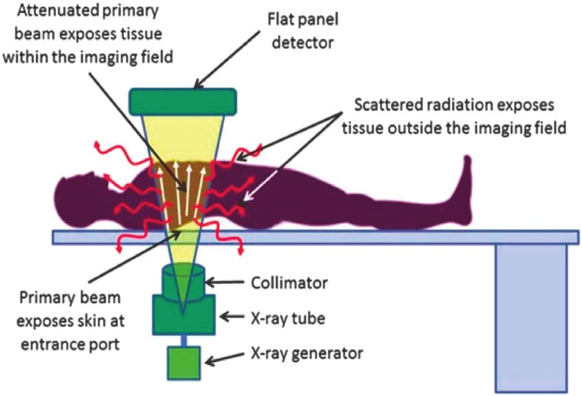
JACC VOL.   -, NO. -, 2018                                                                                                    Hirshfeld et al.   e5
-, 2018:-–-                                                                            ECD on Optimal Use of Ionizing Radiation in CV Imaging

This leads to greater patient radiation exposure and,               Decision Pathways is also available online, as well as the ACC
potentially, to greater exposure for clinical personnel.            disclosure policy for document development.
Although the clinical benefit of these procedures is sub-              The work of the writing committee was supported
stantial, there is concern about the implications of med-           exclusively by the ACC without commercial support.
ical radiation exposure both to patients and to medical             Writing committee members volunteered their time to
personnel. The ACC leadership concluded that it is                  this effort. Conference calls of the writing committee
important to provide practitioners with an educational              were confidential and were attended only by committee
resource that assembles and interprets the current radi-            members and ACC staff.
ation knowledge base relevant to cardiovascular imaging                                           James L. Januzzi, Jr., MD, FACC
procedures that employ ionizing radiation. By applying                                                     Chair, ACC Task Force on
this knowledge base, cardiovascular practitioners will be                                     Expert Consensus Decision Pathways
able to select and perform procedures optimally, and,
accordingly, minimize radiation exposure to patients and
to personnel.                                                       1. INTRODUCTION
  This online published document is a more compre-
hensive treatment of the knowledge base covered in 2                1.1. Document Development Process and Methodology
print published documents published under this docu-
                                                                    1.1.1. Writing Committee Organization
ment’s title with subtitles “Part 1: Radiation Physics and
                                                                    The writing committee consisted of a broad range of
Radiation Biology” and “Part 2: Radiological Equipment
                                                                    members representing 9 societies and the following areas
Operation, Dose-Sparing Methodologies, Patient and
                                                                    of expertise: interventional cardiology, general cardiol-
Medical Personnel Protection.” In addition, this online
                                                                    ogy, pediatric cardiology, nuclear cardiology, nuclear
document contains 3 sections that are not included in
                                                                    medicine,     clinical   electrophysiology,          cardiovascular
the print-published documents: Modality-Specific Oper-
                                                                    computed tomography (CT), cardiovascular imaging, and
ator Education and Certification, Quality Assurance, and
                                                                    the consumer patient perspective. Both a radiation safety
Patient and Medical Personnel Radiation Dose Moni-
                                                                    biologist and a physicist were included on the writing
toring   and    Tracking:      Programmatic      and   Individual
                                                                    committee. This writing committee met the College’s
Considerations.
                                                                    disclosure requirements for relationships with industry
  To avoid actual, potential, or perceived conflicts of in-
                                                                    (RWI) as described in the Preamble.
terest that may arise as a result of industry relationships or
personal interests among the writing committee, all
members of the writing committee, as well as peer re-               1.1.2. Document Development and Approval
viewers of the document, are asked to disclose all current          The writing committee convened by conference call and
healthcare-related relationships, including those existing          e-mail to finalize the document outline, develop the
12 months before initiation of the writing effort. The ACC          initial draft, revise the draft per committee feedback, and
Task Force on Expert Consensus Decision Pathways                    ultimately approved the document for external peer re-
(formerly the ACC Task Force on Clinical Expert Consensus           view. All participating organizations participated in peer
Documents) reviews these disclosures to determine which             review, resulting in 21 reviewers representing 299 com-
companies make products (on the market or in develop-               ments. Comments were reviewed and addressed by the
ment) that pertain to the document under development.               writing committee. A member of the ACC Task Force on
Based on this information, a writing committee is formed            Expert Consensus Decision Pathways served as lead
to include a majority of members with no relevant re-               reviewer to ensure that all comments were addressed
lationships with industry (RWI), led by a chair with no             adequately. Both the writing committee and the task force
relevant RWI. Authors with relevant RWI are not permitted           approved the final document to be sent for ACC Clinical
to draft or vote on text or recommendations pertaining to           Policy Approval Committee. This Committee reviewed the
their RWI. RWI is reviewed on all conference calls and              document, including all peer review comments and
updated as changes occur. Author and peer reviewer RWI              writing committee responses, and approved the docu-
pertinent to this document are disclosed in Appendixes 1            ment in November 2017. The Heart Rhythm Society (HRS),
and 2, respectively. Additionally, to ensure complete trans-        North American Society for Cardiovascular Imaging
parency, authors’ comprehensive disclosure information—             (NASCI), Society for Cardiovascular Angiography and In-
including     RWI   not      pertinent   to   this   document—is    terventions    (SCAI),     and    Society      of    Cardiovascular
available online (see Online Appendix). Disclosure infor-           Computed Tomography (SCCT) endorsed the document in
mation for the ACC Task Force on Expert Consensus                   January 2018. This document is considered current until
e6   Hirshfeld et al.                                                                                                JACC VOL.    -, NO. -, 2018
     ECD on Optimal Use of Ionizing Radiation in CV Imaging                                                                           -, 2018:-–-

     the Task Force on Expert Consensus Decision Pathways                   medical ionizing radiation, accounting for approximately
     revises or withdraws it from publication.                              40% of total medical radiation exposure (exclusive of ra-
                                                                            diation oncology) (1,2). Among occupationally exposed
     2. PURPOSE                                                             healthcare workers, interventional cardiologists and
                                                                            clinical electrophysiologists are among the most highly
     2.1. Document Purpose                                                  exposed, and there is potential for exposure to support
     This document’s purpose is to assist cardiovascular                    personnel as well (in particular, nonphysician staff who
     practitioners to provide optimal cardiovascular care                   work in x-ray fluoroscopy and nuclear cardiology envi-
     when employing ionizing radiation in diagnostic and                    ronments) (3,4).
     therapeutic procedures. It is written to serve as an
     accessible resource that compiles the current radiation                2.3. The Need for Physician Radiation Safety Education
     biology and safety knowledge base applicable to cardio-
                                                                            Cardiovascular specialists have a responsibility to un-
     vascular imaging. The document covers both patient and
                                                                            derstand the radiation safety knowledge base, in partic-
     medical personnel safety issues for the 3 cardiovascular
                                                                            ular, to:
     procedure classes that employ ionizing radiation: x-ray
     fluoroscopy, x-ray CT, and radionuclide scintigraphy.                   1. Apply knowledge of the radiation safety knowledge
     It includes discussions of radiation dosimetry and                       base to make appropriate case selection choices.
     its determinants, radiation harm, basics of equipment                  2. Conduct      radiation-assisted      procedures         optimally,
     operation, strategies to minimize dose, and issues of                    minimizing exposure to patients and personnel.
     radiation monitoring and tracking. The document’s goal
                                                                              There is evidence that many cardiovascular specialists
     is to enable cardiovascular practitioners to select the
                                                                            who order and conduct radiation-employing procedures
     optimal imaging technique for a given clinical circum-
                                                                            are not fully informed about the radiation doses that
     stance while balancing a technique’s risk and benefits,
                                                                            accompany the procedure or the associated health impli-
     and to apply that technique optimally to generate high-
                                                                            cations for their patients and for themselves (5,6).
     quality diagnostic images that deliver the greatest
                                                                            Consequently, there is a need to augment and standardize
     clinical value with minimal radiation exposure.
                                                                            the level of knowledge and competence that cardiovas-
                                                                            cular specialists should hold in radiation safety and
     2.2. The Radiation Safety Issue
                                                                            management. This knowledge base should be incorpo-
     Cardiovascular procedures that employ ionizing radia-
                                                                            rated into training curricula and in physician board cer-
     tion have transformed the practice of cardiovascular
                                                                            tification procedures.
     medicine. These procedures have great value for diag-
                                                                              Cardiovascular      specialists      fall   into    2      categories
     nosis and treatment of appropriately selected patients
                                                                            requiring different levels of knowledge: those who order
     with      known      or    suspected       cardiovascular   disease.
                                                                            cardiac imaging procedures and those who perform them.
     In addition, they enable more refined recognition
                                                                            Training curricula should furnish the level of knowledge
     and characterization of cardiovascular disease. These
                                                                            appropriate for a particular physician’s practice activity.
     procedures are also integral to either planning or
                                                                            Achieving    this   goal requires        collaboration        between
     executing numerous treatment modalities, which can
                                                                            various stakeholders in graduate and postgraduate edu-
     have profound impacts on the outcomes of cardiovas-
                                                                            cation. The blueprints of certification and recertification
     cular disorders.
                                                                            examinations should include specifications of radiation
        However, ionizing radiation has molecular-level detri-
                                                                            safety subject matter. Training programs should configure
     mental effects on exposed human tissue, with potential
                                                                            their   teaching    curricula     to   prepare       their     trainees
     for injury both to patients and to exposed medical
                                                                            appropriately.
     personnel. Consequently, it is desirable to minimize ra-
     diation exposure both to patients and to medical
                                                                            2.4. Appropriateness of Medical Radiation
     personnel in a manner consistent with achieving optimal
     health benefits. This principle requires that clinicians                The balance between a procedure’s risk and benefit
     employ judicious selection of and conduct of radiation-                determines its appropriateness. Although the technical
     employing procedures to achieve an optimal balance of                  hazards that accompany a procedure are well known,
     a procedure’s therapeutic benefit against the incremental               the hazard associated with attendant exposure to ionizing
     risk conferred by the radiation exposure.                              radiation    should   also   be     considered       a     potentially
        Currently, cardiovascular diagnostic and therapeutic                important determinant of a procedure’s risk-benefit
     procedures are a major source of patient exposure to                   relationship.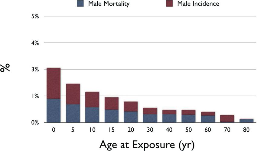
JACC VOL.     -, NO. -, 2018                                                                                                                                    Hirshfeld et al.    e7
-, 2018:-–-                                                                                                        ECD on Optimal Use of Ionizing Radiation in CV Imaging

 TABLE 1         Typical Effective Doses for Cardiac Procedures                             TABLE 1         Continued

                                                                   Typical Effective                                                                        Typical Effective
Modality                              Protocol                       Does (mSv)           Modality                          Protocol                          Does (mSv)

MDCT            Coronary CT angiography:                                  8–30            Fluoroscopy      TAVR, transapical approach                              12–23
                helical, no tube current modulation
                                                                                          Fluoroscopy      TAVR, transfemoral approach                            33–100
MDCT            Coronary CT angiography:                                  6–20
                                                                                          Fluoroscopy      Diagnostic electrophysiological study                  0.1–3.2
                helical, tube current modulation
                                                                                          Fluoroscopy      Radiofrequency ablation of arrhythmia                    1–25
MDCT            Coronary CT angiography:                                  0.5–7
                prospectively triggered axial                                             Fluoroscopy      Permanent pacemaker implantation                        0.2–8
MDCT            Coronary CT angiography:
e8 Hirshfeld et al. JACC VOL. -, NO. -, 2018
ECD on Optimal Use of Ionizing Radiation in CV Imaging -, 2018:-–-
Patient radiation dose ranges (in millisieverts) for the 3 exposure must be weighed in relation to the health status
principal radiation-based cardiovascular imaging studies: benefits achieved by these procedures.
x-ray fluoroscopy, x-ray CT, and nuclear cardiology. In- Physicians who are invasive cardiovascular procedure
dividual procedure categories are further subdivided ac- operators are among the most highly exposed of the
cording to types of image acquisition protocols. Note that occupationally exposed healthcare workers. Measure-
for a particular procedure category, the dose can vary ments of interventional cardiologist operator exposure
considerably depending on image acquisition protocol using current equipment and protection practices
and, within a given image acquisition protocol, procedure demonstrate an exposure range of 0.2 to >100 micro-
conduct and patient characteristics. sieverts ( m Sv) per procedure with a per-procedure average
However, augmented capabilities have led to increased of 8 to 10 m Sv (11). Thus, an active interventional cardi-
utilization levels, resulting in greater radiation exposure ologist performing 500 procedures/year employing cur-
both at the individual and at the population levels. In rent technology may be expected to receive, in addition to
addition, refinement of x-ray fluoroscopic systems, background exposure, a dose of as much as 10 mSv/year
yielding greatly improved image quality, has facilitated or, in a most extreme scenario, 300 mSv over a 30-year
the development of increasingly complex cardiovascular active professional career.
interventional procedures. These procedures often Nonphysician clinical personnel working in an x-ray
require longer fluoroscopic times, resulting in larger ra- environment should receive substantially smaller doses
diation exposures than more basic procedures. than tableside operators, although nuclear cardiology
Increasing radiation exposure has the potential to in- technologists who handle radioactive materials tend to be
crease the risk of adverse effects such as radiation- more highly exposed. Determinants of nonphysician
induced cancer. It is uncertain, however, whether exposure include time spent in an active procedure room,
medical radiation is in actuality increasing cancer inci- location in the procedure room during active procedures,
dence in the population, because a small increase would and exposure handling radioactive materials. There are
be difficult to detect against the large background inci- no data that characterize the total number of exposed
dence of cancer. workers or their exposure values.
During the 2014 calendar year, the U.S. healthcare
system performed, on Medicare beneficiaries, an esti- 3.2. Potential Consequences of Patient and Medical Personnel
mated 925,848 diagnostic cardiac catheterization pro- Radiation Exposure
cedures, 342,675 percutaneous coronary interventions, There are 3 important potential consequences of the
248,234 clinical electrophysiologic procedures, 61,207 growing use of ionizing radiation in cardiovascular med-
cardiovascular x-ray CT scans, and 2,111,558 nuclear car- icine (see Table 2).
diology examinations, for a total of 3,689,522 cardiovas-
1. At the individual patient level, although many in-
cular procedures that use ionizing radiation in Medicare
dividuals receive little or no medical radiation expo-
beneficiaries (8). Medicare beneficiaries are estimated to
sure, some receive lifetime doses in excess of 100 mSv.
consume 30% to 40% of all cardiovascular procedures.
At the population level, such doses are associated with
Natural background radiation averages 3.0 millisieverts
a detectably increased cancer risk (12). Consequently,
(mSv) (see Section 4 for a discussion of the Sievert unit of
such exposures may place individuals at increased
radiation exposure) per person/year in the United States—
personal risk of developing cancer or tissue reactions,
equivalent to 150 posteroanterior chest radiographs (a
including skin injury and cataracts. The actual magni-
posteroanterior chest-x-ray dose is 0.02 mSv; combined
tude of this risk varies substantially with patient
posteroanterior and lateral is 0.06 mSv) (9). At the pop-
characteristics (see Section 5.4).
ulation level, between 1987 and 2006, estimated per
person total medical radiation exposure grew from 0.6
Potential Consequences of Patient and Medical
mSv/year (0.2 background) to 3.2 mSv/year (1.07 TABLE 2
Personnel Radiation Exposure
background) (10). Consequently, patients are currently
Individual Although many individual patients receive little or no medical
receiving, on average, more radiation from medical Patient radiation exposure, some receive lifetime doses in excess of
sources than from natural background sources. 2006 is 100 mSv. Doses in excess of 100 mSv are associated with a
detectable increased cancer risk
the latest year for which compiled data are available. (The
Population Increased total exposure incurred by total population of
National Council on Radiation Protection is currently patients has the potential to increase the population
compiling contemporary data—expected availability incidence of cancer and other radiation-related disorders
2019—and it is likely that current average medical expo- Occupationally Occupationally exposed physicians and support staff may
Exposed receive doses as large as 10 mSv per year over a career that
sure will be found to have increased further). The 2006 Workers may span 30–40 years. The implications of this level of
medical exposure is equivalent to 160 posteroanterior exposure at the level of the individual practitioner are
uncertain.
chest x-rays per person/year. Risks associated with thisJACC VOL. -, NO. -, 2018 Hirshfeld et al. e9
-, 2018:-–- ECD on Optimal Use of Ionizing Radiation in CV Imaging
2. At the population level, the increased total exposure radiation with tissue will be characterized from the
incurred by patients has the potential to increase the perspective of 4 of the previously mentioned inter-related
population incidence of cancer. frames of reference: exposure, absorbed dose, equivalent
3. With respect to medical personnel, occupationally dose, and effective dose. It should be noted that in the
exposed physicians and support staff may receive literature, the terms “exposure” and “dose” are often
doses as large as 10 mSv/year over a career that may used with less specific meanings than those used in this
span 30 to 40 years. The implications of this level of document. For this document’s purpose, these metrics
exposure at the level of the individual practitioner are have specific meanings as defined in the following text.
uncertain. Exposure
Radiation exposure refers to the presence of ionizing ra-
The ongoing magnitude of exposure to the general
diation at the location of the exposed tissue. This is
population and to occupationally exposed healthcare
quantified by standardized measures of a physical quan-
workers has health implications at the population level
tity that represent the amount of radiation present at that
and for individual patients and healthcare workers. It is
location. The typically used measure of radiation quantity
important that physicians and healthcare workers un-
is air kerma (Section 4.4.1), which is the amount of energy
derstand the ionizing radiation knowledge base and apply
released by the interaction of the radiation with a unit
it to protect patients, themselves, and their colleagues
mass of air. Its unit of measure is the gray (Gy). Its units
through judicious case selection and appropriate conduct
are joules (J)/kg. One Gy is the quantity of radiation that
of radiation-assisted procedures.
when interacting with 1 kg of air releases 1 joule of energy.
It should be noted that this is a measure of cumulative
4. THE MANY MEASURES OF RADIATION energy intensity as the energy deposition is normalized to
a quantity of the absorbing material.
4.1. Radiation Exposure and Dose Metrics Absorbed Dose
Ionizing radiation exposure and dosimetry are not easily Absorbed radiation dose is a measure of the energy that
characterized by simple metrics. Radiation exposure and radiation deposits in an exposed tissue through in-
dose may be considered from the perspective of 5 distinct teractions with its molecular constituents. It differs from
but inter-related frames of reference. For this document’s exposure in that the radiation present at a given location
purpose, these metrics have specific meanings as defined does not deposit all of its energy there. The fraction of its
in the following text: energy that a given radiation exposure will deposit in the
exposed tissue varies with the type and energy of the
n Exposure: the quantity of radiation that impinges on a
radiation and the tissue composition.
tissue.
Absorbed dose is also a measure of the intensity of cu-
n Absorbed Dose: the concentration of energy deposited
mulative energy deposition (energy deposited per unit
by radiation in a specific exposed tissue.
mass of tissue) and is expressed in Gy—joules of energy
n Equivalent Dose: the absorbed dose adjusted by a
deposited per kilogram of tissue. In exposure by external
radiation weighting factor to reflect the different de-
radiation beams, dose is not uniform throughout the
grees of biological damage caused by various types of
exposed volume, but varies, typically as a function of
radiation.
depth from the beam entrance port.
n Effective Dose: a metric that reflects the overall bio-
Equivalent Dose
logical effect from radiation on an average subject
Different types of ionizing radiation cause varying de-
from a particular radiation exposure scenario.
grees of tissue injury for a given absorbed dose. Equiva-
n Injected Dose: A metric that describes the quantity of
lent dose is a construct used to account for differences in
radioactivity of a radioactive substance injected into a
tissue injury caused by different radiation types. X-rays
patient for a nuclear scintigraphy study (expressed in
and gamma rays are the benchmarks against which par-
millicuries [mCi]). Injected dose is a determinant of the
ticle radiation types such as protons, neutrons, and beta
4 dose parameters listed previously. However, the
particles are compared. Some particles, in particular,
exact relationships between injected dose and absor-
protons, neutrons and alpha particles, cause greater tis-
bed dose and equivalent dose are complex, and are
sue injury at a given dose than do x-rays, gamma rays,
discussed in depth in Section 6.4.
and electron particles. To adjust for this variability, each
A comprehensive assessment of radiation effects re- radiation type is assigned a radiation weighting factor by
quires consideration of all 5 parameters. The relationships which the absorbed dose (in Gy) is multiplied to yield a
between these metrics are complex and are determined measure of the expected tissue injury caused by that
by the properties of both the radiation and the exposed dose. The unit of measure is the sievert (Sv) which is the
tissue. For clarity in this document, the interaction of absorbed dose in Gy multiplied by the radiation weightinge10 Hirshfeld et al. JACC VOL. -, NO. -, 2018
ECD on Optimal Use of Ionizing Radiation in CV Imaging -, 2018:-–-
factor. Of the different radiation types, x-rays, gamma provides a longer period for radiation-induced illness to
rays, and electron particles (electrons and positrons) are present (13).
assigned a radiation weighting factor of 1. Other particle
radiation types have weighting factors ranging between 2 4.2. Challenges in Relating Radiation Exposure and Dose to
and 20. For medical imaging, which employs x-rays and Risk of Detrimental Effects
gamma rays, absorbed dose and equivalent dose take the Detrimental effects of radiation exposure typically pre-
same value, that is, an exposure with an absorbed dose of sent weeks to years following exposure. In addition,
20 mGy has an equivalent dose of 20 mSv. many detrimental effects, principally cancer, have a large
Effective Dose background frequency that complicates the attribution of
Effective dose is a measure of the estimated potential for an effect in a particular subject to prior radiation
a biological effect on the complete organism caused by a exposure.
particular absorbed radiation dose. The effective dose
construct has been developed as a measure of the esti-
4.3. Types of Ionizing Radiation Used in Medical Imaging
mated potential for a stochastic effect (such as cancer
Radiation in cardiovascular imaging consists of photons
induction) that would be caused by a particular (nonuni-
with energy >10 kiloelectron volts (keV) (x-rays and
form) absorbed radiation dose. It is the sum of the
gamma rays) and positrons. The physical effect of such
equivalent doses received by each organ with each organ
radiation is to eject electrons from atoms that comprise
equivalent dose multiplied by a coefficient that reflects
tissue molecules forming ions and free radicals. This
that organ’s sensitivity to a stochastic effect. The unit of
causes molecular damage, potentially destroying a mole-
effective dose is also the Sv, as discussed in greater depth
cule or altering its function. This is the basis for the term
in Section 4.5. The Sv, like the Gy of the absorbed dose’s
“ionizing radiation” (discussed in detail in Section 5).
unit, is specific to its particular context and is equal to 1
joule/kg. The connection between effective dose and
absorbed dose is that an effective dose of 1 Sv is associ- 4.3.1. X-Rays and Gamma Rays
ated with the same estimated stochastic risk that accom- X-rays and gamma rays are in a class of ionizing radia-
panies a uniform total body exposure with an absorbed tions, which is transmitted by photons. Photons travel at
dose of 1 Gy of radiation that has a radiation weighting the speed of light, and have no mass and no charge. Their
factor of 1. electromagnetic energy ranges from a few electron volts
In medical radiation exposures, absorbed dose is typi- (eV) to millions of electron volts (MeV). The energies
cally not uniform throughout all tissues. For x-ray imag- commonly employed in cardiovascular imaging are tens
ing, dose is concentrated in the body region being to hundreds of keV.
examined and varies with depth from the beam entrance X-ray or gamma photons cause ionization by colliding
port. For nuclear imaging, dose is concentrated in the with and ejecting electrons from atoms of constituent
tissues that most avidly take up the tracer or are involved tissue molecules. Energy is exchanged in the process,
in its elimination. with the ejected electron gaining energy of motion and
Different tissues have different sensitivities to the photon losing energy. The incident photon may or
radiation-induced effects. In the effective dose construct, may not be extinguished by the interaction. After an
each tissue is assigned a tissue-weighting factor that initial interaction with an atom, photons that were not
specifies its sensitivity to radiation effects. To calculate extinguished continue to travel through the exposed
the effective dose in Sv, each exposed tissue’s equivalent medium at a degraded energy. The weakened (scattered)
dose is multiplied by its tissue-weighting factor yielding photon can collide with additional atoms (further
that tissue’s contribution to the overall risk. The contri- exposing the subject), potentially ionizing them as well,
butions to risk from all exposed tissues are summed, until either all of its energy is dissipated and the photon
yielding total risk, expressed as the effective dose in Sv. ceases to exist, or it escapes from the subject (exposing
(How the effective dose is calculated is discussed in the environment).
greater depth in Section 4.5). X-rays used in x-ray fluoroscopy and x-ray CT have a of
It is important to note with regard to childhood and photon energy spectrum between 30 and 140 keV (the
teenage radiation exposure that tissue weighting factors energy spectrum of x-rays generated in typical diagnostic
do not take into account the increased sensitivity of the x-ray tubes includes photon energiesJACC VOL. -, NO. -, 2018 Hirshfeld et al. e11
-, 2018:-–- ECD on Optimal Use of Ionizing Radiation in CV Imaging
68 to 80 keV range, similar to diagnostic x-rays. Tc-99m the exposed tissue. Not all radiation energy that impinges
releases photons primarily in the 140 keV range. on a tissue is absorbed. Some radiation (a variable quan-
tity depending on both radiation and tissue characteris-
4.3.2. Positrons tics) passes through the tissue without interacting with it,
Positrons are positively charged electrons. They have depositing no energy. (This fraction of the radiation is
mass and charge. When positrons travel through a me- what generates the radiological image). Absorbed dose is
dium, their electrostatic charge causes them to interact also an intensity measured in gray (Gy), which represents
readily with electrons in the medium, leaving a trail of deposition of 1 joule of energy per kilogram of irradiated
ionization. Consequently, they have a very short mean tissue.
free path in tissue of 6 to 7 mm with a maximum of 15.2 External beam energy deposition in tissue is not uni-
mm. Positrons continue to cause ionization until their form. X-ray radiation is attenuated as it passes through
energy decreases to a critical level, at which point they are tissue. For diagnostic x-rays, in most tissues, x-ray in-
annihilated by colliding with an electron of a constituent tensity decreases by approximately a factor of 2 for each 5
atom. This annihilation process releases 2 511-keV gamma cm of tissue that it traverses. Thus, tissue exposed to an
ray photons that travel in opposite directions. Because external x-ray beam, as occurs in x-ray fluoroscopy and
the emitted photons have such high energy, they are x-ray CT, is not exposed uniformly—the dose decreases
minimally attenuated in tissue, and the majority reach the exponentially with depth from the beam entrance port.
imaging detector. Rubidium-82 is the most commonly The incident beam air kerma is a good measure of dose at
used positron emitter for myocardial perfusion imaging; the body surface, but structures deeper than the body
nitrogen-13 ammonia is used less frequently for this surface receive smaller doses. Thus, to estimate the dose
purpose. Fluorine-18 deoxyglucose is used in cardiology to a particular body structure within the path of an x-ray
for metabolic imaging and to detect myocardial sarcoid beam but remote from the beam entrance site, adjust-
and other inflammatory conditions. ments have to be made to account for beam absorbance.
4.4. Relationships Between Exposure and Absorbed Dose 4.4.1.2. Kerma-Area Product: Incorporating the Volume of
Medical radiation exposures occur in 2 ways: Exposed Tissue in X-Ray Fluoroscopy
Kerma (measured in Gy) is a measure of dose intensity
1. Exposure from an external radiation beam (x-ray fluo-
(joules of energy deposited per kg of tissue). The risk of
roscopy and x-ray CT)
radiation harm is related both to the intensity of the ra-
2. Exposure from radioactive decay within the subject
diation dose and to the quantity of tissue that receives the
(nuclear scintigraphy).
dose. (The greater the quantity of tissue that receives a
given dose, the greater the risk.) Kerma-area product
4.4.1. Exposure From External Beams (KAP) is the product of the beam’s kerma and its cross-
For external radiation beams, the absorbed dose is sectional area. Thus, this parameter also incorporates
determined by the total incident exposure, the properties the volume of tissue irradiated. This concept is particu-
of the incident radiation, and the volume of tissue larly important in x-ray fluoroscopy, as imaging field sizes
exposed. Exposure from an external beam is measured can vary considerably leading to very different KAPs from
with the parameter air kerma. one examination to another.
Air kerma is the standard unit of measure for x-ray
beam exposure. Kerma is an acronym for “kinetic energy 4.4.1.3. Kerma-Length Product: Incorporating the Volume of
released in material.” Kerma is an energy intensity Exposed Tissue in X-Ray CT
measured in units of joules of energy released per kilo- CT delivers radiation to a patient in a manner quite
gram of absorbing material (J/kg). The kerma unit of different from that of projectional imaging or fluoroscopy.
measure is the gray (Gy), which represents 1 joule of en- Typically, a narrow x-ray beam with a rectangular cross
ergy released per kilogram of absorbing material. The section is used to collect images from multiple angles as it
metric “air kerma” is used in medical x-ray fluoroscopic rotates around the patient. This distributes the dose much
applications because the measurement is made using air more uniformly around the patient compared with pro-
as the absorbing material that is ionized by the incident jectional imaging. Instead of measuring an entrance air
radiation beam. kerma to the patient, “dose” is measured by convention
as an air kerma inside of an acrylic cylinder used to
4.4.1.1. Absorbed Dose in Tissue From an External Beam simulate a patient. Two sizes of cylinders are used: 32-
As described in Section 4.1, radiation absorbed dose, as and 16-cm diameters, often referred to as body and head
distinguished from exposure, is an energy intensity, the phantoms, respectively. Air kerma is measured inside the
concentration of radiation energy actually deposited in phantom using an ionization chamber in the shape of ae12 Hirshfeld et al. JACC VOL. -, NO. -, 2018
ECD on Optimal Use of Ionizing Radiation in CV Imaging -, 2018:-–-
pencil that is placed inside a hole that is appropriately radiation-induced stochastic risk. The calculation of
drilled in the plastic phantom. This yields the dose effective dose involves estimating each organ’s actual
“intensity” analogous to the air kerma measurement for equivalent dose (in Gy). That dose is adjusted by multi-
x-ray fluoroscopy. plying it by the organ’s tissue-weighting factor. The organ
The phantom air kerma is multiplied by the axial scan sensitivity-adjusted individual organ doses are summed
length to incorporate the volume of tissue irradiated. This to yield a total effective dose (in Sv).
method generates a variety of dose metrics for x-ray CT; For a chest exposure, absorbed dose is concentrated in
these are discussed in detail in Section 6. For example, the skin, mediastinal structures, lungs, breast, and
the computed tomography dose index100 (CTDI 100 ) is a thoracic bone marrow. Doses to these organs would
measure of the dose delivered along a 100-mm scan contribute the largest components to the effective dose
length. Computed tomography dose index w (CTDI W calculation. Smaller quantities of scattered radiation
weighted) accounts for the fact that more peripherally would expose the abdominal viscera and upper neck. As
located structures, which are closer to the beam entrance, these organs would receive smaller exposures, their
receive larger doses than deeper structures. contribution to the effective dose calculation would be
smaller. Other types of radiological examinations, such as
4.4.2. Exposure From Radionuclides x-ray CT and cardiac scintigraphy, would have different
Unlike external beam exposures, radionuclide exposures organ exposure distributions yielding different effective
come from radioactive decay within the subject. In nu- dose calculations.
clear cardiology applications, a radiopharmaceutical is Deriving a quantitative measure of a subject’s esti-
administered systemically and distributes throughout the mated increased cancer risk due to a specified effective
body. Distribution may be preferential to particular tis- dose is complex because the risk magnitude is determined
sues depending on the pharmacological properties of the by numerous other variables, including subject age (chil-
radiopharmaceutical. The dose delivered by a radiophar- dren and young adults are more susceptible), gender
maceutical is determined by the activity administered, (women are more susceptible), and natural life expec-
the tracer distribution, the tracer elimination rate, and the tancy (longer natural life expectancy confers a longer time
tracer’s time-activity relationships. These data in combi- available for cancer to present [13]). Statistical models
nation with the tracer radionuclide’s radiation charac- that attempt to quantify the dose-risk relationship have
teristics permit estimation of radiation dose delivered to been developed based on large population exposures.
each organ or tissue. This model is discussed in greater These models are discussed in Section 5.4.
detail in Section 6. The individual tissue weighting factors have been
revised over time as accumulating epidemiological evi-
4.5. Estimating Effective Dose dence permits more precise estimates of organ sensitivity.
The concept of effective dose was formulated to create a The International Commission on Radiation Protection
metric that estimates a given radiation dose’s contribu- published the most recent organ sensitivity estimates in
tion to stochastic health risk—namely the risk of cancer 2007 in ICRP Publication 103 (15). The estimates are listed
induction and of genetic changes (see Section 5.2.2) (14). in Table 3.
The effective dose concept is derived from 2 facts of ra- The tissue weighting factors are measures of the indi-
diation dosimetry: vidual tissue’s intrinsic sensitivities to radiation-induced
cancer. Three applications of the effective dose concept
1. Medical and occupational radiation exposures are
have relevance for medical exposure to patients under-
generally not uniform, with some organs and tissues
going cardiovascular procedures and occupationally
receiving greater exposures than others.
exposed medical personnel. They are:
2. Sensitivity to radiation-induced detrimental effects
varies among different organs and tissues.
1. For cardiac x-ray fluoroscopy and cardiac x-ray CT, the
The effective dose is expressed in units termed Sv radiation is concentrated in the subject’s chest. Thus,
(Section 4.1). The units are a special term for J/kg (the the exposures that contribute the most to stochastic
same as for the Gray). The Sv represents the hypothetical risk are the thoracic red bone marrow; the lung; and, in
uniform whole-body dose that confers the same stochas- females, the breast.
tic risk as the nonuniform dose actually delivered. A 2. Measurements of exposure in phantoms provide
uniform total body absorbed dose of 1 J/kg of radiation models that enable the rough estimation of equivalent
that has a radiation weighting factor of 1 would yield an dose delivered to particular internal structures by the
effective dose of 1 Sv. exposure as measured by the subject exposure param-
The effective dose construct assigns each organ/tissue eters, including KAP product for x-ray fluoroscopy,
a weighting factor that reflects the tissue’s sensitivity to kerma-length product for x-ray CT, and radionuclideJACC VOL. -, NO. -, 2018 Hirshfeld et al. e13
-, 2018:-–- ECD on Optimal Use of Ionizing Radiation in CV Imaging
Tissue Weighting Factors Used to Calculate
effective dose is an estimate that involves a number of
TABLE 3
Effective Dose in Sieverts assumptions, the calculated and reported values should
be accompanied by the actual exposure measurements
Tissue Weighting factors
Organs (ICRP103–2007) and a description of the methodology used for estimation,
Red bone marrow 0.12 that is, a conversion factor.
Colon 0.12
5. HOW RADIATION CAN HARM PEOPLE
Lung 0.12
Stomach 0.12
5.1. Mechanism of Radiation-Induced Biological Effects
Breasts 0.12
Radiation-induced tissue injury is due to molecular al-
Gonads 0.08
terations caused by particles or photons that have suffi-
Bladder 0.04
cient energy to induce ionization. Atoms ionized by
Liver 0.04
radiation are frequently chemically unstable and trans-
Esophagus 0.04
form themselves or their constituent molecules into free
Thyroid 0.04 radicals. A common example is ionization of water, which
Skin 0.01 upon interacting with an x-ray photon, decomposes into a
Bone surface 0.01 free electron, a proton, and a hydroxyl radical. The hy-
Salivary glands 0.01 droxyl radical, because of its unpaired electron, is highly
Brain 0.01 reactive and interacts avidly with biomolecules (proteins
Remainder of body 0.12 or nucleic acids). Similarly, an x-ray photon can ionize an
Total 1.00 atom that is a constituent of a biomolecule. Thus, a
biomolecule can be altered by either reacting with a
Adapted from the International Commission on Radiological Protection (ICRP) (15).
radiation-generated free radical or by being directly
ionized by radiation. The resulting structural change can
alter a molecule’s function.
doses for cardiovascular nuclear cardiology. These in-
Radiation-induced tissue damage from ionizing radia-
dividual organ equivalent doses may then be converted
tion takes many forms that have variable intervals be-
to effective doses and summed to calculate an estimate
tween exposure and clinical presentation. Some harmful
of the subject’s effective dose. As noted in the previous
radiation effects appear within days to months following
text, the absolute effective dose-risk relationship is
the exposure. Other harmful radiation-induced effects
modulated by subject characteristics.
have long latent periods, becoming evident only many
3. Measurements of occupational exposure for healthcare
years following the inciting exposure, or may not present
workers may be used to estimate the worker’s sto-
in the individual’s remaining lifetime.
chastic risk.
Damage to a molecule that is an important tissue con-
stituent, such as a protein, can alter the cell’s function. If
4.6. Synopsis of Measures of Radiation Exposure and Dose a cell incurs sufficient damage to its constituent mole-
The existence of the many different measures of radiation cules it may not be able to maintain basic cellular opera-
exposure and dose has the potential to cause confusion tions and undergo necrosis. If a cell incurs strategic
leading to misapplication of units of measure. Table 4 damage to its deoxyribonucleic acid (DNA), a previously
contains a synopsis of the principal metrics described in normal gene may be transformed into an oncogene or the
this section. In reporting radiation from an individual ionization process may lead to other changes in cellular
procedure to a specific patient, modality-specific param- environment that promote carcinogenesis.
eters should be used. For x-ray fluoroscopic procedures,
air kerma at the interventional reference point and KAP 5.2. Types of Radiation-Induced Health Effects
should both be reported. For CT procedures, the CTDIvol Radiation-induced health effects are divided into 2 broad
and dose-length product (DLP) (discussed in detail in groups that differ in their mechanism, the nature of their
Section 6.3.3) should be reported, along with the size of effects, their relationship to absorbed dose, and the
the CTDI phantom (32- or 16-cm). Effective dose has lim- temporal relationships between exposure and
itations for calculating individual patient dosimetry manifestation.
because, for example, the tissue weighting factors are
gender and age averaged (not accounting for the fact that 5.2.1. Tissue Reactions (Formerly Called Deterministic Effects)
children and females are more sensitive). However, it may Tissue reactions are caused by radiation-induced injury to
be useful to make general comparisons between modal- structural and functional molecules in cells. Cell necrosis
ities, protocols, and imaging strategies. Given that will occur if the amount of molecular alteration incurredYou can also read