Journées de la Diffusion Neutronique - mai Roquebrune sur Argens
←
→
Page content transcription
If your browser does not render page correctly, please read the page content below
Journées de la Diffusion
   Neutronique

           mai
  Roquebrune sur Argens
Program

                          Monday 14th May                                               Tuesday 15th May                                              Wednesday 16th May

                                                             8:30-9:15                   SFN PhD Award

                                                                                     President: Charles Simon

                                                            9:15-10:00                                                       9:15-10:00 Condensed Matter invited speaker: Arnaud Desmedt
                                                                                           Winner's talk

                                                            10:00-10:25                         Break                        10:00-10:25                     Break
                                                            10:25-11:10      Biology invited speaker: Guillaume Tresset      10:25-10:45               Evgeny Clementyev

                                                                                                                             10:45-11:05               Thibault Charpentier

                                                            11:10-11:30                  Ekaterina Iashina                   11:05-11:25             Afonso da Cunha Ferreira

                                                            11:30-11:50                   Guillaume Sudre                    11:25-11:45                  Monica Ceretti

                                                            11:50-12:10                    François Boué                     11:45-12:00              Concluding remarks

12:00-14:00                     Lunch                       12:10-14:00                       Lunch                          12:00-14:00                      Lunch
14:00-15:00                                                 14:00-14:45      Magnetism invited speaker: Romain Sibille
                              Welcome

15:00-15:45   Soft Matter invited speaker: Olivier Sandre   14:45-15:05                  Evgenii Altynbaev

                                                            15:05-15:25                  Françoise Damay

15:45-16:05               Sandrine Lyonnard                 15:25-15:45                     Sylvain Petit

16:05-16:25                 Fabrice Cousin                  15:45-16:05                   Ketty Beauvois

16:25-16:45                 Nicolas Jouault                 16:05-16:30                        Break
                                                            16:30-17:15 Instrumentation invited speaker: Jean-Marc Zanotti
16:45-17:05                 Jacques Jestin

17:05-17:30                     Break
 17:30 …                                                    17:15-17:35                    Martin Boehm

                                                            17:35-17:55                     Frédéric Ott
                           ESS, ILL, LLB
                                                             17:55 …                       Discussions
                                 news                                                 Neutrons in the future

                                                                                              2FDN
 … 19:00                                                                                      ENSA
19:00-19:30                     CLIPs                        …19:30                          AG-SFN
19:30-21:00                      Diner                      19:30-21:00                     Gala Diner
 21:00 …                        Posters                      21:00 …                           Party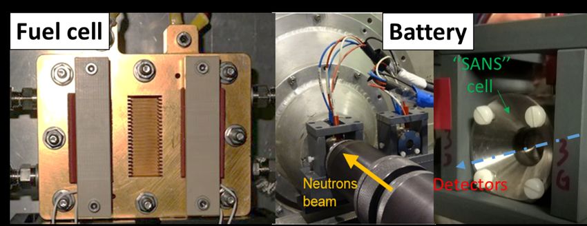
Soft Matter
  Session
SANS studies of polymer chains, “hybrid” core-shells, micelles and vesicles made of magnetic iron oxide
                        nanoparticles, (co)polymers, polypeptides and phospholipids

 Olivier Sandre1*, Gauvin Hémery1, Hélène Meheust1, Etienne Grau1, Henri Cramail1, Elisabeth Garanger1, Sébastien
Lecommandoux1 Annie Brûlet2, Ralf Schweins3, Sarah MacEwan4, Ashutosh Chilkoti4, Thi Phuong Tuyen Dao1, Fabio
             Fernandes5, Manuel Prieto5, Mériem Er-Rafik6, Marc Schmutz6, and Jean-François Le Meins1
  1
    Laboratoire de Chimie des Polymères Organiques (LCPO), UMR5629 CNRS / Université de Bordeaux / Institut
                                Polytechnique de Bordeaux, Pessac 33607 Cedex, France
            2
               Laboratoire Léon Brillouin, UMR12 CNRS/CEA, CEA-Saclay, Gif-sur-Yvette 91191, France
              3
                Institut Laue-Langevin (ILL) CS20156, 71 Avenue des Martyrs, 38042 Grenoble 9, France
                  4
                     Department of Biomedical Engineering, Duke University, Durham, NC27708, USA
                   5
                     Centro de Química-Física Molecular, Instituto Superior Técnico, Lisboa, Portugal
            6
               Institut Charles Sadron (ICS) UPR 22 CNRS / Université de Strasbourg, Strasbourg, France
                                               *olivier.sandre@enscbp.fr

This lecture will cover several soft matter colloidal systems where SANS brings useful information as non-
perturbative method, some of them biology-oriented, of core-shell, micellar or vesicular structure, self-assembled
from inorganic nanoparticles, polypeptides, or block copolymers and phospholipids. With perfectly defined sequence
and chain length, recombinant peptides obtained by protein engineering allow investigating the structure-property
relationships at a level of detail difficult to achieve with synthetic polymers. The temperature-triggered self-assembly
of recombinant elastin-like block peptides (ELP) was studied by SANS for diblock ELPs1,2 and more recently for tri-
block and gradient copolymers.3 On our side, we studied diblock ELP solutions below and above the critical micellar
temperature (CMT) by multi-angle DLS and SANS. Below the CMT, the radius of gyration follows a power law
versus molecular weight similar to Gaussian coils. Above CMT, attractive interactions between the more hydrophobic
block of the ELP copolymers trigger the self-assembly into micelles. Continuing to raise temperature expels more and
more water molecules out of the micelle core, the hydration level decreasing to just few D2O per aminoacid, as
estimated from the contrast in light and neutron scattering. These SANS studies highlight strong similarities between
ELPs and synthetic copolymers: the standard Debye model of Gaussian chains enables fitting the scattering of ELP
unimer chains and other systems. This is also the case for estolide polyesters which are bio-sourced polymers derived
from natural oils. The thermal behavior is however opposite: unlike for ELPs where water switches from a Theta to a
bad solvent, for these polyesters the solvent quality increases with temperature, as also assessed by intrinsic viscosity
measurements. Besides self-assembled micelles, we studied the grafting of the diblock ELPs in the brush regime on
iron oxide cores using a new “convergent” strategy,4 evidencing their response to application of an alternating
magnetic field (AMF) by in situ DLS. SANS also gave us also information on the specific area in the case of
multicore particles (nanoflowers),5 or on magnetic dispersions in crystallized waxes coated by silica shells that melt
under an applied AMF.6 Other kinds of hybrid structures were studied by SANS, i.e. intimately mixed polymer/lipid
vesicles based on poly(dimethyl siloxane) and poly(ethylene oxide) blocks of varying molar masses and architectures
(graft, triblock), mixed with phospholipids (SOPC, DPPC…). Combining respective benefits of liposomes or
polymersomes, Large Unilamellar Hybrid Vesicles (LUHVs) were studied at nanoscale by combining SANS,
fluorescence spectroscopy and electron cryo-microscopy.7 A complete panorama of these LHUVs was obtained,
ranging from wormlike hybrid micelles to either homogeneous or heterogeneous LHUVs, i.e. with membranes
presenting lipid domains which SANS curves can be fitted by a holey vesicle form factor.8

1
  Garanger et al. Macromol. 2015, 48, 6617
2
  Hassouneh et al. Macromol. 2015, 48, 4183
3
  MacEwan et al. Biomac. 2017, 18, 599
4
  Hemery et al. Mol. Syst. Des. Eng. 2017, 2, 629
5
  Hemery et al. Inor. Chem. 2017, 56, 8232
6
  Baillot et al. Part. Part. Syst. Charact. 2017, 1700063
7
  Tuyen Dao et al. ACS Macro Lett. 2015, 4, 182
8
  Tuyen Dao et al. Langmuir 2017, 33, 1705
Probing materials and devices for energy storage and conversion using neutron scattering

                                               S. Lyonnard
                     CEA-INAC, SyMMES, Ionic Conducting Polymer Group, Grenoble, France.

Proton exchange membrane polymer fuel cells (PEMFC) and lithium ion batteries (LiB) are electrochemical devices
used for conversion and energy storage applications. The structure and transport properties of nanomaterials employed
as electrolytes or electrodes in these systems can be probed using different neutron scattering techniques, as SANS,
QENS and imaging. The real-time evolution of the materials during operating can be also monitored in situ, using
home-made neutron-transparent cells capable to operate in representative conditions (Figure). In this talk we will
present the results of our most recent studies on proton conducting polymer membranes [1,2], water management in
fuel cells [3], proton dynamics in fuel cells [4], and nanoscale morphology of silicon-based anodes for lithium-ion
batteries [5]. In general, we will focus on highlighting the interest of neutrons to probe advanced materials and devices
for energy.

[1] H-D. Nguyen et al, ACS applied materials & interfaces, 2017, 9 (2), pp 1671–1683
[2] Q. Berrod et al, Sci. Rep. 2017 7(1):8326. doi: 10.1038/s41598-017-08746-9
[3] A. Morin et al, J. Electrochem. Soc. 2017, 164 (2), 9-21
[4] N. Martinez et al, J. Phys. Chem C 2018, 122 (2), pp 1103–1108
[4] S. Tardif et al, ACS Nano 2017 11(11):11306-11316. doi: 10.1021/acsnano.7b05796
The « Extrapolation to zero concentration method »: a robust way for the determination of polymer
                                       conformation in nanocomposites

                               Anne-Sophie Robbes 1,2, Fabrice Cousin1 and Jacques Jestin1
                 1
                     Laboratoire Léon Brillouin (CEA-CNRS), CEA Saclay, 91191 Gif-sur-Yvette Cedex
                          2
                            Synchrotron Soleil, Saint-Aubin, BP 48, 91191 Gif-sur-Yvette Cedex

One of the main goal towards the understanding of the enhanced mechanical properties of nanocomposites, i.e.
polymer melts filled with hard nanoparticles, is the determination of the conformation of the chains, whether they are
free in the matrix or grafted on the nanofillers. To this aim, the only technique to access it is Small Angle Neutron
Scattering (SANS) using labeling tricks based on mixture of hydrogenated and deuterated polymers that enable to
cancel the contribution to the scattering of the filler. The method that is generally used is Zero Averaged Method
(ZAC) that enables the determination of the chain form factor in a single measurement. Although very elegant, such
ZAC method does not allow to check that the scattering of the filler is effectively matched. Moreover, it is only
suitable to nanocomposite made of “naked” nanoparticles but not the ones where the fillers are grafted by the same
chains as used in the matrix. To circumvent such drawbacks, we will show how to use an alternative and robust
method known as “the extrapolation to zero concentration method”. Briefly, the idea is to use a system for which the
neutron Scattering Length Density (SLD) of the filler is either similar to the SLD of the deuterated polymer or to the
SLD of the hydrogenated one in order to match the scattering of the filler by the matrix. Then, several samples are
made with various contents in hydrogenated polymers, which enables to decouple the contribution of the form factor
and the structure factor within the chain scattering signal.
We will illustrate it by showing a series of experiments based on the same experimental model system made of
polystyrene and magnetic nanoparticles of Fe2O3 that have the same SLD as deuterated PS where we probe various
organizations of the nanofillers [1]: (i) homogeneous isotropic dispersion aggregates of naked nanoparticles [2]; (ii)
chains of naked nanoparticles aligned over the whole sample [3]; (iii) perfect dispersion of grafted nanoparticles [4];
(iv) homogeneous isotropic dispersion of large aggregates of grafted nanoparticles [4]; (v) chains of large aggregates
of grafted nanoparticles objects aligned over the whole sample [4]. We highlight small change of conformation of PS
chains with respect to those of the pure melts: in case of naked particles, I.e. attractive interactions between fillers and
polymer chains, the radius of gyration is swollen while it if reduced in the athermal case of grafted nanoparticles.

[1] A.-S. Robbes, F. Cousin et al, Macromolecules, 2018, DOI: 10.1021/acs.macromol.7b02318.
[2] A.-S. Robbes, J. Jestin et al, Macromolecules, 2010, 43(13), 5785–5796.
[3] A.-S. Robbes, J. Jestin et al, Macromolecules, 2011, 44(22), 8858–8865.
[4] A.-S. Robbes, F. Cousin et al, Macromolecules, 2012, 45, 9220−9231.
Unperturbated Polymer Chain Conformation in the Presence of Very Small Nanoparticles

                                N. Jouault 1, M.K. Crawford 2 and S.K. Kumar 3
                        1
                       Sorbonne Université, Laboratoire PHENIX, F-75005, Paris, France
 2
   DuPont Central Research and Development, E400/5424, Wilmington, and Department of Physics and Astronomy,
                                         University of Delaware, USA
                  3
                    Department of Chemical Engineering, Columbia University, New York, USA

There has been a great deal of recent attention on the effects of nanoparticles (NPs) on polymer chain conformations
in polymer nanocomposites (PNCs). In addition to its intrinsic fundamental interest, changes in polymer conformation
may significantly influence the practically important mechanical properties of the PNCs. The foundational question of
whether the presence of well-dispersed NPs changes the host polymer conformation, characterized by the radius of
gyration Rg, has unfortunately been the source of considerable controversy. Here we focus on the case of PNCs
containing very small NPs (VSNPs, diameters of 1-2 nm) where large chain expansions were observed for two
different polymer-NP systems [1,2]. Small-Angle Neutron Scattering (SANS) measurements have been performed on
PMMA mixed with weakly attractive 1.0 nm diameter polyhedral oligomeric silsesquioxane (POSS, Fig.1). Our
results show no observable changes in the Rg, regardless of the PMMA molecular weight, the amount of residual
solvent, or the POSS loading (from 0 to 20 % by volume). In retrospect, these results are not surprising since scaling
arguments imply that chain size in the concentrated region of the phase diagram of a polymer solution is ideal and
independent of the polymer volume fraction ϕ, and only as the semi-dilute region is entered with decreasing
concentration does the chain size for a good solvent begin to scale as ϕ-⅛. For typical NP concentrations (less than 50
% v/v), PNCs are well within the concentrated polymer regions of their phase diagrams, where ideal chain
conformations are observed for small molecule solvents. By combining the present results with previous results from
literature, we conclude that NPs apparently have little effect on the polymer conformations, especially in typical PNCs
that only incorporate moderate amounts of NPs.

                                                                        106
                                                                                          193K
                                                                          5
                                                                                          193K + 5%POSS
                            a                                           10                193K + 10%POSS
                                                     M .P(q) (g/mol)

                                                                        104

                                                                       1000
                                                                w

                                                                        100
                                1 nm POSS NP
                            b
                                                                         10
                                                                          0,001   0,01         0,1
                                                                                         -1
                                                                                     q (A )
Figure 1: Schematic representation of the POSS molecule. SANS scattering intensities without and with (5% or 10 %
v/v) POSS NPs.

[1] Nakatani, A. I.; Chen, W.;
                            c  Schmidt, R. G.; Gordon, G. V.; Han, C. C., Polymer, 42 (2001) 3713-3722.
[2] Tuteja, A.; Duxbury, P. M.; Mackay, M. E., PRL, 100 (2008) 077801.
Organisation multi-échelle contrôlée de nanoparticules dans une matrice polymère semi-cristallin par
                                           cristallisation isotherme

               Dan Zhaoǂ, Vianney Gimenez-Pintoǂ, Andrew M. Jimenezǂ, Jacques Jestin§*, Sanat K. Kumarǂ
       ǂ
           Department of Chemical Engineering, Columbia University, New York, New York 10027, United States
              §
                Laboratoire Léon Brillouin UMR12 CEA/CNRS, CEA Saclay 91191 Gif/Yvette Cedex France
                                                *jacques.jestin@cea.fr

Mots-clés: Nanocomposites, Conformation, Nanoparticles dispersion, Scattering Methods.

Nous présentons une nouvelle méthode pour organiser hiérarchiquement des nanoparticules dans une matrice
polymère semi-cristallin à différentes échelles caractéristiques (du nanomètre au micron) par le biais de la
cristallisation isotherme du polymère. Le système expérimental consiste une matrice de Poly-oxyéthylène PEO
(Mw=100kg/mol) au sein de laquelle sont dispersées des nanoparticules de silice (R=14nm, 3% vol.) greffées de
chaines de Polyméthacrylate de méthyle PMMA (Mw=24 kg/mol). Ce processus multi-échelle est piloté par la
morphologie des cristaux à partir d’une situation ou les particules sont initialement dispersée individuellement dans la
matrice (i) une fraction des particules est incorporée dans le cristal et reste bien dispersée à l’échelle locale 10nm, (ii)
une fraction des particules s’organisent en chaines de particules anisotropes de part et d’autre de la lamelle cristalline à
une échelle de taille supérieur 100 nm (iii) une fraction de particules forment des agrégats compacts entre les fibrilles
cristallines à l’échelle du micron. La répartition des différentes fractions est contrôlée par la cinétique de cristallisation
du polymère. A partir du bilan des forces répulsives et attractives entre la particule et le front de cristallisation, on
définit un taux de croissance critique Gc typiquement compris entre 0.01 et 1micro-m/s pour une particule de rayon
R=10nm et ajustable en fonction de la température de cristallisation isotherme Tc. Comme illustré sur la Figure 1,
lorsque le taux de croissance des cristaux est supérieur au taux de croissance critique (Tc=52°C), l’ensemble des
nanoparticules sont incorporées dans le cristal et dispersion initiale n’est pas modifiée. Au contraire, lorsque le taux de
croissance est inférieur au taux de croissance critique (Tc=58°C), les nanoparticules sont repoussées par le front de
cristallisation, s’alignent le long des lamelles cristallines et s’agrègent entre les fibrilles. Le taux de croissance et la
structure des cristaux sont déterminés par calorimétrie et microscopie optique. Les différentes organisations sont
caractérisées par microscopie électronique et par une combinaison de mesures de diffusion de rayonnement aux petits
angles (rayons x et neutrons) dont la modélisation permet de quantifier les fractions volumiques des différentes
populations de particules (Figure 2). Ces résultats expérimentaux sont confirmés par des simulations numériques. Les
structures obtenues ressemblent à des organisations naturelles telles que la nacre (alternance de couches organiques
dans une phase inorganique) et lui confère comme pour cette dernière des propriétés mécaniques remarquables : un
gain de près d’une décade sur le seuil de percolation mécanique de renforcement (obtenu ici avec 1% vol. de
particules alignées contre typiquement 10% vol. de particules dispersées de façon aléatoire) sans perte de module de
résistance à la fracture. Cette approche nouvelle permet donc d’envisager des voies de fabrication de matériaux à base
de polymères semi-cristallins à la fois renforcés mécaniquement, légers et résistants.
Biology
Session
Genome and polymer-filled icosahedral viruses

      G. Tresset 1, M. Chevreuil 1, J. Chen 1, M. Tatou 1, C. Le Cœur 2, M. Zeghal 1, S. Combet 2, and L. Porcar 3
                1
                  Laboratoire de Physique des Solides, CNRS, Université Paris-Saclay, Orsay, France
             2
               Laboratoire Léon Brillouin, CEA, CNRS, Université Paris-Saclay, Gif-sur-Yvette, France
                                       3
                                         Institut Laue Langevin, Grenoble, France

The simplest icosahedral viruses can be viewed as nanometer-scaled protein shells called capsids encasing the genome
in the form of nucleic acids. The cowpea chlorotic mottle virus (CCMV) is a nonenveloped single-stranded (ss)RNA
plant virus, whose capsid (28 nm in diameter) is made up of 90 dimeric, chemically identical subunits arrayed onto an
icosahedral lattice. Its multipartite genome consists of four ssRNA segments distributed in three indistinguishable
particles in such a way that all particles package more or less the same mass of RNA, i.e., about 2,800 nucleotides.
The virus self-assembles in its host cell and therefore captures the required segments of its genome with a remarkable
selectivity. Whether such a level of selectivity is due to intricate molecular recognitions or to nonspecific interactions
between genome and capsid is a much-debated question.

We showed that the thermal dissociation of CCMV virions proceeds through a two-dimensional first-order phase
transition and the melting temperature could be related to the interaction energies between viral components via a
mean field theory [1]. By using the contrast variation method in small-angle neutron scattering (SANS), we observed
a slight shrinkage (~2 nm for the radius) of the genome-filled capsid upon an increase of temperature, prior to
dissociation. This counterintuitive result can be ascribed to an enhanced hydrophobic interaction between subunits due
to the increased temperature.

CCMV virions can be assembled in vitro from purified subunits and genome. We observed that the subunit-subunit
and subunit-genome can be tuned via the pH and the ionic strength respectively. By contrast-matching the genome at
neutral pH, we measured by SANS that no subunit was bound on the genome at high ionic strength (0.5 M), while
75±31 subunits were bound on each genome segment at low ionic strength (0.1 M). Upon lowering the pH, we
obtained particles comprising about 90 subunits, structurally identical to native virions.

Deuterated poly(styrene sulfonic acid) (d-PSS) with various molecular weight was packaged into viral capsids in order
to assess the selectivity of subunits. The contrast variation method in SANS allowed us to accurately estimate the
mean mass of packaged polymer !p and that of the surrounding capsid !cap . Remarkably, the mass ratio !p /
 !cap was invariant for molecular weights of polymer spanning more than two orders of magnitude [2]. Capsids
either packaged several chains simultaneously of selectively retained the shortest chains that could fit the capsid
interior. These data are in qualitative agreement with theoretical predictions based on free energy minimization and
emphasize the importance of subunit self-energy. These findings suggest a nonspecific origin for the genome
selectivity, at least for certain viral systems.

[1] J. Chen, M. Chevreuil, S. Combet, Y. Lansac, G. Tresset, J. Phys.: Condens. Matter, 29 (2017) 474001.
[2] G. Tresset, M. Tatou, C. Le Cœur, M. Zeghal, V. Bailleux, A. Lecchi, K. Brach, M. Klekotko, L. Porcar, Phys.
Rev. Lett., 113 (2014) 128305.
Logarithmic fractal structure of the large-scale chromatin organization in the nuclei of the HeLa cell
E. G. Iashina1,2, M. V. Filatov2, R. A. Pantina2, E. Y. Varfolomeeva2, W. G. Bouwman3, C. P. Duif3, D. Honecker4, V.
Pipich5 and S. V. Grigoriev1,2
1
Saint Petersburg State University, Ulyanovskaya 1, Saint Petersburg, 198504, Russia
2
Petersburg Nuclear Physics Institute NRC "Kurchatov institute", Gatchina, St. Petersburg, 188300, Russia
3
Delft University of Technology, Mekelweg 15, 2629 JB Delft, The Netherlands
4
Institut Laue Langevin, F-38042 Cedex 9, Grenoble, France
5
Heinz Maier-Leibnitz Zentrum, Lichtenbergstrasse 1, 85747 Garching, Germany
The majority of eukaryotic cells spend most of their time in interphase. Interphase is the metabolic phase of cells, in
which cells obtain nutrients and metabolize them, grow, read their DNA and synthesize proteins. Cells should do all
these functions in a short time (nano or micro seconds). This fact requires very quick unpacking of DNA and finding a
specific gene site, in order to read the genetic information. To achieve the extreme density of the DNA packing and
high accessibility of enzymes to specific gene site nature uses the hierarchical principle of the structural organization.
However, the question of how a meter-long DNA strand is packed into a micron nucleus is not completely resolved.
Using Small Angle Neutron Scattering and Spin-Echo Small Angle Neutron Scattering methods we have shown that
the HeLa nucleus has the two-scale fractal structure of the chromatin organization: volume fractal structure on
nanometer scales and logarithmic fractal structure on micron scales. Previously, we had proven the existence of the
logarithmic fractal structure in the large scale organization in chicken erythrocyte nuclei [1]. Apparently, such
structure is characteristic for interphase nuclei. We point out the difference between scattering patterns from the
chicken erythrocyte and HeLa nuclei. Moreover the ability of HeLa nuclei to interpenetrate into each other upon
agglomeration process was found and explained in this study.
[1] E. G. Iashina, E. V. Velichko, M. V. Filatov, W. G. Bouwman, C. P. Duif, A. Brulet, S. V. Grigoriev, Physical
Review E vol. 96 N. 1 (2017).Chitosan supported films: physicochemical characterizations and biological responses
M. Diallo 1, J. Tréguier 2, S. Trombotto 1, A. Montembault 1, O. Théodoly 3, T. Mignot 2, L. David 1, T. Delair 1, G.
Sudre 1*
1
Ingénierie des Matériaux Polymères, CNRS-Université Claude Bernard Lyon 1, UMR 5223, 15 bd. A. Latarjet,
69100 Villeurbanne, France.
2
Laboratoire de Chimie Bactérienne, CNRS-Aix Marseille University, UMR 7283, 31 Ch. J. Aiguier, 13009 Marseille,
France.
3
Adhesion and Inflammation Laboratory, INSERM-CNRS-Aix Marseille University, UMR 7333, 163 av. de Luminy,
13009 Marseille, France.
Chitosan is a copolymer belonging to the class of polysaccharides. It is obtained by partial deacetylation of chitin,
which is generally extracted from shrimp shells or squid pens. Chitosan is composed of D-glucosamine and N-acetyl-
D-glucosamine units linked by β (1→4) glycosidic bonds (Fig. 1). [1]
Because of its biocompatibility, biodegradability and non-toxicity, this polysaccharide has received considerable
attention for biomedical applications, e.g. for the preparation and the formulation of biologically compatible in vivo
and ex vivo materials. [2] A recent study has shown that surfaces covered with chitosan can modulate bacteria
adhesion, [3] from allowing the immobilization of bacteria (Fig. 2) without altering their reproduction, to being
antibacterial. These variations depend on the molecular parameters of chitosan, and the control of the biological
response could be used for the engineering of implant surfaces or the development of new devices for bacterial
studies.
The aim of this work is to evaluate the physicochemical properties (thickness, wetting, surface energy, film
morphology, swelling) and the biological responses of various surfaces modified with different chitosans. To this end,
we have prepared a library of chitosans of various molar mass and DA, which have been dissolved in mild acidic
conditions to form thin solid films by spin-coating. We will briefly discuss the wetting results and describe the
swelling determined by neutron reflectivity of chitosan submicronic films as a function of DA (see Fig. 3) and of pH.
We will then present some biological results to illustrate the difficult correlation between the physicochemical
properties of chitosan films to the adhesion response of bacteria.
1
10 Chitosane 180 ; pH 7,1 7
0 DA : 1 % ; Ep. 241 Å
10 DA : 9 % ; Ep. 280 Å
DA : 14,4 % , Ep. 284 Å 6
Reflectivity
-1
R é fle c tiv ité
10
)
Fits
-2
5
Å
-2
10
-6
Figure 1: Chemical structure of chitosan.
N b (1 0
10
-3 4
-4
10 3
-5
10
2
0,01 0,02 0,03 0,04 0,05 0,06 0,07 0 20
q (Å ) -1
1
10 Chitosane 600 ; pH 7,1 7
Figure 3: 0 Reflectivity curves DAof : 2,4
chitosan films
% ; Ep. 454 Å (dry
thickness10of about 250 Å) of various
DA : 8DA,
% ; Ep. 426 Å in D O
swollen 2 6
Figure 2: E. Coli adhesion on chitosan. DA : 12,2 % ; Ep. 466 Å
at pH 7.1. -1From top to bottom: DA 1%, DA 9% and DA
)
10 Fits
R é fle c tiv ité
-2
14%. 5
Å
-2
-6
10
N b (1 0
-3 4
10
[1] P. K. Dutta, J. Dutta, & V. S. Tripathi. Journal of Scientific and Industrial Research, 63 (2004) 20-31.
[2] C. Peniche, W. Argüelles-Monal, H. Peniche, & N. Acosta.
-4
10 Macromolecular Bioscience, 3 (2003) 511–520. 3
[3] L. M. Faure1, J.B. Fiche, L. Espinosa1, et al. Nature, 539 (2016)
-5 530-535.
10 2
0,01 0,02 0,03 0,04 0,05 0,06 0,07 0 200
V e c t e u r d 'o n d e q ( Å )Monitoring food structure in plant protein solutions and gels during digestion
J. Pasquier1, 2, A. Boire4, F. Jamme2, J. Perez2, E. Lutton3, F. Boué1,3, A. Brûlet1
1
Laboratoire Léon Brillouin, CEA-Saclay, F-91191 Gif-sur-Yvette, France
2
Synchrotron SOLEIL, F-91192 Gif-sur-Yvette, France
3
Génie et Microbiologie des Procédés Alimentaires, UMR0782 INRA - AgroParisTech, F-78850 Thiverval-Grignon,
France
4
Biopolymères, Interactions Assemblages, UR1268, INRA, F-44316 Nantes, France
Leaning on experimental evidences that nutritional quality of food not only depends on its molecular composition but
also on its structure, several measurements have been conducted on kinetics of dairy food matrices (milks and gels)
during in vitro digestion through UV fluorescence imaging and SAXS (DISCO and SWING beam lines respectively
at Synchrotron SOLEIL). SANS has also been used at LLB (PAXY, TPA) in order to study mixtures of proteins dairy
gels with fat (from whole milk) by the way of contrast variation.
Through Jade Pasquier’s PhD (LLB-SOLEIL-INRA), we have completed these studies and introduced new food
models, plant proteins solutions/gels coming from rapeseed, which are part of the Global Food Security challenge
highlighting the sustainable agriculture to fight against malnutrition: http://www.globalfoodsecurityconference.com/.
In order to do this, two rapeseed storage proteins have been extracted and purified, i.e. napin and cruciferin, in
collaboration with INRA-BIA. First experiments with same imaging and diffusion techniques have been led on in
vitro digestion of these rapeseed proteins in solutions or in networks after thermally induced gelation, each of them at
high alkaline pH (i.e. 11). Now, we aim to monitor their degradation kinetics at different pH, from 7 to 11, with an in-
depth study of intestinal phase and finally in interaction with fat in a mixed food system.Magnetism Session
Signatures of quantumness in rare-earth pyrochlore oxides

                                                 Romain Sibille
        Laboratory for Neutron Scattering and Imaging, Paul Scherrer Institut, 5232 Villigen PSI, Switzerland

Magnetic systems with competing interactions often adopt exotic ground states, which can be relevant to study new
physics in quantum matter [1]. A recurrent ingredient to stabilize such phases is geometrical frustration, such as in
pyrochlore oxides where rare-earth magnetic moments decorate a lattice of corner-sharing tetrahedra. An unusual spin
liquid appears for example in the pyrochlore Ho2Ti2O7, which features a classical ‘spin ice’ short-range correlated
state [2,3]. A local constraint – the 2-in-2-out ‘ice rule’ acting on each tetrahedron – leads to a manifold of degenerate
ground states in which the spin correlations give rise to emergent magnetostatics [4]. Spin flips violating the ice rule
generate magnetic monopole excitations [5], a mobile magnetic charge regarded as a quasiparticle carrying half of the
dipole moment. A quantum analogue of the spin ice state is predicted to be a special type of quantum spin liquid
formed through the coherent superposition of spin ice configurations [6,7]. Remarkably, the low-energy physics of
this quantum spin ice state is predicted to be a lattice analogue of quantum electrodynamics.

In Pr2Hf2O7, the single-ion ground state doublet [10] has a dipole Ising-like moment of ~ 2.4 µB, and electric
quadrupoles that formally allow quantum tunneling between the in and out states of the dipole [11]. A correlated
ground state with indications of spin ice correlations forms below 0.5 K [10]. Neutron scattering experiments
demonstrate that the experimental structure factor has pinch points – a signature of a classical spin ice – that are
partially suppressed, as expected in the presence of quantum dynamics [12]. Moreover, a continuum of magnetic
excitations is observed in inelastic neutron scattering, which relates to the monopoles of spin ices that become
quantum-coherent fractionalized excitations – akin to the spinons found for instance in quantum spin chains. Taken
together, these two signatures strongly suggest that the low-energy physics of Pr2Hf2O7 can be described by emergent
quantum electrodynamics.

In Tb2Hf2O7, neutron scattering experiments also provide strong arguments for the realization of a quantum spin liquid
at low temperature [13]. However, the microscopic mechanism that brings quantum fluctuations into play is probably
different here. Indeed, the detailed study of this material demonstrates that disorder can play a crucial role in
preventing long-range magnetic order. This observation relates to a theoretical model of disorder-induced quantum
fluctuations due to quenched random transverse fields acting on non-Kramers rare-earth ions [14].

[1] Balents, L. Nature 464, 199 (2010).
[2] Bramwell, S. T. et al. Phys. Rev. Lett. 87, 047205 (2001).
[3] Castelnovo, C., Moessner, R. & Sondhi, S. L. Annu. Rev. Condens. Matter Phys. 3, 35–55 (2012).
[4] Fennell, T. et al. Science 326, 415 (2009).
[5] Castelnovo, C., Moessner, R. & Sondhi, S. L. Nature 451, 42–45 (2008).
[6] Hermele, M., Fisher, M. P. A. & Balents, L. Phys. Rev. B 69, 064404 (2004).
[7] Gingras, M. J. P. & McClarty, P. A. Rep. Prog. Phys. 77, 056501 (2014).
[8] Curnoe, S. H. Phys. Rev. B 78, 094418 (2008).
[9] Ross, K. A., Savary, L., Gaulin, B. D. & Balents, L. Phys. Rev. X 1, 021002 (2011).
[10] Sibille, R. et al. Phys. Rev. B 94, 024436 (2016).
[11] Onoda, S. & Tanaka, Y. Phys. Rev. Lett. 105, 047201 (2010).
[12] Sibille, R. et al. Nature Physics http://dx.doi.org/10.1038/s41567-018-0116-x (2018).
[13] Sibille, R. et al. Nature Commun. 8:892 (2017).
[14] Savary, L. & Balents, L. Phys. Rev. Lett. 118, 087203 (2017).
Evolution of the magnetic structure of Mn1-xFexGe with x < 0.35 under external magnetic field
E. V. Altynbaev1,2,3, K. A. Pschenichnyi1,2,3, A. Heinemann4, G. Chaboussant5, N. Martin5,
A. Tsvyashenko3, S. Grigoriev1,2,3
1
Petersburg Nuclear Physics Institute, Gatchina, 188300 St-Petersburg, Russia
2
Faculty of Physics, Saint-Petersburg State University, 198504 Saint Petersburg, Russia
3
Institute for High Pressure Physics, 142190, Troitsk, Moscow Region, Russia.
4
Helmholtz Zentrum Geesthacht, 21502 Geesthacht, Germany
5
Laboratoire Leon Brillouin, CEA Saclay, 91191 Gif-sur-Yvette Cedex, France
We have grown Mn1−xFexGe compounds with x = 0.0, 0.3 and 0.35. These compounds can be synthesized under high
pressure only [1]. All samples have been tested with the x-ray diffraction and show B20 type structure without any
other phases. It is well known that the magnetic system of Mn1-xFexGe B20-type solid solutions with x < 0.4 orders
into helical structure with a wave vector k ≈ 2 nm−1 at low temperatures [2, 3]. The temperature evolution of the
magnetic structure of Mn1-xFexGe with x < 0.4 was already studied using small angle neutron scattering (SANS). Two
quantum phase transitions at xC1 ≈ 0.25 and xC2 ≈ 0.4 have been found which system undergoes with increase of x [4].
Here we present comprehensive SANS measurements of the evolution of the magnetic structure of Mn1-xFexGe (with x
= 0.0, 0.3 and 0.35) under magnetic field. As the result the (H-T) phase diagrammes have been plotted for each
compound. The critical temperatures TN, Th and TSR found earlier for Mn1-xFexGe compounds (x < 0.4) [3, 4] are
better specified under applied field. The skyrmion lattice (A-phase) has been found for the Mn1-xFexGe compounds
with x = 0.3 and 0.35, but it has not been detected for pure MnGe compound in the temperature range 90 K < T < 200
K and up to field H = 10 T.
We suggest that the effective Ruderman-Kittel-Kasuya-Yosida interaction is the fundamental interaction resulting in
the helical structure in MnGe. The increase of x modifies and (highly probable) destabilizes the effective RKKY
interaction within the Heisenberg model of magnetism. The Dzyaloshinskii-Moriya interaction can be considered as
an instrument for destabilization of the helical structure upon increase of x and serves as the main reason for the
appearance of the skyrmion lattice (A-phase) in Mn1-xFexGe solid solutions with x > xC1 ≈ 0.25.
Authors thank for support the Russian Scientific Foundation (Grant No 17-12-01050).
[1] A. V. Tsvyashchenko, J. Less-Common Met. 99, (1984) L9.
[2] S.V. Grigoriev, et al., Phys. Rev. Lett., 110, (2013) 207201.
[3] E.V. Altynbaev, et al., Phys. Rev. B, 90, (2014) 174420.
[4] E.V. Altynbaev, et al., Phys. Rev. B, 94, (2016) 174403.Coupling magnetic sublattices in heterometallic ludwigite Fe3-xMnxBO5
F. Damay1, L.Chaix1, F. Lainé2, A. Maignan2, A. Guesdon2, P. Beran3, F. Fauth4, J. Sotmann2, L. Nataf4,S. Petit1, and
C. Martin2
1
Laboratoire Léon Brillouin, CEA-CNRS UMR 12, 91191 GIF-SUR-YVETTE CEDEX, France
2
Laboratoire CRISMAT, CNRS UMR 6508, 6 bvd Maréchal Juin, 14050 CAEN CEDEX, France
3
Nuclear Physics Institute, Academy of Sciences of the Czech Republic, 25068 Rez near Prague, Czech Republic
4
Synchrotron Soleil, Saint-Aubin BP 48, 91192 GIF-sur-YVETTE Cedex, France
5
Synchrotron Alba, Carretera BP 1413, 08290 Cerdanyola del Vallès, BARCELONA, Spain
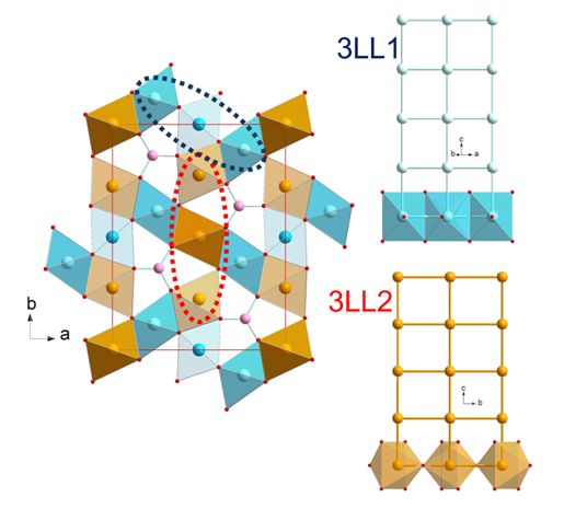
Ludwigite oxyborates M2M’BO5, where M and M’ are divalent and trivalent 3d metal ions, respectively, have an
intriguing orthorhombic crystal structure made of interconnected low dimensional units in the form of three-leg (3LL)
ladders, 3LL1 and 3LL2 [1] (Figure 1).
The existence of two crystallographically distinct sublattices is not anodyne : in Fe3BO5, Mössbauer and X-ray
diffraction studies at room temperature have evidenced that Fe3+ species occupy preferentially 3LL1, while 3LL2 is
occupied by Fe2+ [2]. In addition, a charge ordering transition has also been observed at TCO = 283 K [1], [4],
resulting from the ordering on 3LL1 of the extra itinerant electron within each Fe3+ triad. This also impacts the
magnetic properties : according to neutron diffraction results, 3LL1 and 3LL2 magnetically order, but independently,
at TN1 = 112 K and TN2 = 70 K, respectively, with orthogonal propagation vectors.
In the presented work, the isostructural system Fe3-xMnxBO5 has been studied by electron microscopy, neutron
scattering, and Mn and Fe K-edge X-ray absorption, combined with physical properties measurements. . A decrease
of both TN’s, more pronounced for TN2, along with a reduced ordered moment, is observed with increasing x, up to x =
1. For x = 1, only short-range magnetic ordering is observed on 3LL2, leading to a superparamagnetic ac
susceptibility response [3], in agreement with the preferred substitution of Mn2+ on 3LL2. Surprisingly, however, for
x = 1.5, 3D long-range ordering below TN = 100 K is observed, which couples both 3LLs within a new collinear k = 0
0 0 structure, without any sign of magnetic disorder.
Figure 1 : Ludwigite crystal structure and its two square ladders sublattices.
[1] M. MIR et al., Phys. Rev. Lett. 87, 147201 (2001) ; P. BORDET and E. SUARD, Phys. Rev. B 79, 144408 (2009).
[2] A. P. DOUVALIS, et al., J. Phys. Cond. Matter 14, PII S0953 (2002).
[3] A. MAIGNAN et al., J. Solid State Chem. 246, 209 (2017).Effets quantiques dans les systèmes magnétiques frustrés de type «pyrochlore»
Sylvain Petit1, Elsa Lhotel2, Julien Robert2
1
LLB, CEA Saclay, F-91191 Gif-sur-Yvette
2
Institut Néel, F-38000 Grenoble
La frustration magnétique, c'est à dire l'incapacité d'un système à satisfaire simultanément l'ensemble de ses
interactions, fait l'objet de nombreuses recherches en physique de la matière condensée. Ce phénomène, qui peut être
lié à la topologie du réseau cristallin ou aux compétitions entre interactions, constitue la source de nouveaux états
exotiques de la matière, dont la description va au-delà des modèles classiques. Les «glaces de spin» et leurs analogues
quantiques, constituent un exemple emblématique de cette physique. La structure cristallographique de ces matériaux
est basée sur un réseau de type «pyrochlore», formé d’un ensemble de tétraèdres connectés par leurs sommets, chaque
nœud étant occupé par un ion de terre rare magnétique (Tb, Dy, Ho, Pr, etc.). Dans ces composés, les orbitales
électroniques pertinentes ont une forme d’aiguille très fine, allongée en direction du centre de chaque tétraèdre. Le
moment magnétique de chaque ion ne peut alors pointer que vers l’intérieur ou vers l’extérieur d’un tétraèdre, à
l’instar des états \pm1 d’une variable Ising. L’état fondamental classique d’un tel système est très particulier car
infiniment dégénéré. En effet, la seule prescription pour le construire est de suivre une règle locale qui stipule que
chaque tétraèdre doit comporter deux spins «in» qui pointent vers l’intérieur et deux spins «out» qui pointent vers
l’extérieur. Ces dernières années, les physiciens théoriciens ont proposé une vision nouvelle du problème, remarquant
que la règle «two in-two out» est en fait analogue à la loi de conservation d'un flux magnétique fictif div B=0 en
électromagnétisme [1]. L’analogie est complète dès lors qu’on incorpore les fluctuations quantiques. En effet, les
fluctuations du champ magnétique fictif B, créent en vertu de la loi de l’induction rot E= dB/dt un champ électrique
«émergent» E. Selon les prédictions théoriques, une glace de spin quantique devrait comporter un spectre d’excitation
particulier caractérisé par un mode analogue au photon de l’électromagnétisme.
A l'aide d'exemples tirés de la littérature, nous souhaitons montrer dans cet exposé qu'en dépit de nombreux travaux,
les expériences réalisées jusqu'à aujourd'hui dans cette famille de composés n'ont pas encore permis de mettre en
évidence cette dynamique particulière, à l'exception possible de Pr2Hf2O7. Toutefois, l'influence des effets quantiques
a très clairement été observée, mettant en lumière une très grande richesse de comportements. Nous discuterons en
particulier le cas de Tb2Ti2O7, l'influence des défauts dans Pr2Zr2O7, la fragmentation magnétique dans Nd2Zr2O7, ainsi
que, au-delà de la physique propre aux spins Ising, l'ordre par le désordre dans Er2Ti2O7 et la compétition
d'interactions dans Yb2Ti2O7.
[1] Quantum spin ice: a search for gapless quantum spin liquids in pyrochlore magnets, M.J.P. Gingras and P.A.
McClarty, Rep Prog Phys 77 (2017) 056501.Magnetic interactions in the frustrated pentagonal compound Bi2Fe4O9
K. Beauvois1, V. Simonet2, E. Ressouche1, S. Petit3, M. Gospodinov4 and V. Skumryev5
1
CEA, INAC/SPSMS-MDN, Grenoble, France
2
Institut Néel-CNRS, Grenoble, France
3
CEA-CNRS, LLB Saclay, France
4
Institute of Solid State Physics, Bulgarian Academy of Sciences, Bulgaria
5
Universitat Autònoma de Barcelona, Spain
The Fe3+ ions in Bi2Fe4O9 materialize the first analogue of a magnetic pentagonal lattice [1]. The unit cell
contains two different sites of four iron atoms each, which have different connectivities with the other irons (three or
four neighbours for Fe1 and Fe2 respectively), and that form a lattice of pentagons. Because of its odd number of
bonds per elemental brick, this lattice is prone to geometric frustration. The compound magnetically orders around
240 K: the resulting spin configuration on the two sites is the same, i.e. two orthogonal pairs of antiferromagnetic
spins in a plane, with a global rotation between the two sites Fe1 and Fe2. This peculiar magnetic structure, which is
the result of the complex connectivity, has opened new perspectives in the field of magnetic frustration.
Here, we present the work in progress concerning the understanding and the consequences of the peculiar
magnetic interactions in this original system. The magnetic excitations have been investigated by inelastic neutron
scattering using thermal neutron triple axis spectrometers at the LLB and the ILL. The confrontation of the
experimental results with spinwave calculations shows that there is a hierarchy of the interactions between the iron
sites in the lattice and suggest that this system can be alternatively understood as a frustrated lattice of classical spin
dimers. This transforms to a paramagnetic state of classical dimers above the Néel temperature as suggested by
preliminary magnetization distribution maps measured at ILL using polarized neutrons under an applied magnetic
field.
Our new experimental results on Bi2Fe4O9 open interesting perspectives in the field of frustrated pentagonal
lattices.
[1] E. Ressouche, V. Simonet, B. Canals, M. Gospodinov, V. Skumryev, Phys. Rev. Lett. 103, 267204 (2009).Instrumentation
SessionIN6-SHARP: towards a new cold neutron spectrometer at ILL.
Illustration of the potentialities of QENS to probe the dynamics of Ionic liquids in bulk and under
1D nanometric confinement.
F. Ferdeghini1, Q. Berrod1, P. Judeinstein1, J. Dijon2, J.-M. Zanotti1
1.
Laboratoire Léon Brillouin (CEA-CNRS), Université Paris-Saclay, 91191 Gif-sur-Yvette, France
2.
CEA/LITEN/DTNM, 38054 Grenoble, France
Following the agreement to strengthen the Franco-Swedish cooperation in the field of neutron
scattering, the Laboratoire Léon Brillouin (LLB) is involved in the construction of an inelastic time-of-
flight spectrometer. After the announcement of the Orphée reactor shutdown in 2020, the project
originally planned at Saclay has been be transferred to the Laue Langevin Institute (ILL). This
renaissance takes the form of an A type CRG contract concluded on September 29th 2017 between the
DRF of the CEA, the INP of the CNRS, and the ILL. This new project SHARP (Spectromètre Hybride
Alpes Région Parisienne) consists of a complete rebuilding of the IN6 secondary spectrometer: sample
environment, time-of-flight chamber and detection. This seminar will start by an update on the project.
We will then illustrate the potentialities of Quasi-Elastic Neutron Scattering (QENS) in the study of
Ionic liquids (ILs). ILs are pure solutions of charged organic molecules with no solvent. These
molecular electrolytes show a property original for a pure liquid: they self-organize in nanometric
fluctuating aggregates [1]. When probed at the macroscopic scale, ILs behave as highly dissociated (i.e.
strong) electrolytes [2] while, at the molecular scale, they show clear characteristics of weak ionic
solutions [3]. In this talk, we report a multi-scale analysis that sheds new light on these apparently at
odd behaviors [4,5].
Due to their remarkable chemical and electrochemical stability, ILs have been identified as prime
candidates electrolytes for the development of new safe and sustainable energy storage systems. We
show [6] a noticeable enhancement (by a factor 3) of the transport properties of a neat IL under CNT
(Carbon NanoTube) confinement in a 1D situation. Such CNT membranes are a possible route to boost
the transport properties and hence the specific power of lithium batteries [7]. We then address the
conductivity of electrolytes directly relevant to the field of electrochemical storage systems: ILs
charged with lithium salts. We show that these electrolytes confined in 1D CNT membranes show a
drastic and unprecedented increase in ionic conductivity. Compared to the bulk analogues, we indeed
report conductivity gains by a factor up to 50 upon macroscopic 1D CNT confinement.
Such a disruptive concept of a 1D CNT based separator laying at the cross-road of basic science is
probably of interest for future technological outcomes.
[1] Hayes, R., Warr, G. G. & Atkin, R. Structure and Nanostructure in Ionic Liquids. Chem. Rev. 115,
6357–6426 (2015).
[2] Lee, A. A., Vella, D., Perkin, S. & Goriely, A. Are Room-Temperature Ionic Liquids Dilute
Electrolytes? J. Phys. Chem. Lett. 6, 159–163 (2015).
[3] Gebbie, M. A., Dobbs, H. A., Valtiner, M. & Israelachvili, J. N. Long-range electrostatic
screening in ionic liquids. Proc. Natl. Acad. Sci. 112, 7432–7437 (2015).
[4] Ferdeghini, F. et al. Nanostructuration of ionic liquids: impact on the cation mobility. A multi-
scale study. Nanoscale 9, 1901–1908 (2017).
[5] Quentin Berrod et al. Ionic Liquids: evidence of the viscosity scale-dependence. Sci. Rep. 7,
(2017).
[6] Berrod, Q. et al. Enhanced ionic liquid mobility induced by confinement in 1D CNT
membranes. Nanoscale 8, 7845–7848 (2016).
[7] Berrod, Q., Ferdeghini, F., Judeinstein, P. & Zanotti, J.-M. Nanocomposite membranes for
electrochemical devices. Patent WO 2016151142 A1. (2016).Augmented Reality in Neutron Experiments
M. Boehm 1, Y. Le Goc 1, T. Weber1 and P. Mutti 1
1
Institut Laue-Langevin, 71 avenue des Martyrs, 38042 Grenoble, France
Today’s user community appreciates the active hand-on participation in experiments and the scientific exchange at the
facilities, which act as international crossings for experts of very different scientific fields. Nevertheless, this way of
access also imposes long missions on the users and puts time constraints on participants and instrument schedules,
which hinders flexible handling of experiments or rapid integration of hot scientific topics. In future, a major severe
constraint to the present user access arises from changing legislation, which has to assure increasing security standards
for protecting nuclear installations and/or for protecting against misappropriation of materials on-site. Especially this
last point might completely modify the future way of performing experiments and might have severe impact on the
attractiveness of the technic as such.
Here, we suggest exploring state-of-the-art computing technology and develop an alternative, virtual, access mode for
neutron experiments (NEVA). We suggest merging existing instrument control software, computing and scientific
software tools, state-of the art communication technology and modern 3D animation into a new virtual platform. This
platform would not concurrence existing tools for instrument control, data sorting and data analysis, but it will
complement them. It will provide a generic interface for importing, illustrating and sharing information relevant to the
experiments. In parallel to the virtual platform we envisage testing the usefulness of augmented reality at the
instrument and experimental areas, for scientists and technical staff on-site. First concepts of a virtual platform have
been successfully tested based on experience with existing single crystal inelastic neutron scattering software (TAKIN
[1], vTAS [2]). NEVA will be open source and platform independent and usable with different instrumental
techniques at the different facilities. The software will be a modern web application written in fast, just-in- time
compiled Javascript.
[1] T. Weber, R. Georgii, P. Böni, SoftwareX 5 (2016), p. 121-126.
[2] M. Boehm, A. Filhol, Y. Raoul, J. Kulda, W. Schmidt, K. Schmalzl, E. Farhi, Nucl. Instr. and Meth. A 697 (2013),
p. 40-44.Quelle suite instrumentale sur une source compacte?
F. Ott
Laboratoire Léon Brillouin, CEA, CNRS, Université Paris-Saclay, 91191 Gif sur Yvette
Les développements technologiques permettent d’envisager la construction de sources de neutrons compactes dont le
coût d’investissement est significativement plus faible que la construction d’un réacteur nucléaire ou d’une source à
spallation. Diverses stratégies peuvent être envisagées, de la construction d’une source aux performances modestes en
lien avec l’université avec une vocation de formation et une utilisation pour des expériences de caractérisation simples
à une source à haute brillance dont les performances se comparent à une source à spallation ou un réacteur de
puissance moyenne.
Les performances de différents types d’instruments seront présentées. Une discussion sur les différentes suites
instrumentales envisageables en fonction de la puissance et des caractéristiques temporelles de la source sera ouverte.Condensed Matter
SessionPhysical-chemistry of gas hydrates : from astrophysics to new opportunities for energy technologies
A. Desmedt
Groupe Spectroscopie Moléulaire – Institut des Sciences Moléculaires UMR5255 CNRS – Univ. Bordeaux
Gas hydrates are ice-like systems made of a network of hydrogen-bonded water molecules (forming host cages) that is
stabilized by the presence of foreign guest molecules [1]. The natural existence of large quantities of hydrocarbon
hydrates in deep oceans and permafrost is certainly at the origin of numerous applications in areas such as energy and
geophysics sciences and technologies [2]. Their hypothetical occurrence in extraterrestrial objects (planets, comets
and planetesimal) is also the subject of numerous researches in astrophysics [3]. At a fundamental level, their
nanostructuration confers on these materials specific properties (e.g. molecular selectivity, transport properties) for
which the host-guest interactions play a key role [4,5]. These interactions occur on a broad timescale and thus require
the use of multi-technique approach (Neutron scattering, Raman, NMR, Classical and ab-initio Molecular Dynamics
Simulations). The presentation will review recent results obtained on the physical chemistry of clathrate hydrates
towards two main issues - for which neutron scattering brings significant contributions: gas selectivity and structural
metastability on one hand, and super-protonic conduction on the other hand.
Recent theoretical works suggest that the nitrogen depletion observed on the Jupiter family comet 67P/Churyumov-
Gerasimenko might be due to preferential encapsulation of carbon monoxide with respect to nitrogen inside mixed gas
hydrate [6]. The presentation will report the first experimental investigations of such a preferential trapping, together
with unusual structural metastability, by means of Raman scattering and Neutron diffraction in various mixed gas
(CO, CO2, N2) hydrates [7,8,9,10,11].
In addition to gaseous species, clathrate hydrates may encapsulate strong acids. Such supramolecular assembly leads
to generate super-protonic conductors (i.e. with protonic conduction of the order of 0.1S/cm) [12]. Quasi-elastic
neutron scattering is a unique technique for disentangling the proton transport mechanism involved in such ice-like
systems [13,14]. This issue will be reviewed by outlining the contributions of Neutron scattering together with
complementary techniques such as ab-initio Molecular Dynamics, Raman imaging or pulsed-field gradient proton
NMR. Moreover, new opportunities in the area of energy (electrochemical energy production [15] and hydrogen
storage [16,17,18]) are offered thanks to the strong acidic character of clathrate hydrates. These points will be
outlined.
[1] E. D. Sloan and C. A. Koh, Clathrate Hydrates of natural gases, Taylor & Francis-CRC Press, Boca Raton, FL, 3rd
edn, 2008.
[2] L. Ruffine, D. Broseta, A. Desmedt, Eds, Gas Hydrates 2: Geoscience Issues and Potential Industrial Applications,
Wiley: London (2018).
[3] e.g. G. Tobie et al, Nature 2006, 440, P.61 // Nature 2015, 519, p.162.
[4] D. Broseta, L. Ruffine, A. Desmedt, Eds, Gas hydrates 1: Fundamentals, Characterization and Modeling, Wiley:
London (2017).
[5] A. Desmedt, et al. Eur. Phys. J. Special Topics 213 (2012) 103-127
[6] S. Lectez, et al, Astrophys. J. Lett., 2015, 805: L1.
[7] C. Petuya, et al, J. Phys. Chem. C 121(25) (2017) 13798–13802
[8] C. Petuya, et al, J. Phys. Chem. C 122(1) (2018) 566 –573
[9] C. Petuya, et al, Crystals 8 (2018) 145(1-13)
[10] C. Petuya, et al, Chem. Comm. 54 (2018) 4290-4293.
[11] C. Petuya, et al. Submitted to PCCP (2018).
[12] J. Cha, et al. J. Phys. Chem. C 2008, 112, 13332−13335.
[13] L. Bedouret, et al, J. Phys. Chem. B 118 (2014) 13357−13364.
[14] A. Desmedt, et al, Solid State Ionics, 252 (2013) 19-25
[15] S. Desplanche et al, in preparation.
[16] E. Pefoute, et al, J. Phys. Chem. C 116(32) (2012) 16823
[17] A. Desmedt, et al, J. Phys. Chem. C 119 (2015) 8904-8911
[18] T.T. Nguyen, et al, in preparation.Anomalous crystal field splitting in antiferro-quadrupolar compound TmTe and isostructural Tm0.1Yb0.9Te
E. Clementyev 1,2 and J.-M. Mignot 3
1
I. Kant Baltic Federal University, Kaliningrad, Russia
2
Petersburg Nuclear Physics Institute, Gatchina, St-Petersburg, Russia
3
Laboratoire Léon Brillouin, CEA-CNRS, CEA/Saclay, France
The Tm monochalcogenide family displays exotic physical properties including valence instabilities and quadrupolar
ordering. In particular TmTe was reported to undergo a phase transition at TQ=1.7K above the magnetic ordering
temperature (TN=0.4K). One of the most crucial questions is the origin of the ground state of the Tm2+ 4f multiplet in
TmTe (see [1] and references therein). A lack of experimental consensus regarding the the detailed level sequence of
the crystal field level scheme is a significant problem. Experimental data collected on pure TmTe show relatively
broad crystal field transitions below 1 meV (INS measurements have been performed on the 4F1 triple-axis
spectrometer (LLB, Saclay). To avoid the broadening due to the quadrupolar correlations the measurements on a
diluted compound, namely 10% Tm impurity in the nonmagnetic YbTe matrix have been performed on the time-of-
flight spectrometer FOCUS (SINQ, PSI). The incoming neutron energy was 3.27 meV yielding the energy resolution
at the elastic position of about 0.1 meV. For the first time two distinct crystal field transitions have been observed (at
E~0.3 meV and E~0.9 meV) in TmTe-based system. Such a spectrum is unique since the total magnitude of the
crystal field splitting is one or even two orders of magnitude smaller than in the majority of the 4f systems and in
isostructural metallic TmxLa1-xTe systems [2]. The observed effects are opposite to the predictions of the point charge
and related models. The puzzling small magnitude of the total crystal field splitting in TmTe is discussed.
[1] R. Shiina and N. Tatsuya, J. Phys. Soc. Jpn., vol: 77, num: 12 (2008) 124715/1-124715/6.
[2] T. Matsumura et al., Phys. Rev. B 66 (2002) 104410.You can also read