Mandibular sawing in a snail eating snake - Nature
←
→
Page content transcription
If your browser does not render page correctly, please read the page content below
www.nature.com/scientificreports
OPEN Mandibular sawing in a snail‑eating
snake
Yosuke Kojima1*, Ibuki Fukuyama2, Takaki Kurita3, Mohamad Yazid Bin Hossman4 &
Kanto Nishikawa2,5
The jaws of vertebrates display a striking diversity in form and function, but they typically open and
close like a trapdoor rather than sliding like a saw. Here, we report unique feeding behaviour in the
blunt-headed snail-eating snake, Aplopeltura boa (family Pareidae), where the snake cuts off and
circumvents the indigestible part (the operculum) of its prey in the mouth using long sliding excursions
of one side of the mandible, while the upper jaws and the mandible on the other side maintain a
stable grasp on the prey. This behaviour, which we call ‘mandibular sawing’, is made possible by
extraordinarily independent movements of the jaw elements and is a surprising departure from usual
feeding behaviour in vertebrates.
Talos, the Greek mythological inventor, invented a saw inspired by a serpent jawbone or a fish s keleton1, but none-
theless, the jaws of snakes or other vertebrates usually do not act like a saw due to anatomical constraint on the
jaw movements (but s ee2). Because the diversity in vertebrate jaws represents modifications of the homologous
apparatus, their form and function are strongly constrained by the phylogeny3. Advanced snakes differ from most
other vertebrates in their ability to move the left and right jaws virtually independently and swallow their prey
using alternate movements of the two sides of the j aws4. Snail-eating snakes of Southeast Asia (Pareidae) or the
Neotropics (Dipsadinae) retain the unilateral mobility of the jaws, but their feeding apparatus is further modi-
fied; these snakes have lost the articulation between the upper and lower jaws in contrast to most other snakes,
and this makes the lower jaws of the snail-eating snakes extensively mobile and allows the mandibles to perform
independent sliding excursions4–9. Several species use asynchronous retractions of the mandibles to extract snails
from their shells or ingest s lugs10–16. Feeding behaviors of most species of those peculiar snakes, however, have
never been described perhaps due to limited access to these tropical, nocturnal, and secretive animals.
The blunt-headed snail-eating snake, Aplopeltura boa, is a pareid species that feeds on snails, including oper-
culate species17. Gastropodan opercula are indigestible for s nakes18, like their shells, and therefore are potentially
harmful when consumed (e.g., they may cause intestinal obstruction). Indeed, feeding experiments showed that
a snail-specialist pareid did avoid eating all of three species of sympatric operculate snails, whereas it consumed
most of sympatric non-operculate snail species19. We collected A. boa and a syntopic, abundant operculate snail,
Leptopoma sp. (Cyclophoridae) in a rainforest in Borneo and observed the snake feeds on the tough prey. Also,
we investigated the relative abundance of operculate and non-operculate snails in the habitat of A. boa.
Results and discussion
We found that operculate snails were more abundant than were non-operculate snails: 34 and 22 individuals
were encountered, respectively. Among snails of moderate size (shell width, 10–20 mm), which were often con-
sumed by A. boa, operculate snails were more than 10 times as abundant as were non-operculate snails (25 and
2 individuals were encountered, respectively).
Individuals of A. boa (n = 8) readily preyed upon Leptopoma sp. (n = 30) in our feeding trials. Upon capture,
the snakes immediately inserted their mandibles into the aperture of the shell and then extracted the operculate
soft body using the mandibles. After extraction, the snakes regurgitated the extracted snail and precisely repo-
sitioned it so that the operculum protruded out of the mouth and the junction of the operculum and the soft
body came to lie along the mandible on the right (n = 24, 80%) or the left (n = 6, 20%) side. From this position,
the snakes moved the side of the relevant mandible backward and forward, while the snail was held in the stable
1
Department of Biology, Toho University, Funabashi, Chiba 274‑8510, Japan. 2Graduate School of Human and
Environmental Studies, Kyoto University, Sakyo‑ku, Kyoto 606‑8501, Japan. 3Chiba Biodiversity Center, Aoba‑cho
955‑2, Chuo‑ku, Chiba 260‑8682, Japan. 4Research, Development and Innovation Division, Sarawak Forest
Department, 93250 Sarawak, Malaysia. 5Graduate School of Global Environmental Studies, Kyoto University,
Sakyo‑ku, Kyoto 606‑8501, Japan. *email: yosukekojima1@outlook.jp
Scientific Reports | (2020) 10:12670 | https://doi.org/10.1038/s41598-020-69436-7 1
Vol.:(0123456789)www.nature.com/scientificreports/
Figure 1. Infrared images illustrating mandibular sawing by the blunt-headed snail-eating snake, Aplopeltura
boa. (a) The extracted operculate snail in the snake’s mouth. (b) The snail has been accurately repositioned. (c)
The operculum being sliced off. (d) The soft parts of the snail being ingested while the operculum is discarded. A
white and black arrow indicates the position of the operculum and the tip of the mandible, respectively. Videos
showing this behavior are available with supplementary information of this article (Videos S1, S2).
position by the upper jaws and the mandible on the other side. These mandibular movements were especially
vigorous when the mandible was being retracted. Six to 51 (median, 14) strokes of these sliding movements
resulted in the removal of the operculum (Fig. 1, Supplementary Information, Videos S1, S2). Snakes consumed
solely the soft part of the snails, while discarding the shell and the operculum (n = 30, 100%). Discarded oper-
cula retained little soft tissue. The duration of extraction, repositioning, and sawing processes ranged 36–274
(median, 96), 9–459 (median, 66), and 15–428 (median, 30) seconds, respectively (Supplementary Information,
Table S1). Thus, the snakes spent considerable time for handling of the extracted snail (reposition and sawing).
The sequence of feeding was highly consistent among all cases, and the routine operculum-removing behaviour
presumably allows A. boa to regularly consume the otherwise hazardous prey, which is abundant in its habitat.
The feeding apparatus of A. boa is illustrated in Fig. 2. Aplopeltura boa exhibits a set of derived morphological
features known in other pareids and dipsadines, including short snout, short pterygoids, reduced supratemporals,
long mandibles, and comb-like mandibular teeth. The skull of A. boa is short and tall, in which the snout is very
short, and the orbits are exceptionally large. The pterygoids are greatly shortened, and their posterior ends are
completely detached from the quadrato-mandibular joint. The quadrates are remarkably long and stout, extend-
ing ventrally rather than ventrolaterally. The mandibles are long and carry dense teeth, which are more robust
than the maxillary teeth. The size and interval of mandibular teeth gradually change along the mandible; the
anterior teeth are larger and sparser. The lower jaw unit (the mandible and the quadrate) is L-shaped, and the
mandible travels anteroposteriorly when the quadrate swings backward and forward, as anticipated or observed
in other pareids or dipsadines. This mechanism enables independent, substantial anteroposterior excursions of
the mandible (Fig. 3), which is used for the extraction and the sawing processes during feeding on the operculate
snails. The elongated quadrates are likely to contribute to long mandibular e xcursion18.
Most of > 3,700 species of snakes swallow their prey whole, and prey-breaking behaviours are known only
from a few species that feed either on c rabs20–23 or termites24–26, which may be relatively easily broken into seg-
ments. These snakes grasp their arthropod prey with their jaws and break it apart usually using the movements
of the head or the trunk. In contrast, A. boa cuts its mollusk prey using independent movements of the lower jaw.
The mandibular sawing is, therefore, a surprising evolutionary solution for the limbless animals to utilize new
food. This dexterous behaviour is especially surprising given that few other vertebrates, if any, are able to sever
food in the mouth using unilateral sliding movements of a jaw element like A. boa. The evolutionary invention
of sawing was evidently made possible by the unique feeding mechanism in the snail-eating snakes. Extensive
mobility of the mandibles is a convergent trait in the two distinct lineages of snakes (pareids and dipsadines)
that feed on slugs or snails, suggesting it is an adaptation to feeding on their slippery p rey4–9. It is likely that
acquisition of the free mandibular apparatus promoted the subsequent evolution of the novel behaviour and has
resulted in functional versatility of the free-moving jaw elements.
Most pareids and some dipsadines have a larger number of teeth on the right mandible than on the left as
feeding specialization to extract dextral (clockwise-coiled) s nails13,15. There is a cline in the degree of the denti-
tional asymmetry in correlation with diets, where snail-specialist species have highly asymmetrical mandibles,
whereas slug-specialist species have symmetrical mandibles13,15. However, A. boa is an exception of this pattern
Scientific Reports | (2020) 10:12670 | https://doi.org/10.1038/s41598-020-69436-7 2
Vol:.(1234567890)www.nature.com/scientificreports/
Figure 2. Feeding apparatus of the blunt-headed snail-eating snake, Aplopeltura boa. CT images of the skull
and the jaws from left lateral (a), posterior (b), ventral (c), and dorsal (d) views and the quadrates and the
mandibles on the left (e) and right (f) sides. m, mandible; p, pterygoid; q, quadrate. These images are from the
specimen KUHE59285.
because individuals exhibit only weak mandibular asymmetry despite its snail diet13. In the phylogeny, A. boa is
nested within the derived clade with mandibular a symmetry27, suggesting the presence of additional selective
forces toward the mandibular symmetry in this species. By showing the additional role of its mandibles (cutting
the prey), our results suggest functional trade-offs in A. boa (typical comb-like teeth in snail-eating snakes are
expected to facilitate extraction by providing a firm grip on the prey but probably are not optimal to cut the prey
tissue), highlighting the importance of behavioural studies to understand selective forces on functional units.
Methods
This study was conducted in the Gunung Mulu National Park, Sarawak, Malaysia (4.0440°N, 114.8144°E). We
collected A. boa and Leptopoma sp. by visual search and brought them to the Research Centre in the national
park for the feeding trials. All but three snakes that were used for other studies were released at the site of capture
after trials.
Feeding trials were conducted between 2100 and 0400 h. We fed A. boa with Leptopoma sp. and video-
recorded feeding events with infrared video cameras (HC-VX985M, Panasonic; FDR-AX30, Sony).
We conducted census survey on snails by walking the habitats of A. boa carefully looking for snails and
counting all snails larger than 5 mm in shell width. This survey was conducted on two nights in August and
December, respectively.
We conducted micro CT-scanning on six specimens of A. boa held at Kyoto University or Sarawak Forest
Department (specimen nos: KUHE27025, KUHE56016, KUHE57425, KUHE59285, SRC01008, and SRC01009).
All procedures followed the Animal Experiment Guideline of Kyoto University and were approved by the
ethical review committee of the Graduate School of Human and Environmental Studies of Kyoto University and
Scientific Reports | (2020) 10:12670 | https://doi.org/10.1038/s41598-020-69436-7 3
Vol.:(0123456789)www.nature.com/scientificreports/
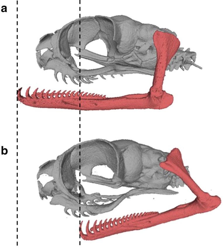
Figure 3. Movements of the lower jaw in the blunt-headed snail-eating snake, Aplopeltura boa. CT images of
the skull and the jaws from left lateral view with the mandible protracted (a) and slightly retracted (b). These
images and the videos on feeding behaviour demonstrate that the mandibles slide about half the length of the
skull. These mandibular movements can be performed unilaterally. Images (a) and (b) represent the specimen
SRC01008 and SRC01009, respectively.
the Research, Development and Innovation Division of the Sarawak Forest Department (approval no. 30-A-7).
All fieldwork was permitted by the State Government of Sarawak, the Sarawak Forest Department, and the
Gunung Mulu National Park (permission nos. (133)JHS/NCCD/600–7/2/107, WL72/2018, WL103/2018, and
WL68/2019).
Received: 27 March 2020; Accepted: 6 July 2020
References
1. Graves, R. The Greek Myths (Penguin Books, London, 1960).
2. Gorniak, G. C., Rosenberg, H. I. & Gans, C. Mastication in the tuatara, Sphenodon punctatus (Reptilia: Rhynchocephalia): structure
and activity of the motor system. J. Morphol. 171, 321–353 (1982).
3. Schwenk, K. Feeding: Form, Function and Evolution in Tetrapod Vertebrates (Academic Press, New York, 2000).
4. Cundall, D. & Greene, H. W. Feeding in snakes in Feeding: Form, Function, and Evolution in Tetrapod Vertebrates (ed. Schwenk,
K.) 293–333. (Academic Press, New York, 2000).
5. Dunn, E. R. The status of the snake genera Dipsas and Sibon, a problem for “quantum evolution”. Evolution 5, 355–358 (1951).
6. Brongersma, L. D. Some features of the Dipsadinae and Pareinae (Serpentes, Colubridae). Proc. K. Ned. Akad. Wet. (Ser. C) 61,
7–12 (1958).
7. Peters, J. A. The snakes of the subfamily Dipsadinae. Misc. Publ. Mus. Zool. Univ. Mich. 114, 1–224 (1960).
8. Kofron, C. P. A review of the Mexican snail-eating snakes, Dipsas brevifacies and Dipsas gaigeae. J. Herpetol. 16, 270–286 (1982).
9. Savitzky, A. H. Coadapted character complexes among snakes: fossoriality, piscivory, and durophagy. Am. Zool. 23, 397–409 (1983).
10. Gans, C. Reptiles of the World. (Ridge Press/Bantam Books, New York, https://carlgans.org/reptiles-of-the-world/, 1975).
11. Sazima, I. Feeding behavior of the snail-eating snake, Dipsas indica. J. Herpetol. 23, 464–468 (1989).
12. Götz, M. The feeding behavior of the snail-eating snake Pareas carinatus Wagler 1830 (Squamata: Colubridae). Amphibia-Reptilia
23, 487–493 (2002).
13. Hoso, M., Asami, T. & Hori, M. Right-handed snakes: convergent evolution of asymmetry for functional specialization. Biol. Lett.
3, 169–173 (2007).
14. Danaisawadi, P., Asami, T., Sutcharit, C. & Panha, S. Predatory behavior of the snail-eating snake Pareas carinatus (Boie, 1828)
(Squamata: Pareidae): an ethogram study. Trop. Nat. Hist. 16, 21–31 (2016).
15. Hoso, M. Asymmetry of mandibular dentition is associated with dietary specialization in snail-eating snakes. PeerJ 5, e3011 (2017).
16. Laporta-Ferreira, I. L., Salomão, M. G. & Sawaya, P. Biologia de Sibynomorphus (Colubridae, Dipsadinae)—Reprodução e hábitos
alimentares. Rev. Brasil. Biol. 46, 793–799 (1986).
17. Stuebing, R. B. & Inger, R. F. A Field Guide to the Snakes of Borneo, 84–85 (Natural History Publications (Borneo), Kota Kinabalu,
1999).
18. Ray, J. M., Montgomery, C. E., Mahon, H. K., Savitzky, A. H. & Lips, K. R. Goo-eaters: diets of the neotropical snakes Dipsas and
Sibon in Central Panama. Copeia 2012, 197–202 (2012).
Scientific Reports | (2020) 10:12670 | https://doi.org/10.1038/s41598-020-69436-7 4
Vol:.(1234567890)www.nature.com/scientificreports/
19. Hoso, M. & Hori, M. Identification of molluscan prey from feces of Iwasaki’s slug snake, Pareas iwasakii. Herpetol. Rev. 37, 174–176
(2006).
20. Shine, R. & Schwaner, T. Prey constriction by venomous snakes: a review, and new data for Australian species. Copeia 1985,
1067–1071 (1985).
21. Jayne, B. C., Voris, H. K. & Ng, P. K. L. Snake circumvents constraints on prey size. Nature 418, 143 (2002).
22. Voris, H. K. & Murphy, J. C. The prey and predators of homalopsine snakes. J. Nat. Hist. 36, 1621–1632 (2002).
23. Noonloy, T. et al. Crab-ripping: An unusual feeding behavior newly recorded in freshwater snakes. Bull. Chicago Herpetol. Soc. 53,
53–56 (2018).
24. Smith, H. M. Curious feeding habit of a blind snake, Leptotyphlops. Herpetologica 13, 102 (1957).
25. Reid, J. R. & Lott, T. E. Feeding of Leptotyphlops dulcis dulcis (Baird and Girard). Herpetologica 19, 141–142 (1963).
26. Mizuno, T. & Kojima, Y. A blindsnake that decapitates its termite prey. J. Zool. 297, 220–224 (2015).
27. Figueroa, A., McKelvy, A. D., Grismer, L. L., Bell, C. D. & Lailvaux, S. P. A species-level phylogeny of extant snakes with description
of a new colubrid subfamily and genus. PLoS ONE 11, e0161070 (2016).
Acknowledgements
Funding was provided by the Asahi Glass Foundation (to K.N. and Y.K.), the Japan Society for the Promotion
of Science (Grant No. 18J00809 to Y.K.), the Shikata Memorial Trust for Nature Conservation (to I.F.), and JST/
JICA, SATREPS (to K.N.). We thank the State Government of Sarawak and the Sarawak Forest Department for
permitting us to conduct the research, the Gunung Mulu National Park for permission and cooperation in the
field, R. Fukuyama for assistance with field research, N. Morimoto, M. Nakatsukasa, and S. Hara for cooperation
in CT scanning, and A. M. Durso for valuable comments on the manuscript.
Author contributions
Y.K. and I.F. conceived/designed the study and collected data. K.N., M.Y.H., and T.K. provided research support.
Y.K. analyzed data and wrote the manuscript. All authors reviewed and edited the manuscript and approved
the final version.
Competing interests
The authors declare no competing interests.
Additional information
Supplementary information is available for this paper at https://doi.org/10.1038/s41598-020-69436-7.
Correspondence and requests for materials should be addressed to Y.K.
Reprints and permissions information is available at www.nature.com/reprints.
Publisher’s note Springer Nature remains neutral with regard to jurisdictional claims in published maps and
institutional affiliations.
Open Access This article is licensed under a Creative Commons Attribution 4.0 International
License, which permits use, sharing, adaptation, distribution and reproduction in any medium or
format, as long as you give appropriate credit to the original author(s) and the source, provide a link to the
Creative Commons license, and indicate if changes were made. The images or other third party material in this
article are included in the article’s Creative Commons license, unless indicated otherwise in a credit line to the
material. If material is not included in the article’s Creative Commons license and your intended use is not
permitted by statutory regulation or exceeds the permitted use, you will need to obtain permission directly from
the copyright holder. To view a copy of this license, visit http://creativecommons.org/licenses/by/4.0/.
© The Author(s) 2020
Scientific Reports | (2020) 10:12670 | https://doi.org/10.1038/s41598-020-69436-7 5
Vol.:(0123456789)You can also read