Nausea and Vomiting in Pregnancy Dr C Griffiths Wexham Park Hospital
←
→
Page content transcription
If your browser does not render page correctly, please read the page content below
16/04/2019

                   Nausea and Vomiting in Pregnancy
                                       Dr C Griffiths
                              Wexham Park Hospital
Oxford

   • 45 y/o female
   • P0+1 (Miscarriage at 9 weeks in 2017)
   • BMI 22

   • Background: Hypothyroidism, Gestational Diabetes,
     Coeliac Disease

   • DCDA twins – IVF Therapy

   • Routine scans and care normal

                                                                 1
16/04/2019
• Presented with Projectile vomiting and dizziness at 31+4/40
• 3-4 episodes a day for 1 week
• Reports ‘black-out episodes’
• Dizziness, feeling faint
• Good Fetal movements
• No abdominal pain
• No PV symptoms, No urinary or bowel symptoms
• No Pruritus, no SOB or chest pain
• DH – Levothyroxine 75mcg, Aspirin 75mg, Metformin 500mg
• SH – Ex Smoker
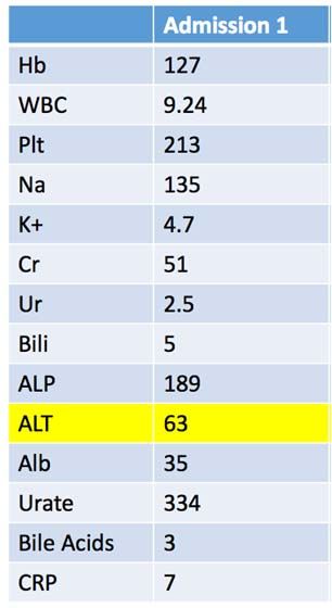
Investigations
• Urine Dip 3+ Ketones
• Urine culture NAD
• Bloods:
• ALT – 63
• CRP – 7
Plan
1. Admit for monitoring
2. IV Fluids
3. IV Antiemetics
4. TEDs and VTE prophylaxis
216/04/2019
Following day
• No further vomits
• ? Rise in ALT due to dehydration and vomiting
• Growth Scan
• Normal Liquor
• Fetal Heart rate visualised
• Twin 1 Cephalic, Twin 2 Breech
• EDF Positive
• Reassuring CTG
• Discharged Home
• With advice to return if symptoms return or new symptoms develop
• Represented 5 days later
• Now 32+2/40
• Pc: Vomiting for 2 days + Reduced oral intake
• Husband reported that pt had been acting ‘odd’ on and off
• No PV bleed, No abdominal pain
• No urinary or bowel symptoms, normal fetal movements
Temperature 36.5
Investigations
O2 Sats 100%
• Urine dip 4+ Ketones, 2+ Protein
RR 17
HR 72
BP 110/74
316/04/2019
Admission 1 (31+4) Admission 2 (32+2)
• Bloods: Hb 127 123
WBC 9.24 13.09
Plt 213 181
Na 135 134
K+ 4.7 4.7
Cr 51 53
Ur 2.5 2.3
Bili 5 6
ALP 189 229
ALT 63 97
Alb 35 33
Urate 334 307
Bile Acids 3 2
CRP 7 4
Plan
1. Admit for monitoring
2. IV Fluids
3. IV Antiemetics
4. TEDs and VTE prophylaxis
Day 2:
• Ongoing dizziness and tiredness
• No further vomiting
• Reassuring CTGs
• Rise in ALT US liver:
• Normal, No gallstones seen, 5mm polpy in the gall bladder
• BMs stable
• ?Medical Review
• ?Psych Review – due to Husband’s concerns over mood
Day 3:
• Midwife reports multiple episodes of confusion
• Patient now very withdrawn (found to be sitting in her own vomit)
• Monotonous voice, lethargic, denies hearing voices
• AMTS 9/10 – said the year was 1918 repeatedly
• No nystagmus, no focal neurology
416/04/2019
• Blood tests – FBC: Normal
Admission 1 Admission 2 Day 2
Bili 5 6 ‐
ALP 189 229 204
ALT 63 97 100
Alb 35 33 ‐
Urate 334 307 ‐
Bile Acids 3 2 2
CRP 7 4 ‐
• Medical Review
• ECG – Sinus Rhythm
• Full Confusion Screen - sent
• Urine MSU – sent
• Psych Review
• Presented with low mood and anxiety
• Expressed concerns over twins and likelihood of having an emergency section
• Worried kids may be taken away
• Psych suggested to mobilise off ward and planned to review in 2 days
Day 5
• Urine Dip – 3+ Ketones
• No clinical evidence of PET/Fatty Liver/OC Medics impression gastroenteritis
• CTG remains reassuring
Day 6
• Feeling better but struggling to remember the day of the week
• Fetal movements normal
• Urine dip now NAD
• Negative liver screen – ANA, Mitochondiral Abs, Gastric Parietal cells, Smooth
muscle abs
• MDT Discussion– ongoing confusion, tiredness, rise in ALT ?cause MRI Head
Admission 1 Admission 2 Day 2 Day 4 Day 5 Day 6
Bili 5 6 ‐ 4 4 4
ALP 189 229 204 220 207 231
ALT 63 97 100 152 163 178
Bile Acids 3 2 2 ‐ 4 ‐
CRP 7 4 ‐ 2 ‐ ‐
516/04/2019
Day 7
• MRI Head Report
• Cerebellar Haemangioblastoma
• 4.5cm x 3 x 3.2cm cystic mass in right cerebella hemisphere
• Seen to contain a peripheral solid nodule in its inferior cyst wall
• No obvious intrinsic haemorrhage or nodule calcification
• Marked surrounding vasogenic oedema
• 4th ventricle compressed and right side of brainstem compressed
• Transferred to Neurosurgical department for MDT management with
Obstetrics, Neurology and Neonatal
616/04/2019
• Tumours of vascular origin
• Grade I WHO tumours
• Usually well circumscribed and Benign
• With a highly vascular mural nodule and a peripheral cyst which has similar contents as
blood plasma
• Occur in the Central Nervous System (also the kidneys, liver and pancreas)
• Account for 1-2% of all intracranial tumours
• Peak Incidence 30-60 years old
• ~25% of Posterior fossa Haemangioblastomas became symptomatic in pregnancy
and required surgery
• Bulet et al. (2002) reported a case of Haemangioblastoma with worsening
symptoms in the third trimester
• Exacerbation of symptoms in pregnancy due to:
1. Rapid expansion or engorgement of vascular bed, which is presumably the
result of generalised increased in blood volume in pregnancy
2. Direct hormonal effect on tumor growth rate, mediated by hormonal receptors.
3. Several metabolic and hemodynamic changes associated with pregnancy
may be responsible for increase in vascularity
4. Arterial Hypertension/Pre-eclampsia due to retention of fluid both extracellular
and intracellular
5. Cardiac output rises by 20%
6. Increased trophoblastic activity increased production of Oestrogen and
Progesterone
716/04/2019
Clinical Presentation
1. Headaches
2. Nausea
3. Persistent Vomiting
4. Altered Mental State
5. Cerebellar dysfunction
6. Neurological deficit
• Can often be mistaken for Hyperemesis Gravidarum (Satyarthee et al. 2016)
• This case difficulty to diagnose – lack of neurological deficit, diagnosis delayed due
to deranged LFTs.
Management
• Most often SURGICAL resection
Delivery
• 33+6 weeks by Emergency Caesarean Section under GA
• Twins both well – required feeing assistance for 2 weeks
• Tumour Resected
• Confirmed Haemangioblastoma
• Patient doing well!
• Ongoing management and follow up with Neurosurgeons
816/04/2019
• Cerebellar Haemangioblastoma is a rare condition however NEEDS to be
considered when persistent nausea and vomiting is present +/-
neurological deficit
• Early diagnosis is key
• Treatment with resection is often curative
• Associated with good fetal and maternal outcome
• Bulent E, Orhan S, Volkan AM, Tayfun B, Murad B. Cerebellar
haemangioblastoma in pregnancy: A case report. J Reprod Med 2002; 47:
864-866.
• Satyarthee G, Kumar S. Cerebellar Hemangioblastoma Symptomatic
During Pregnancy: A Short Review. American Journal of Clinical
Neurology and Neurosurgery 2016; Vol. 2, No. 1, pp. 25-28
• Karskis EJ, Tibbs AP, Lee C. cerebellar haemangioblastoma symptomatic
during pregnancy. Neurosurg 1988; 22: 770-772.
• Vaquereo J, Martinez R. Progesterone receptor proteins in ceebellar
hemangioblastoma. Surg Neurol 1984; 21: 99.
916/04/2019
Dr Charlotte Griffiths
Wexham Park Hospital
Oxford
10You can also read