NOVEL APPROACH OF DERMATOPHYTOSIS ERADICATION IN SHELTERS: EFFECT OF PYTHIUM OLIGANDRUM ON MICROSPORUM CANIS IN FIV OR FELV POSITIVE CATS
←
→
Page content transcription
If your browser does not render page correctly, please read the page content below

Načeradská et al. BMC Veterinary Research           (2021) 17:290
https://doi.org/10.1186/s12917-021-03001-w

 RESEARCH                                                                                                                                           Open Access

Novel approach of dermatophytosis
eradication in shelters: effect of Pythium
oligandrum on Microsporum canis in FIV or
FeLV positive cats
Martina Načeradská1*, Michaela Fridrichová2, Martina Frühauf Kolářová1 and Tereza Krejčová1

  Abstract
  Background: Shelters and similar facilities with a high concentration and fluctuation of animals often have
  problems with various infections, which are usually difficult to solve in such environments and are very expensive
  to treat. This study investigated the eradication of Microsporum canis, the widespread cause of zoonotic
  dermatophytosis in shelters, even in immunosuppressed feline leukaemia virus or feline immunodeficiency virus
  positive cats.
  Results: Our study showed the increased effectiveness of an alternative topical therapy for affected animals using
  the mycoparasitic fungus Pythium oligandrum, which is gentler and cheaper than the standard systemic treatment
  with itraconazole, and which can also be easily used as a preventative treatment. A decrease in the number of M.
  canis colonies was observed in cats treated with a preparation containing P. oligandrum 2 weeks after the start of
  therapy (2 cats with P-1 score, 2 cats with P-2 score, 5 cats with P-3 score) compared with the beginning of the
  study (9 cats with P-3 score = massive infection). The alternative topical therapy with a preparation containing P.
  oligandrum was significantly more effective compared with the commonly used systemic treatment using
  itraconazole 5 mg/kg in a 6-week pulse. After 16 weeks of application of the alternative topical therapy, the clinical
  signs of dermatophytosis were eliminated throughout the whole shelter.
  Conclusion: The complete elimination of the clinical signs of dermatophytosis in all cats indicates that this therapy
  will be useful for the management and prevention of zoonotic dermatophytosis in animal shelters.
  Keywords: Dermatophytosis, FeLV, FIV, Itraconazole, Microsporum canis, Pythium oligandrum, Animal shelter

Background                                                                             number and fluctuation of animals. This disease is
Dermatophytosis, one of the most widespread zoonoses,                                  caused by more than 30 different species, especially
is a fungal infection affecting the surface layers of the                              Microsporum canis, M. gypseum, and Trichophyton men-
skin and nails in humans as well as hair and claws in an-                              tagrophytes in animals [3].
imals [1–5]. Its elimination is difficult and very expen-                                Cats are primarily affected by M. canis. The typical
sive, especially in large farms or shelters with a high                                clinical symptoms are regular and circular alopecia, with
                                                                                       hair breakage, desquamation, and sometimes an ery-
* Correspondence: naceradska@af.czu.cz
1
                                                                                       thematous margin with a central healing zone. Second-
 Department of Veterinary Sciences, Faculty of Agrobiology, Natural and
                                                                                       ary bacterial infections occur very often, especially in
Food Resources, Czech University of Life Sciences in Prague, Kamýcká 129,
165 21 Prague 6, Czech Republic                                                        immunosuppressed animals [6]. This dermatophyte can
Full list of author information is available at the end of the article

                                        © The Author(s). 2021 Open Access This article is licensed under a Creative Commons Attribution 4.0 International License,
                                        which permits use, sharing, adaptation, distribution and reproduction in any medium or format, as long as you give
                                        appropriate credit to the original author(s) and the source, provide a link to the Creative Commons licence, and indicate if
                                        changes were made. The images or other third party material in this article are included in the article's Creative Commons
                                        licence, unless indicated otherwise in a credit line to the material. If material is not included in the article's Creative Commons
                                        licence and your intended use is not permitted by statutory regulation or exceeds the permitted use, you will need to obtain
                                        permission directly from the copyright holder. To view a copy of this licence, visit http://creativecommons.org/licenses/by/4.0/.
                                        The Creative Commons Public Domain Dedication waiver (http://creativecommons.org/publicdomain/zero/1.0/) applies to the
                                        data made available in this article, unless otherwise stated in a credit line to the data.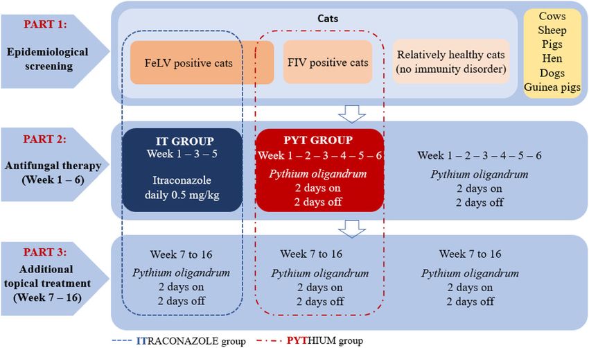
Načeradská et al. BMC Veterinary Research   (2021) 17:290                                                       Page 2 of 10

also cause subclinical infections, where infected individ-     prevent the conversion of lanosterol to ergosterol, which
uals, especially long-haired cats living in a contaminated     maintains cell wall integrity and activity [22]. Solutions
environment, termed asymptomatic carriers, might not           and shampoos with active substances (lime sulfur, enil-
present with clinical symptoms [7]. Predisposing factors       conazole, miconazole, chlorhexidine) and their combina-
include immunosuppression caused by disease or im-             tions can be used for topical therapy. Due to the nature
munosuppressive treatment, other diseases, nutritional         of these substances, it is necessary to adhere to the pre-
deficits (especially proteins and vitamin A), high             scribed concentrations, prevent possible contact with the
temperature, and high humidity [8–10]. In healthy cats,        cat’s mucous membranes during their application, and
dermatophytosis is a self-limiting disease where the clin-     to prevent possible ingestion by the cat, which can cause
ical symptoms usually disappear within 4 months if the         severe side effects [23–25]. Standard systemic and top-
infection is mild and its source is removed [11, 12]. Shel-    ical therapies are relatively expensive, require a long dur-
ters are often inhabited with sick, immunocompromised          ation of application, and can cause side effects in the
animals that are unable to cope with the infection on          affected animals because of the nature of their active in-
their own, and thus such animals often remain in shel-         gredients. In recent years, the mycoparasitic fungus
ters for a long time because they are difficult to adopt.      Pythium oligandrum has been used for the topical treat-
Examples of cat-related infections include feline              ment of dermatophytosis in humans [26] as well as vet-
leukemia virus (FeLV) and feline immunodeficiency              erinary medicine [27] and is non-pathogenic for animals
virus (FIV). These are serious, incurable retroviral dis-      [27, 28] and humans [26]. It is a non-pathogenic soil
eases that can be easily transmitted between cats [13–         oomycete that colonises the root system of many plant
15]. Country recommendations for shelters regarding            species [29]. P. oligandrum shows strong mycoparasitism
the handling of FIV and FeLV positive cats are different.      against more than 50 species of fungi and oomycetes. It
Some countries favour euthanasia [16]; however, there is       is commonly used in plant protection, for example
a no-kill policy in the Czech Republic, where there is an      against Fusarium spp., P. spinosum, P. nunn, P. ultimum,
effort to place sick animals in suitable conditions. FIV       and P. irregulare [30]. In vitro, it has also been shown to
and FeLV positive cats in shelters have a very weakened        be effective against dermatophytosis agents such as M.
immune system and they find it difficult to cope with          canis, M. gypseum, and T. mentagrophytes [1, 3, 26]. P.
dermatophytosis and therefore require therapy [17, 18].        oligandrum produces fast-growing hyphae, round oogo-
The treatment of dermatophytosis is recommended for            nies with spikes, and a large number of enzymes (chiti-
all affected animals (not only FIV and FeLV positive           nases, cellulases, proteases, and glucanases) collectively
cats) to shorten the disease course and reduce the risk of     referred to as oligandrin, which breaks down the cell
spreading the infection [12].                                  wall of host cells leading to complete cytoplasmic de-
   Therapy of dermatophyte infections should include a         struction and host death [29, 31–33].
combination of topical and systemic antifungal medica-           Although a combination of topical and systemic ther-
tions as well as environmental decontamination [3].            apy is currently recommended for the treatment of
Many active substances can be used for systemic therapy        dermatophytosis [3, 12], this study focused on the use of
(allylamines, azoles, echinocandins, polyenes, griseoful-      gentle topical therapy, which is not otherwise burden-
vin, lufenuron), but these substances also cause relatively    some for cats. The effectiveness of topical therapy using
serious side effects including teratogenicity, embryotoxi-     P. oligandrum was compared with the recommended
city, liver toxicity, anorexia, vomiting, and diarrhoea [6].   and standard systemic itraconazole therapy by pulsed
Currently, the use of itraconazole in cats is preferred for    administration for 6 weeks, according to a previous study
the systemic treatment of dermatophytosis, despite its         [12]. In this study, the experimental scheme reported by
relatively high cost. Itraconazole is better tolerated by      Puls et al. [12] was followed and adapted for the topical
cats than ketoconazole or griseofulvin, but it also causes     application of the preparation containing P. oligandrum.
side effects such as hypersalivation, anorexia, vomiting,      To demonstrate the effect of our proposed therapy, we
and hepatotoxicity. Although its teratogenicity and            used immunosuppressed cats (FeLV or FIV positive) of
embryotoxicity are lower than ketoconazole, it is still        different ages (young to very old), which were not ex-
contraindicated for pregnant females, animals less than        pected to be spontaneously cured.
6 months of age, or animals with liver or kidney disease
[3, 6, 19, 20]. Its usual dose is 5 mg/kg by pulse therapy     Methods
for 4–6 weeks (1 week on, 1 week off) [12, 21], this           This pilot study was conducted in collaboration with a pri-
course of therapy is a registered way of use of itracona-      vate asylum for animals in need in the Tibet Shelter (Mar-
zole (Itrafungol, Elanco, Greenfield, USA). Itraconazole       efy 44, Bučovice, Czech Republic). The study design,
is a first generation triazole that works by inhibiting fun-   planned sampling, and clinical examination methodology,
gal cytochrome P450 enzyme 14α demethylase to                  as well as the proposed treatment protocols, were
Načeradská et al. BMC Veterinary Research   (2021) 17:290                                                    Page 3 of 10

approved in advance by the owners of the Tibet Shelter         guinea pigs, housed in the shelter and cared for by the
and by the Animal Ethics Committee of the Czech Uni-           owners and volunteers.
versity of Life Sciences in Prague. The presented study
fully complies with the legislative regulations of the Czech   Experimental design
Republic and the Act on the Protection of Animals against      Part 1
Cruelty No. 246/1992 Coll.                                     Initial epidemiological screening of all animals present
                                                               in the shelter to determine the overall prevalence of
Animals and shelter facility                                   dermatophytosis and to assess potential contamination
A shelter with well-adjusted animal management was se-         in the living environment.
lected for this study. All animals underwent veterinary
examination on admission. Cats placed in a modern              Part 2
quarantine area with fully disinfectable cages were            Study comparing the effectiveness of topical therapy
dewormed, vaccinated (vaccine Purevax RCP, Merial,             with P. oligandrum and systemic itraconazole therapy in
Saint Priest, France, according to the scheme recom-           FeLV or FIV positive cats. This part lasted 6 weeks and
mended by the manufacturer), neutered, and tested for          followed a previously reported methodology [12]. During
retroviral infections (FIV and FeLV). Antigen Rapid FIV        this time, other animals in the shelter, in addition to
Ab/FeLV Ag Test Kit (Bionote, Gyeonggi-do, South               cats, were treated with a preparation containing P. oli-
Korea; Sensitivity: FIV 96.8%, FeLV 94.7%, Specificity:        gandrum to prevent further transmission of dermato-
FIV 99.6%, FeLV 99.7%) was used for testing of the cats.       phytes between animals in the shelter.
FeLV antigen test positive cats were verified by PCR in
the Idexx laboratory for FeLV proviral DNA and all of          Part 3
them tested positive, further named as “FeLV positive”.        Continuation of topical therapy with P. oligandrum in
FIV antibody test positive cats were repeatedly tested by      all sheltered animals for 16 weeks (4 months). Otherwise
the same method, further mentioned as “FIV positive”.          healthy cats were able to recover on their own if the
Cats not included in the selected group were offered for       source of infection was removed. Further design of per-
adoption.                                                      manent measures for the management of the shelter and
  The cats were kept in small groups housed separately         subsequent follow-up was performed. The whole study
in this shelter. Most groups had access to separate out-       flow is summarized in Scheme 2.
door runs. Housing facilities were enriched with play el-
ements, places suitable for climbing, as well as suitable      Sampling method (Mackenzie method)
beds and cat trees. The shelter premises were regularly        At the beginning and end of the study (after 16 weeks),
cleaned and disinfected. A plan of the shelter is shown        the full screening of all animals was performed to detect
in Scheme 1.                                                   dermatophytes (epidemiological screening). A sample
  At the beginning of the study, there were 111 cats, 13       was taken from all animals in the shelter, in addition to
dogs, 2 cows, 3 pigs, 6 sheep, 10 hens, 1 rabbit, and 2        cats, for culture to detect dermatophytes. Sampling was

 Scheme 1 Plan of the shelter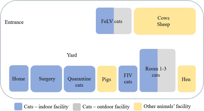
Načeradská et al. BMC Veterinary Research   (2021) 17:290                                                     Page 4 of 10

 Scheme 2 Study flow in the shelter

performed from the whole body by combing the hair            dermatophytes were selected and divided into 2 groups
with a sterile toothbrush [34, 35], which was then imme-     that received different treatments:
diately placed in a sterile resealable plastic bag with a
label and shipped to an accredited diagnostic laboratory     IT group 10 FeLV positive cats that were easy to handle
(Sevaron, Brno, Czech Republic).                             (see Supplementary data 1). This group of cats was
  Samples were taken in the same way for cats included       treated by the oral administration of itraconazole (Itra-
in the study (groups PYT and IT; more detailed informa-      fungol, Elanco) according to the schedule: week of treat-
tion in Treatment protocols and Scheme 2). Sampling          ment (1x daily administration of the drug, at a dose of 5
was performed at 2, 4, and 6 weeks. In these groups,         mg/kg), 1 week break (all repeated 3 times) as previously
samples were taken from affected areas and areas with        reported [12, 37].
fluorescence under Wood’s lamp.
                                                             PYT group 9 FIV or FeLV positive cats (see Supplemen-
Fungal cultures                                              tary data 1). This group of cats was treated with a prep-
All samples were examined at an accredited diagnostic        aration containing P. oligandrum Ecosin (BARD s.r.o.,
laboratory (Sevaron s.r.o., Brno, Czech Republic). Derm-     Prague, Czech Republic) with the solution applied dir-
atophyte Test Medium (DTM) (OXOID CZ; Thermo                 ectly to the fur (three effervescent tablets were diluted in
Fisher Scientific, Brno, Czech Republic) was used to cul-    six litres of lukewarm water per one application for the
ture the samples. Samples were incubated at 25–29 °C         whole facility). Dermasin oil (BARD s.r.o., Prague, Czech
for 14 days. The results were evaluated under a micro-       Republic) was applied to the affected areas of the head
scope by an experienced mycologist. The results were         on each cat (cca 1 ml pec cat). P. oligandrum was ap-
interpreted using the P-score system: P-0 = negative, P-     plied according to the Scheme 2: 2 days application of
1 = 1–4 colony forming units (cfu)/plate, P-2 = 5–9 cfu/     the solution to the fur, 2 days break (for a total of 6
plate, and P-3 = ≥10 cfu/plate.                              weeks). The owner of the shelter applied the product
                                                             gently, by stroking the cats with a glove soaked in the
Diagnostic criteria for dermatophytosis                      product or by applying the oil directly to the affected
Positive cats were those with positive clinical examin-      areas of the head.
ation with use of scoring system [12, 36], Wood’s lamp         All animals that were not included in the selected
fluorescence and positive mycological culture (P-score       groups were treated in the same way as the PYT group
P1 and higher).                                              during the study to treat their symptoms and prevent
                                                             the spread of dermatophytosis among the animals in the
Treatment protocols                                          shelter. Substances soaked in the product, which were
Regarding the very gentle approach to cats, their loca-      placed in the entrance to the outdoor areas, were also
tion, and the statistical evaluation of the study results,   used to apply the product containing P. oligandrum
FeLV or FIV positive cats with a positive finding of         (Ecosin, BARD). In this way, cats were in contact with
Načeradská et al. BMC Veterinary Research (2021) 17:290 Page 5 of 10
the product when passing through the entrance, includ- (100%) guinea pigs. The other animals were negative
ing non-socialized cats for which normal handling was (see Supplementary data 5 and 6). Scoring system for le-
impossible. Therefore, timid cats did not have to be sions and Wood’s lamp examination was adjusted ac-
bathed in the product, which is usually a very stressful cording to the literature [12, 36]. This screening also
procedure. showed a significantly greater detection of dermatophyte
fungi in cats with retroviral infection: 100% of FIV posi-
Examination of cats tive cats (8/8) and 50% of FeLV positive cats (14/28)
Every 14 days, swabs were taken from the fur of cats in were affected compared with other more or less healthy
both groups to examine the presence of dermatophytes. cats and cats accepted for quarantine, where the detec-
At the beginning (day 0) and end (after 6 weeks) of the tion of dermatophyte positivity was 33% (25/75).
study, biochemical analysis of blood was performed for
all cats in the IT and PYT groups (Idexx Catalyst one
analyser) (see Supplementary data 2). Part 2
At all times, the cats were under veterinary supervision In cats, the dominant cause of dermatophytosis is M.
and their health was regularly checked, using a method canis. In our study, the cultivation of samples taken from
to minimize stress. areas positive by Wood’s lamp examination, confirmed a
massive infection (i.e., ≥10 cfu) of M. canis in all cats
Environmental cleaning from the IT and PYT groups. After the first 2 weeks of
Extensive cleaning of the shelter was performed before treatment, there was a significant decrease in the num-
the start of the study. All areas, including the walls, were ber of M. canis cfu in the PYT group compared with the
washed and treated with disinfectant. Incidin plus (1%) IT group. Regarding P-scores, in the PYT group, 2 cats
(Ecolab s.r.o., Prague, Czech Republic), which has no an- were scored as P-1, 2 as P-2, and 5 cats as P-3. In the IT
tifungal properties, was used as a disinfectant. This dis- group, all cats were scored as P-3 (see Fig. 1) and they
infection was used throughout the study and was showed high levels of salivation and loss of appetite after
alternated with Incidin OxyDes (1%) (Ecolab s.r.o., the administration of itraconazole. Furthermore, these
Prague, Czech Republic). cats began to refuse this preparation and their treatment
To reduce the fungal contamination of the environ- was more difficult. No adverse reactions were observed
ment, the areas where the animals were kept were also in the group of cats treated with topical P. oligandrum.
regularly treated during the study: 1x weekly application This was due to the nature of the active substance and
of a solution with P. oligandrum in a preparation the way it was applied: the cats were not subjected to
intended for surface treatment (Biorepel, Biopreparáty any potentially stressful manipulations during its
s.r.o., Prague, Czech Republic). A detailed breakdown of application.
cleaning is provided in Supplementary data 3. After 4 weeks of treatment, a course similar to that
after 2 weeks of treatment was observed (see Fig. 1). In
Statistical analysis the IT group, all cats were scored as P-3. Two cats died
The method of least squares – linear model GLM with one of which was diagnosed with feline infectious peri-
fixed effects at a level of significance α ≥ 0.05 (SAS soft- tonitis, FIP (post-mortem autopsy and ascitic fluid was
ware) was used to demonstrate statistically significant coronavirus PCR-positive). Autopsy of the second cat re-
differences. vealed a rupture of the liver and a diagnosis of amyloid-
The model equation for the calculation was: Y = type osis was confirmed by histopathology examination. Four
of treatment + weeks of treatment + type of treatment * weeks after the administration of itraconazole, the enor-
weeks of treatment + e. mous salivation and loss of appetite remained in cats in
Y – is a manifestation (P – score), type of treatment – the IT group, they began to show signs of stressed be-
fixed effect (pythium, itraconazole), weeks of treatment haviour before itraconazole administration and their
– fixed effect (0, 2, 4, and 6 weeks). A table providing in- treatment was problematic. In the PYT group, 4 cats
formation on the evidence of effects (independent vari- were scored as P-1 and 5 as P-3. Of note, the cats were
ables) on disease manifestation (dependent variables) is not afraid of the treatment, which was applied by petting
provided in Supplementary data 4. with a wet glove, and they showed better socialization by
approaching the person of their own free will during the
Results treatment application.
Part 1 After 6 weeks of treatment, a similar course was still
The results of the initial screening of all animals present evident. In the IT group, all cats were scored as P-3
in the shelter confirmed a massive dermatophyte infec- whereas in the PYT group, 5 cats were scored as P-1
tion in 47/111 (42%) cats, 3/13 (23%) dogs, and 2/2 and 4 cats as P-3 (see Fig. 1).Načeradská et al. BMC Veterinary Research (2021) 17:290 Page 6 of 10 Fig. 1 Graphs comparing the two treatments for dermatophytosis at the beginning of the study, and after 2, 4, and 6 weeks of treatment. Cats in the IT group were treated with standard itraconazole and cats in the PYT group were treated with topical P. oligandrum (see Materials and Methods, Treatment protocols). *Significantly more effective therapy evaluated with linear GLM model is marked with asterisk (see Methods, Statistical analysis) Fig. 2 Graphs comparing the two treatments for dermatophytosis at the beginning of the study and after 16 weeks of treatment. Cats in the IT group were treated with standard itraconazole for 6 weeks and then for 10 weeks with topical P. oligandrum. Cats in the PYT group were treated with topical P. oligandrum (see Materials and Methods, Treatment protocols). Statistically significant differences were observed between the start of the study and after 16 weeks of treatment. No statistically significant differences were observed between the chosen method of therapy after 16 weeks of treatment
Načeradská et al. BMC Veterinary Research         (2021) 17:290                                                                              Page 7 of 10

Part 3                                                                         application of P. oligandrum. At the beginning of the
The study continued for another 10 weeks (16 weeks in                          study, of 111 cats present in the shelter, 64 (58%) were
total = 4 months). All animals present in the shelter, as                      negative and 47 (42%) were M. canis positive with a P-3
well as cats in the original IT group, were treated with P.                    score. During the study, 22 (20%) cats that were initially
oligandrum topical therapy (see Material and Methods)                          negative were adopted or died, and 15 (14%) cats that
during this time. Itraconazole systemic therapy was not                        were originally positive (P-3), died or had to be eutha-
continued due to the occurrence of adverse reactions.                          nised due to poor health (a panleukopenia infection oc-
   After 16 weeks and 10 weeks, respectively, of topical                       curred in quarantine cats during the study). After 16
therapy with P. oligandrum, 3 cats were scored as nega-                        weeks (4 months) of treatment, 17 of the original 111
tive (P-0), 3 as P-1, and 1 as P-2. One cat died of FIP in-                    cats remained positive (16%). Eleven of these positive
fection (confirmed by autopsy and PCR examination) in                          cats were scored as P-1 (10% of the original 111 cats),
the former IT group. In the PYT group, 5 cats were eval-                       which can be considered to be caused by contamination
uated as negative (P-0), 1 as P-1, 1 as P-2, and 2 as P-3                      from the environment. These cats did not show clinical
(see Fig. 2). Two cats scored as P-3 suffered from an-                         signs and were negative by Wood’s lamp examination.
other disease. One was a very old FeLV positive blind                          Of the other 6 cats (6%), 3 were scored as P-2 and 3 as
cat with suspected Conn’s syndrome. The second was an                          P-3. These cats had no clinical signs of dermatophytosis
old FeLV positive cat with acute inflammation of the                           but all of them suffered from other acute illnesses at that
oral cavity, which could not take care of itself and which                     time. One died due to multiple neoplasia, one had to
might have been a source of contamination to other cats.                       undergo enucleation of the eye due to massive inflam-
These cats were further treated with topical P. oligan-                        mation, and the other four underwent intensive
drum. When examined by Wood’s lamp after 16 weeks,                             treatment.
no FIV or FeLV cats were positive and none showed                                The results of the other tested animal species in the
clinical signs of dermatophytosis (Supplementary data 6                        shelter are shown in Supplementary data 5.
and 7).                                                                          As mentioned previously, the important task of the
   After 16 weeks (4 months), a final epidemiological                          study was setting a sustainable management plan for the
screening of all shelter animals was performed. A signifi-                     shelter in order to prevent new outbreak of the dermato-
cant improvement in the epidemiological situation (see                         phytosis. At the end of the study, we suggested recom-
Fig. 3, Supplementary data 5 and 6) in the shelter was                         mendations summarised in scheme 3. At follow-up 1
observed in terms of the occurrence of dermatophytosis                         year after the start of the study, no clinical signs of
in all animals when compared with the beginning of the                         dermatophytosis were observed in cats that had stayed
study (see Fig. 3). All new animals admitted to the shel-                      in the shelter for a long time. The shelter still follows an
ter during the study were quarantined, tested for derma-                       established procedure during admission; cats with skin
tophytes, and prophylactically treated with a topical                          lesions are tested for dermatophytes (fungal culture,

 Fig. 3 Comparison of the incidence of dermatophytosis in cats at the beginning of the study and after 16 weeks of treatment with topical P.
 oligandrum. P-0: M. canis negative, P-1: 1–4 cfu of M. canis, P-2: 5–9 cfu of M. canis, P-3: ≥10 cfu of M. canis, O/N: out of study cats (adopted or
 death), which were M. canis negative at the beginning of the study, O/P: out of study cats (death), which were M. canis positive (P-3) at the
 beginning of the study
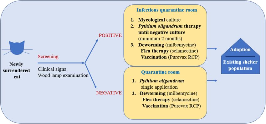
Načeradská et al. BMC Veterinary Research    (2021) 17:290                                                        Page 8 of 10

 Scheme 3 Overview of measures for cats newly admitted to a shelter

Wood’s lamp testing) and prophylactically treated with                weeks of therapy. In a group of cats treated with
P. oligandrum (see Scheme 3). Shelter spaces are treated              standard itraconazole, side effects potentially related
at regular intervals.                                                 to this preparation were observed [6]. Puls et al. [12]
                                                                      reported the occurrence of hypersalivation and a
Discussion                                                            slightly increased frequency of anorexia and vomiting
In this study, we verified a gentle and inexpensive pro-              in cats, but did not describe behavioural changes in
cedure for the eradication of dermatophytosis in ani-                 cats treated with itraconazole. In our study, we ob-
mals, targeted at the needs of shelters, asylums, or large            served changes in the behaviour of cats. With an in-
animal farms, using the mycoparasitic fungus P. oligan-               creasing duration of itraconazole administration, cats
drum. Our study was based on a previous report show-                  showed signs of stressful behaviour, fear of its appli-
ing the beneficial effect of the mycoparasitic fungus P.              cation, and aggression. Two cats treated with itraco-
oligandrum on M. canis [1, 27], one of the most com-                  nazole died during the 6 weeks of therapy. Although
mon causes of dermatophytosis in cats and dogs.                       it is not possible to prove a causal relationship be-
   The results of the initial epidemiological screening               tween these deaths and the active substance used
showed a relatively high overall prevalence of dermato-               (itraconazole), we concluded that the increase in
phytosis in cats in our shelter. A total of 42% of cats               stress to which cats were exposed during this applica-
were positive, despite the quality of the management of               tion may have significantly contributed to the acute
the shelter. Of these cats, 23% of otherwise healthy cats             phase of both diseases and the death of the cats.
were dermatophytosis positive, which is about 10 times                   Puls et al. [12] described the rapid onset of action
higher than previously reported for other countries                   of itraconazole. By the second week of treatment, they
(2.4% USA, 3.6% Canada, 1.3% UK) [3].                                 observed a clinical cure and after 4 weeks they ob-
   As data on the average prevalence of dermatophytosis               served a mycological cure (2 consecutive negative cul-
in the Czech Republic have not been published, it is not              tures). In our study, itraconazole therapy outcome
possible to objectively assess the cause of such a high               was different. After 6 weeks of therapy, none of the
prevalence in the monitored shelter. In our opinion, the              groups was completely mycologically cured of derm-
environmental load of dermatophytes in this locality                  atophytosis, but 1 cat out of 8 from IT group and 5
might have a significant role on the results of this study.           cats out of 9 from PYT group did not exhibit any
The massive occurrence of M. canis and other common                   clinical signs (supplementary data 6 and 7). Six weeks
keratinophilic fungi species comparable to the occur-                 is the standard recommended duration of treatment
rence of dermatophytes in similar studies, was confirmed              with itraconazole in otherwise healthy cats [3, 12].
here [38, 39].                                                        Previous studies usually exclude immunosuppressed
   After 2 weeks of the recommended therapy, a statis-                cats from the shelter population prior to treatment
tically significant difference was seen between the two               for    dermatophytosis     [40].   These   cats   were
treatment groups. Our topical therapy with P. oligan-                 intentionally included in our study. To date, the
drum had a statistically significant beneficial effect                higher incidence of fungal diseases in retrovirus-
compared with the standard systemic therapy using                     affected cats has not been clearly established [3, 39];
itraconazole. A similar effect was seen after 4 and 6                 however, in our study, there was a significant
Načeradská et al. BMC Veterinary Research (2021) 17:290 Page 9 of 10
difference in the incidence of dermatophytosis be- Acknowledgements
tween sick cats (FIV 100%, FeLV 52%) and healthy We thank Prof. Markéta Sedmíková from Czech University of Life Sciences in
Prague for her valuable advice and kind support of our work. We thank Prof.
cats (23%). A previous study reported a similar find- Karen Moriello from University of Wisconsin-Madison for her valuable advice
ing, where dermatophytosis was observed in 74% of and help with study design.
FIV positive cats and in 25% of FIV seronegative cats We thank John Croxford for language editing.
from shelters and households with access outside Authors’ contributions
[34]. MN created the concept of the work, MN and TK collected the data and
When comparing the effect of both therapies, after 2 prepared the manuscript. TK and MFK processed the data and prepared
tables for Supplementary materials. MFK and MF prepared the figures and
weeks a statistically significant decrease in the number contributed to the manuscript. MN did the final check and is corresponding
of M. canis cfu was evident in cats treated with topical author. All authors read and approved the manuscript.
P. oligandrum compared with systemic itraconazole. Of
Authors’ information
note, the topical therapy had a faster treatment onset Martina Načeradská
and cats with declining M. canis cfu were potentially ORCID iD: 0000-0002-2404-7060
much less dangerous in terms of transmitting and Researcher ID: D-6472-2019
Michaela Fridrichová
spreading this infection to other cats, animals, and ORCID iD: 0000-0002-3636-723X
humans with whom they came into contact. Researcher ID: A-7017-2013
After 16 weeks of therapy, there was no statistically Scopus Author ID: 54986064800
Tereza Krejčová
significant difference in P-scores between the IT and ORCID iD: 0000-0001-5840-9499
PYT groups. Of note, dermatophytosis was almost eradi-
cated in both groups (cats exclusively receiving topical Funding
P. oligandrum and cats receiving systemic therapy with Not applicable.
itraconazole followed by topical P. oligandrum) in the Availability of data and materials
shelter. Apart from 3 cats that had a poor clinical condi- Detailed data available in attached Supplementary file.
tion requiring intensive treatment, the other cats were
Declarations
cured of dermatophytosis despite them being FeLV and
FIV positive and very old. Of the original 42% of positive Ethics approval and consent to participate
cats, at the end of the study 3% of cats were scored as P- The study design, planned sampling, and clinical examination methodology,
as well as the proposed treatment protocols, were approved in advance by
3 (clinically very sick requiring intensive treatment), 3% the owners of the Tibet Shelter (informed consent signed) and by the
were scored as P-2, and 10% were scored as P-1. These Animal Ethics Committee of the Czech University of Life Sciences in Prague.
results might be partly related to contamination from The study was conducted in compliance with the ARRIVE guidelines. (The
Explanation and Elaboration for the ARRIVE guidelines 2.0 were originally
the environment (see Fig. 3). published in PLOS Biology doi:https://doi.org/10.1371/journal.pbio.3000411).
The whole study was conducted in accordance with local and EU law and
Conclusion with principles of good practice. Preparations with P. oligandrum are
registered veterinary cosmetic preparations (Ecosin and Dermasin, USKVBL
In this study, we demonstrated the comparable efficacy registration under No.057–09/C).
of an alternative to itraconazole for dermatophytosis
treatment using the topical application of a product con- Consent for publication
Not applicable.
taining P. oligandrum. Of note, the alternative treatment
with P. oligandrum had a faster beneficial effect com- Competing interests
pared with itraconazole. Therefore, this topical therapy The authors declare that they have no competing interests.
is recommended as a cheaper and gentler alternative to
Author details
itraconazole for the treatment and prevention of derm- 1
Department of Veterinary Sciences, Faculty of Agrobiology, Natural and
atophytosis, especially in animal shelters or large farms, Food Resources, Czech University of Life Sciences in Prague, Kamýcká 129,
where the classic method of therapy is expensive and 165 21 Prague 6, Czech Republic. 2Department of Inorganic Chemistry,
Faculty of Science, Charles University, Hlavova 8, 128 43 Prague 2, Czech
difficult to manage, even in ill animals (FeLV or FIV Republic.
positive).
Received: 16 May 2021 Accepted: 23 August 2021
Abbreviations
FeLV: feline leukaemia virus; FIV: feline immunodeficiency virus;
DTM: dermatophyte test medium; cfu: colony forming units References
1. Naceradska M, Fridrichova M, Kellnerova D, Pekova S, Lany P. Antifungal
effects of the biological agent Pythium oligandrum observed in vitro. J
Supplementary Information Feline Med Surg. 2017;19(8):817–23. https://doi.org/10.1177/1098612X1
The online version contains supplementary material available at https://doi. 6658690.
org/10.1186/s12917-021-03001-w. 2. Moriello KA, DeBoer DJ. Fungal flora of the coat of pet cats. Am J Vet Res.
1991;52(4):602–6.
Additional file 1. 3. Moriello KA, Coyner K, Paterson S, Mignon B. Diagnosis and treatment of
dermatophytosis in dogs and cats.: clinical consensus guidelines of theNačeradská et al. BMC Veterinary Research (2021) 17:290 Page 10 of 10
world Association for Veterinary Dermatology. Vet Dermatol. 2017;28(3):266– 27. Naceradska M. Control of dermatophytosis in the cat shelter with use of
e68. https://doi.org/10.1111/vde.12440. mycoparasite Pythium oligandrum and vaccinaction. Mycoses. 2018;61:31.
4. Gräser Y, Kuijpers AF, El Fari M, Presber W, de Hoog GS. Molecular and 28. Tan Y, Peng L, Yuan L, Wang S. Toxicity of Pythium oligandrum broth to
conventional taxonomy of the Microsporum canis complex. Med Mycol. animal and its control effect on rot diseases caused by Penicillium italicum
2000;38(2):143–53. https://doi.org/10.1080/mmy.38.2.143.153. and Penicillium digitatum in orange fruit storage. Wei Sheng Wu Xue Bao.
5. Weitzman I, Summerbell RC. The dermatophytes. Clin Microbiol Rev. 1995; 2015;55(11):1418–26.
8(2):240–59. https://doi.org/10.1128/CMR.8.2.240-259.1995. 29. Gerbore J, Benhamou N, Vallance J, Le Floch G, Grizard D, Regnault-Roger C,
6. Frymus T, Gruffydd-Jones T, Pennisi MG, Addie D, Belák S, Boucraut-Baralon et al. Biological control of plant pathogens: advantages and limitations seen
C, et al. Dermatophytosis in cats: ABCD guidelines on prevention and through the case study of Pythium oligandrum. Environ Sci Pollut Res Int.
management. J Feline Med Surg. 2013;15(7):598–604. https://doi.org/10.11 2014;21(7):4847–60. https://doi.org/10.1007/s11356-013-1807-6.
77/1098612X13489222. 30. Brozova J. Exploitation of the mycoparasitic fungus Pythium oligandrum in
7. Foster A, Foil C. BSAVA Manual of Small Animal Dermatology. 2nd ed. plant protection. Plant Prot Sci. 2002;38:29–35.
Gloucester 2003. 169–174. 31. Picard K, Tirilly Y, Benhamou N. Cytological effects of cellulases in the
8. Cafarchia C, Romito D, Sasanelli M, Lia R, Capelli G, Otranto D. The epidemiology of parasitism of Phytophthora parasitica by Pythium oligandrum. Appl Environ
canine and feline dermatophytoses in southern Italy. Mycoses. 2004;47(11–12):508– Microbiol. 2000;66(10):4305–14. https://doi.org/10.1128/aem.66.10.43
13. https://doi.org/10.1111/j.1439-0507.2004.01055.x. 05-4314.2000.
9. Polak KC, Levy JK, Crawford PC, Leutenegger CM, Moriello KA. Infectious 32. Benhamou N, Belanger R, Rey P, Tirilly Y. Oligandrin, the elicitin-like protein
diseases in large-scale cat hoarding investigations. Vet J. 2014;201(2):189–95. produced by the mycoparasite Pythium oligandrum, induces systemic
https://doi.org/10.1016/j.tvjl.2014.05.020. resistence to Fusarium crown and root rot in tomato plants. Plant Physiol
10. Greene CE. Infectious diseases of the dog and cat. St. Louis: Elsevier/ Biochem. 2001;39(7):681–96.
Saunders; 2012. 33. Mohamed N, Lherminier J, Farmer M, Fromentin J, Béno N, Houot V, et al.
11. DeBoer DJ, Moriello KA. Inability of two topical treatments to influence the Defense responses in grapevine leaves against Botrytis cinerea induced by
course of experimentally induced dermatophytosis in cats. J Am Vet Med application of a Pythium oligandrum strain or its Elicitin, Oligandrin, to
Assoc. 1995;207(1):52–7. roots. Phytopathology. 2007;97(5):611–20. https://doi.org/10.1094/PHYTO-97-
12. Puls C, Johnson A, Young K, Hare J, Rosenkrans K, Young L, et al. Efficacy of 5-0611.
itraconazole oral solution using an alternating-week pulse therapy regimen for 34. Mancianti F, Giannelli C, Bendinelli M, Poli A. Mycological findings in feline
treatment of cats with experimental Microsporum canis infection. J Feline Med Surg. immunodeficiency virus-infected cats. J Med Vet Mycol. 1992;30(3):257–9.
2018;20(10):869–74. https://doi.org/10.1177/1098612X17735967. https://doi.org/10.1080/02681219280000321.
13. Hardy WD, Hess PW, MacEwen EG, McClelland AJ, Zuckerman EE, Essex M, 35. Mackenzie DW. "hairbrush diagnosis" in detection and eradication of non-
et al. Biology of feline leukemia virus in the natural environment. Cancer fluorescent scalp ringworm. Br Med J. 1963;2(5353):363–5. https://doi.org/1
Res. 1976;36(2 pt 2):582–8. 0.1136/bmj.2.5353.363.
14. Ishida T, Washizu T, Toriyabe K, Motoyoshi S, Tomoda I, Pedersen NC. Feline 36. Moriello KA, Deboer DJ, Schenker R, Blum JL, Volk LM. Efficacy of pre-
immunodeficiency virus infection in cats of Japan. J Am Vet Med Assoc. treatment with lufenuron for the prevention of Microsporum canis infection
1989;194(2):221–5. in a feline direct topical challenge model. Vet Dermatol. 2004;15(6):357–62.
15. Lutz H, Addie D, Belák S, Boucraut-Baralon C, Egberink H, Frymus T, et al. Feline https://doi.org/10.1111/j.1365-3164.2004.00406.x.
leukaemia. ABCD guidelines on prevention and management. J Feline Med Surg. 37. Colombo S, Cornegliani L, Vercelli A. Efficacy of itraconazole as a combined
2009;11(7):565–74. https://doi.org/10.1016/j.jfms.2009.05.005. continuous/pulse therapy in feline dermatophytosis: preliminary results in
16. Moriello KA, Newbury S. Recommendations for the management and nine cases. Vet Dermatol. 2001;12(6):347–50. https://doi.org/10.1046/j.
treatment of dermatophytosis in animal shelters. Vet Clin North Am Small 0959-4493.2001.00274.x.
Anim Pract. 2006;36(1):89–114, vi. https://doi.org/10.1016/j.cvsm.2005.09.006. 38. Caretta G, Mancianti F, Ajello L. Dermatophytes and keratinophilic fungi in
17. Hofmann-Lehmann R, Hartmann K. Feline leukaemia virus infection: a cats and dogs. Mycoses. 1989;32(12):620–6.
practical approach to diagnosis. J Feline Med Surg. 2020;22(9):831–46. 39. Sierra P, Guillot J, Jacob H, Bussiéras S, Chermette R. Fungal flora on
https://doi.org/10.1177/1098612X20941785. cutaneous and mucosal surfaces of cats infected with feline
immunodeficiency virus or feline leukemia virus. Am J Vet Res. 2000;61(2):
18. Little S, Levy J, Hartmann K, Hofmann-Lehmann R, Hosie M, Olah G, et al.
158–61. https://doi.org/10.2460/ajvr.2000.61.158.
2020 AAFP Feline Retrovirus Testing and Management Guidelines. J Feline
40. Newbury S, Moriello K, Coyner K, Trimmer A, Kunder D. Management of
Med Surg. 2020;22(1):5–30. https://doi.org/10.1177/1098612X19895940.
endemic Microsporum canis dermatophytosis in an open admission shelter:
19. Foy DS, Trepanier LA. Antifungal treatment of small animal veterinary
a field study. J Feline Med Surg. 2015;17(4):342–7. https://doi.org/10.1177/1
patients. Vet Clin North Am Small Anim Pract. 2010;40(6):1171–88. https://
098612X14543854.
doi.org/10.1016/j.cvsm.2010.07.006.
20. Mawby DI, Whittemore JC, Genger S, Papich MG. Bioequivalence of orally
administered generic, compounded, and innovator-formulated itraconazole in Publisher’s Note
healthy dogs. J Vet Intern Med. 2014;28(1):72–7. https://doi.org/10.1111/jvim.12219. Springer Nature remains neutral with regard to jurisdictional claims in
21. Mignon B. Dermatophytosis. In: Guaguère ÉAM, Prélaud P, Craig JM, editors. published maps and institutional affiliations.
A practical guide to canine dermatology. Paris: Kalianxis; 2008. p. 151–64.
22. Vanden Bossche H, Koymans L, Moereels H. P450 inhibitors of use in
medical treatment: focus on mechanisms of action. Pharmacol Ther. 1995;
67(1):79–100. https://doi.org/10.1016/0163-7258(95)00011-5.
23. Guillot J, Malandain E, Jankowski F, Rojzner K, Fournier C, Touati F, et al.
Evaluation of the efficacy of oral lufenuron combined with topical
enilconazole for the management of dermatophytosis in catteries. Vet Rec.
2002;150(23):714–8. https://doi.org/10.1136/vr.150.23.714.
24. Hnilica KA, Medleau L. Evaluation of topically applied enilconazole for the
treatment of dermatophytosis in a Persian cattery. Vet Dermatol. 2002;13(1):
23–8. https://doi.org/10.1046/j.0959-4493.2001.00282.x.
25. Moriello K, Coyner K, Trimmer A, Newbury S, Kunder D. Treatment of shelter
cats with oral terbinafine and concurrent lime Sulphur rinses. Vet Dermatol.
2013;24(6):618–20, e149-50. https://doi.org/10.1111/vde.12069.
26. Gabrielová A, Mencl K, Suchánek M, Klimeš R, Hubka V, Kolařík M. The
Oomycete Pythium oligandrum can suppress and kill the causative agents
of Dermatophytoses. Mycopathologia. 2018;183(5):751–64. https://doi.org/1
0.1007/s11046-018-0277-2.You can also read