Rocky Mountain spotted fever Epidemiology - Arizona ...
←
→
Page content transcription
If your browser does not render page correctly, please read the page content below
Rocky Mountain spotted fever
                                                                       Epidemiology
   A. General
         • Rocky Mountain spotted fever (RMSF) is the most severe tick-borne rickettsial
            illness in the United States1,2,3,4. Since its emergence in tribal lands in Arizona in
            2003, over 400 cases have been identified, with nearly 30 fatalities. The case
            fatality rate for Arizona (6%) is higher than the U.S. rate (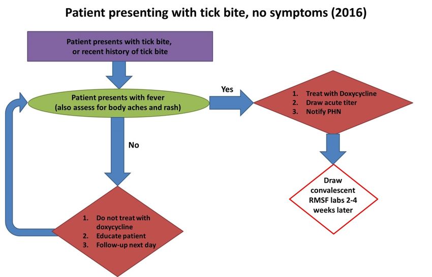
5 days after symptom onset as small, flat, non-itchy, pink macules on the wrists,
              forearms, and ankles. This rash may then spread to the truck of the body.1,2
          •   About 35–60% of cases develop a petechial rash around day 5 or 6 of illness. This
              can progress to a purpuric type of rash (reddish-purple non-blanching spots),
              which often indicates severe disease.1,2
          •   Clinical laboratory findings indicative of RMSF may include thrombocytopenia,
              anemia, leukopenia, and/or elevated liver enzymes.1-4,9,13-15
          •   Severe illness and vascular damage can lead to prolonged hospitalization and
              long-term health problems.1,2,4,9

   E. Modes of Transmission
        • RMSF is spread by the bite of an infected tick. In Arizona, the vector reservoir is
            R. sanguineus (brown dog tick).1,2,5
        • Transmission occurs from the bite of an infected tick. At least 4–6 hours of tick
            attachment are required before the bacteria can be transmitted and cause
            disease in people.1,2 Contamination of cuts, scrapes, or wounds in the skin or
            mucous membranes with crushed tissues or feces of the tick may also lead to
            infection. Laboratory-acquired infection has resulted from accidental inoculation
            and aerosol contamination.1,2 Transmission has occurred on rare occasions by
            blood transfusions.1,2
        • RMSF is not directly transmitted from person-to-person.1,2

   F. Incubation Period
          • Symptoms of RMSF usually occur 2–14 days after the bite of an infected tick.1,2

   G. Period of Communicability
         • Ticks remain infected for life, and female ticks can pass the bacteria
             transovarially to offspring. Life span is commonly as long as 18 months.1,5

   H. Susceptibility and Resistance
      • Susceptibility is universal. After infection, immunity is believed to be life-long.
         Antibodies can persist in the body for months to years after initial infection.1,2

   I. Treatment
      • Doxycycline is the first line treatment for adults and children.1-4,9,13-15 This should be
         prescribed whenever RMSF is suspected. Chloramphenicol is an alternative when
         there are contraindications to tetracyclines, such as life-threatening allergies.2

Laboratory Diagnostics1,2
   A. Diagnosis
          • Serology is the best diagnostic option and is widely used for detecting antibodies
              against RMSF. However, paired samples (acute and convalescent) are essential

RMSF
Last Revised: 3/25/2020
for confirmation because antibody responses are rarely detectable in acute
samples.1,2,3
o Always recommend doxycycline as soon as RMSF is suspected!
• Polymerase chain reaction (PCR) or immunohistochemical (IHC) testing methods
are appropriate only for cases of severe illness, or from post-mortem specimens
before doxycycline has been given.1,2
o Biopsies of the rash are appropriate when present; however, the rash
may not be present until late in disease progression and should always be
coupled with serology (negative PCR does not rule out disease).1,2
B. Laboratory Criteria for Diagnosis17
Confirmatory Laboratory Evidence
• Detection of Spotted Fever Group Rickettsiae (SFGR) nucleic acid in a clinical specimen via
amplification of a Rickettsia genus- or species-specific target by polymerase chain reaction
(PCR) assay; OR
• Serological evidence of a fourfold increase in IgG-specific antibody titer reactive with SFGR
antigen by immunofluorescence assay (IFA) between paired serum specimens (one taken in the
first two weeks after illness onset and a second taken two to ten weeks after acute specimen
collection)*; OR
• Demonstration of SFGR antigen in a biopsy or autopsy specimen by immunohistochemical
methods (IHC); OR
• Isolation of SFGR from a clinical specimen in cell culture and molecular confirmation (e.g., PCR
or sequence).
*A four-fold rise in titer should not be excluded (as confirmatory laboratory criteria) if the acute and
convalescent specimens are collected within two weeks of one another.
Presumptive Laboratory Evidence
• Serologic evidence of elevated IgG antibody at a titer ≥1:128 reactive with SFGR antigen by
IFA in a sample taken within 60 days of illness onset.**
**This includes paired serum specimens without evidence of fourfold rise in titer, but with at
least one single titer ≥1:128 in IgG-specific antibody titers reactive with SFGR antigen by IFA.
The 60-day cut-off is especially important for probable cases with a single IgG titer to better
capture real acute infection.
Suspect Laboratory Evidence
• Serologic evidence of elevated IgG antibody at a titerresponse may be persistent (making it harder to identify how recent the infection was).
Complement fixation (CF) tests and other older test methods are neither readily available nor
commonly used. CDC uses in-house IFA IgG testing (cutoff of ≥1:64), preferring simultaneous
testing of paired specimens, and does not use IgM results for routine diagnostic testing.
C. Interpreting serology results1,3,17
a. Paired serology is the most common diagnostic test for RMSF to look for
increasing levels of RMSF-specific IgG antibodies. This suggests recent infection.
b. Early in any tick-borne rickettsial disease, most of the acute tests will be
negative. This is because it takes typically 7–10 days after symptom onset for the
body to make enough antibodies to reach detectable levels.
c. Ideally, the first (acute) sample should be taken early (within the first week of
symptoms) to provide a recent baseline antibody level and the second
(convalescent) sample should be taken 2–4 weeks later after the body has a full
antibody response.
d. Antibody levels may remain high for months following illness.
D. Clinical Evidence
Fever as reported by the patient or a healthcare provider, AND one or more of the
following:
• rash
• eschar
• headache
• myalgia
• anemia
• thrombocytopenia, or
• any hepatic transaminase elevation.
E. Exposure
• Exposure is defined as having been in potential tick habitats within the past 14 days
before onset of symptoms. Occupation should be recorded if relevant to exposure. A
history of a tick bite is not required.17
F. Case Classification3,17
CASE DEFINITION DESCRIPTION
Confirmed A clinically compatible case (meets clinical evidence
criteria) that meets the confirmatory laboratory criteria.
Probable A clinically compatible case (meets clinical evidence
criteria) that meets the presumptive laboratory criteria
Suspect A case with confirmatory or presumptive laboratory
evidence of infection with no clinical
information available;
RMSF
Last Revised: 3/25/2020A person who meets the clinical description and has
supportive laboratory evidence
Criteria to Distinguish a New Case from an Existing Case
A person previously reported as a probable or confirmed case-patient may be counted as a new
case-patient when there is an episode of new clinically compatible illness with confirmatory
laboratory evidence.
For the purposes of entering new laboratory information for an existing case, the timeframe of
6 months can be used as a rule of thumb for creating a new case, until evidence is obtained to
determine whether there is a new episode of clinically compatible illness.*
G. Bioterrorism Potential: None
H. Outbreak Definition:
• An outbreak in endemic areas is defined as a significant increase in cases above the
level normally seen in the area during the time period in question. In non-endemic
areas, an outbreak is one or more case(s) of confirmed RMSF with no history of
travel to endemic areas.
Rocky Mountain spotted fever
Case Investigation
A. Reporting
• RMSF is a nationally notifiable condition and should be reported within 5
working days to the local health jurisdiction.
• Local county public health is responsible for conducting an investigation on any
reported cases. Use the rickettsial disease investigation form:
http://www.cdc.gov/ticks/forms/2010_tbrd_crf.pdf
1. Confirm the diagnosis with an appropriate medical provider/laboratory.
Before contacting the patient or family, determine what information is available from
medical records and healthcare providers.
I. Obtain information that supports clinical findings in the case definition and information
on the onset date and order of the symptoms, including:
a. Hospitalizations
b. Life threatening complications
c. Clinical symptoms of fever, headache, myalgia, anemia, thrombocytopenia,
leukopenia, elevated hepatic transaminases or others.
d. Underlying immunosuppressive condition.
II. Outcome status: survived or date of death
III. Area of residence and tribal affiliation (if any)
RMSF
Last Revised: 3/25/2020IV. Obtain information on any laboratory tests performed and results or date results are
expected.
a. If laboratory tests have not been run to test for RMSF IgG, coordinate testing of
an acute specimen with ADHS. IgM results are not reliable and should not be
considered.
b. Arrange to have a convalescent sample drawn at the hospital, public health clinic
or primary care providers 2–6 weeks after symptom onset
V. Obtain date when doxycycline treatment was initiated
a. If doxycycline has not been initiated, arrange for doxycycline to be administered
with the provider.
2. Conduct case investigation –
Epidemiological investigation report should be submitted in MEDSIS by filling out the full
DSO and Travel Table. Interview case to determine source, risk factors and transmission
settings.
I. Interview the case or proxy to determine source and risk factors; focus on the 2 week
incubation period prior to illness onset. Consider:
a. Recent travel to endemic areas or history of possible exposure to ticks. List
geographic location(s) and date(s).
b. Ownership or contact with a dog
c. Exposure to animals or pets with ticks.
d. Other people who might have been exposed
i. If case is resident in an area with free roaming dogs assume other
household members are at risk
e. Outdoor activities.
f. Occupational risks (e.g., forestry worker, landscape worker, etc.). History of tick
bites, include geographic location of bite and date.
g. Time spent in any area with free roaming dogs
3. Conduct contact investigation – Locate additional cases and/or contacts
Contacts are those with possible exposure to the source of infection. Contacts are not
persons in close proximity to a case; the disease is not transmitted person-to person.
Consideration should be given to any higher than normal incidence in reports of spotted
fever symptoms in individuals that were in the same geographic location as the case’s
suspected exposure. For suspected outbreaks, refer to the Managing Special Situations
section. Follow up with symptomatic contacts as suspect cases.
4. Isolation, Work and Child Care Restrictions– None
5. Case Management – Ensure all suspect cases receive doxycycline as soon as possible. Do not
wait for laboratory results to begin treatment! Delaying treatment can result in death.
6. Contact Management, Including Susceptible Contacts
Instruct those companions of the case who had similar exposures (i.e., contact with ticks,
RMSF
Last Revised: 3/25/2020contact with free roaming dogs, travel to endemic areas, etc.) to monitor themselves for
symptoms. Preventive treatment is not warranted. Treatment is necessary only if symptoms
develop. Those who exhibit any signs or symptoms compatible with tick-borne illness should
be referred to their medical provider for evaluation.
7. Environmental Measures
In cooperation with the Department, a local health agency or another local agency responsible
for vector control within a jurisdiction shall:
1. Conduct an assessment of the environment surrounding each spotted fever rickettsiosis case
or suspect case and implement vector control measures as necessary.
Community-based integrated tick management strategies may reduce the incidence of tick-
borne infections. Limiting exposure to ticks is presently the most effective prevention method.
Tick management approaches include the following:
• Community-wide application of an acaricide (i.e., chemicals that kill ticks and mites)
and control of tick habitats (e.g., tall grass, leaf litter, and brush, as well as non-
traditional tick-breeding sites such as objects with fabric that ticks might try to breed
in such as rugs, couches, or clothing around the outside of a home)
• Use of a topical or systemic tick-control treatment such as permethrin, fipronil,
seasonal dips, or the use of impregnated collars containing amitraz or propoxur to
prevent tick attachment to dogs
• In areas with free roaming dogs, effective animal control programs and tick
collaring/tick treatment of owned and stray dogs is critical to the reduction of
Rhipicephalus sanguineus populations
8. Education and Prevention:
As opportunities allow, the following general messages should be distributed:
• In tick-infested areas, the highest risk for bites occurs from March–July, but due to the
warmth of the Arizona climate, ticks can be found year-round and tick prevention
measures must also continue year-round3,5,11.
• The use of protective clothing, including light-colored garments, long pants tucked into
socks, long-sleeved shirts, hats, as well as tick repellents (particularly those containing
DEET), may reduce risk.
• Outdoor activities in tick-infested areas present many opportunities for exposure.
• Keep yards clear of excessive leaves, brush, tall grasses, and non-traditional tick-
breeding sites such as objects with fabric that ticks might try to breed in such as rugs,
couches, or clothing around the outside of a home
• Keep pets free of ticks.
• Remove attached ticks intact, do not leave embedded head parts. Use gentle, direct
traction with tweezers or hemostat. Other methods, such as application of a hot match
or petroleum products to the tick, are not effective. Do not crush ticks.
RMSF
Last Revised: 3/25/2020o Tick Removal Procedure:
§ Do not handle the tick with bare hands because infectious agents may
enter through mucous membranes or breaks in the skin. This precaution
is particularly directed to individuals who remove ticks from domestic
animals with unprotected fingers. Children, the elderly and
immunocompromised persons may be at greater risk of infection and
should avoid this procedure.
§ Use fine-tipped tweezers or shield fingers with a tissue, paper towel, or
rubber gloves.
§ Grasp the tick as close to the skin surface as possible and pull upward
with steady, even pressure. Do not twist or jerk the tick; this may cause
the mouthparts to break off and remain in the skin. If this happens,
remove mouthparts with tweezers.
§ Do not squeeze, crush, or puncture the body of the tick because its fluids
(e.g., saliva, hemolymph and gut contents) may contain infectious
organisms.
§ After removing the tick, thoroughly disinfect the bite site and wash hands
with soap and water.
§ Dispose of the tick by flushing down the toilet. Do not throw in garbage
as the tick can escape and lay eggs in the home
• Since it is easy to miss ticks on a pet’s body if they are small or due to a large amount of
fur, it is important to follow up manual tick removal with the use of a topical or oral
tick treatment to ensure all ticks will be controlled. If a pet has a tick burden that makes
tick removal not practical, the use of topical or oral tick treatments as the main
treatment is warranted.
9. Notifications:
• All confirmed and suspect cases of RMSF should be reported within five working days to
the local health jurisdiction.
• Report all confirmed, probable and suspect cases to ADHS Office of Infectious Disease
Services Vector-borne and Zoonotic Disease program, within five working days of initial
report.
Outbreak Investigations:
There are no formal outbreak definitions; however, the investigator may consider the
possibility of an outbreak if an RMSF case with no history of travel to an endemic area is
detected in a non-endemic area, particularly if the case resides in an area with a large
population of free roaming dogs. Under these circumstances:
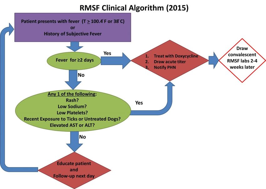
1. Notify the local jurisdiction and request that they alert providers and hospitals to
implement the RMSF algorithm (Appendix I)
2. Request that the hospital submit serum specimens to ASPHL. If the patient is critically ill,
request whole blood and (if possible) rash punch biopsy specimens for PCR testing at
CDC.
3. ADHS will notify CDC Rickettsial Zoonoses Branch and request PCR testing of whole
RMSF
Last Revised: 3/25/2020blood and IHC of punch biopsy.
4. Advise the local jurisdiction to conduct a home and community assessment. The case’s
home should be treated for ticks, and all dogs in the home should be on tick-prevention
products.
5. Educate household members about the symptoms of RMSF and request that they
immediately seek care if they experience fever.
6. The local jurisdiction can consider conducting chart reviews or a canine serosurvey to
determine the RMSF risk in the community.
RMSF
Last Revised: 3/25/2020REFERENCES
1. Rocky Mountain spotted fever (Internet). Centers for Disease Control and Prevention;
2017 (cited 2017 April). Available from http://www.cdc.gov/rmsf
2. Diagnosis and Management of Tick-borne Rickettsial Diseases: Rocky Mountain Spotted
Fever and Other Spotted Fever Group Rickettsioses, Ehrlichioses, and Anaplasmosis —
United States: A Practical Guide for Health Care and Public Health Professionals. MMWR
Recommendations and Reports May 13, 2016 / 65(2); 1-44:
https://www.cdc.gov/mmwr/volumes/65/rr/rr6502a1.htm?s_cid=rr6502a1_w
3. Arizona Rocky Mountain spotted fever Handbook—2015 (Internet). Arizona Department
of Health Services. (cited 2017 April). Available from:
http://www.azdhs.gov/documents/preparedness/epidemiology-disease-control/rocky-
mountain-spotted-fever/rmsf-handbook.pdf
4. Dahlgren, F.S., Holman, R.C., Paddock, C.D., Callinan, L.S., McQuiston, J.H. (2012). Fatal
Rocky Mountain spotted fever in the United States, 1999-2007. American Journal of
Tropical Medicine and Hygiene. 86(4):713-719.
5. Demma, L., Traeger, M., Nicholson, W., Paddock, C., Blau, D., Eremeeva, M., Dasch, G.,
Levin, M., Singleton, J., Zaki, S., Cheek, J., Swerdlow, D., and McQuiston, J. (2005). Rocky
Mountain spotted fever from an unexpected tick vector in Arizona. New England
Journal of Medicine. 353, 587–594.
6. Demma, L., Traeger, M., Blau, D., Gordon, R., Johnson, B., Dickson, J., Ethelbah, R.,
Piontkowski, S., Levy, C., Nicholson, W., Duncan, C., Heath, K., Cheek, J., Swerdlow, D.,
and McQuiston, J. (2006). Serologic evidence for exposure to Rickettsia rickettsii in
eastern Arizona and recent emergence of Rocky Mountain spotted fever in this region.
Vectorborne and Zoonotic Disease. 6, 423–429.
7. Drexler, N., Traeger, M., McQuiston, J., Williams, V., Hamilton, C., Regan, J. (2015).
Medical and indirect costs associated with Rocky Mountain spotted fever epidemic in
Arizona, 2002-2011. American Journal of Tropical Medicine and Hygiene.
http://www.ajtmh.org/content/early/2015/05/28/ajtmh.15-0104.full.pdf
8. Drexler N., Miller, M., Gerding, J., Todd, S., Adams, L., et al. (2014) Community-Based
Control of the Brown Dog Tick in a Region with High Rates of Rocky Mountain Spotted
Fever, 2012–2013. PLoS ONE 9(12): e112368. doi:10.1371/journal.pone.0112368
9. Folkema, A.M., Holman, R.C., McQuiston, J.H., Cheek, J.E., (2012). Trends in Clinical
Diagnoses of Rocky Mountain spotted fever among American Indians, 2001–2008. The
American Journal of Tropical Medicine and Hygiene. 86, 152-158.
RMSF
Last Revised: 3/25/202010. McQuiston, J.H., Guerra, M.A., Watts, M.R., Lawaczeck, E., Levy, C., Nicholson, W.L.,
Adjemian, J., and Swerdlow, D.L. (2011). Evidence of Exposure to Spotted Fever Group
Rickettsiae among Arizona Dogs outside a Previously Documented Outbreak Area.
Zoonoses and Public Health. 58(2), 85-92.
11. McQuiston, J.H., Drexler. N.A., Massung, R.M., Gerding, J., Miller, M., Levy, C., Komatsu,
K., Herrick, K., Weis. E., Traeger, M., Villar, R., Reidhead, T., Williams. V., Hamilton, C.,
and Piontkowski, S. Rocky Mountain spotted fever on Tribal Lands in Arizona, 2003-
2012: The Story from Emergence of a New Epidemic to Control and Prevention. Not
published, White Paper for Use in Arizona.
12. Nicholson, W.L., Gordon, R., and Demma, L. J. (2006). Spotted fever group rickettsial
infection in dogs from eastern Arizona: how long has it been there? Annals of the New
York Academy of the Sciences. 1078, 519–522.
13. Regan, J., Traeger, M. , Humpherys, D., Mahoney, D., Martinez, M. et al. (2015). Risk
factors for fatal outcome from Rocky Mountain spotted fever in a highly endemic area:
Arizona, 2002-2011. Clinical Infectious Diseases. Available online February 19, 2015.
14. Traeger, M. , Regan, J., Humpherys, D., Mahoney, D., Martinez, M. et al. (2015). Rocky
Mountain spotted fever characterization and comparison to similar illness in a highly
endemic area: Arizona, 2002-2011. Clinical Infectious Diseases. Available online
February 19, 2015.
15. Todd, S., Dahlgren, S., Traeger, M., Beltrán-Aguilar, E., Marianos, D., et al. (2015, May).
No Visible Dental Staining in Children Treated with Doxycycline for Suspected Rocky
Mountain spotted fever. Journal of Pediatrics. 166(5), 1246-1251.
16. ASPHL laboratory submission form: http://azdhs.gov/documents/preparedness/state-
laboratory/public-health-microbiology/clinical-microbiology-submission-form.pdf
17. ADHS Case Definitions for Reportable Communicable Morbidities, 2017, available at:
http://www.azdhs.gov/documents/preparedness/epidemiology-disease-
control/disease-investigation-resources/case-definitions.pdf
RMSF
Last Revised: 3/25/2020APPENDIX I RMSF Last Revised: 3/25/2020
RMSF Protocol

Protocol Reviewed/Approved by:

 Heather Venkat, Vector-borne and Zoonotic Disease Program Manager, 3/25/2020
 _______________________________                _________________ ___________
 Signature                                      Title                     Date

Last Revised:

       Date                          Revised by                 Epi Manager Signature
  1/8/2015           Hayley Yaglom
  4/26/2017          Hayley Yaglom                            Heather Venkat

RMSF
Last Revised: 3/25/2020
You can also read