Ultrasound use in the ICU for interventional pulmonology procedures
←
→
Page content transcription
If your browser does not render page correctly, please read the page content below
Review Article on Interventional Pulmonology in the Intensive Care Unit

Ultrasound use in the ICU for interventional pulmonology
procedures
Ivana Milojevic1, Kewakebt Lemma1, Rahul Khosla2
1
Department of Pulmonary, Critical Care and Sleep Medicine, George Washington University Medical Faculty Associates, Washington, DC, USA;
2
Department of Pulmonary and Critical Care Medicine, US Department of Veterans Affairs, Washington, DC, USA
Contributions: (I) Conception and design: All authors; (II) Administrative support: R Khosla, I Milojevic; (III) Provision of study materials or patients:
R Khosla; (IV) Collection and assembly of data: All authors; (V) Data analysis and interpretation: All authors; (VI) Manuscript writing: All authors;
(VII) Final approval of manuscript: All authors.
Correspondence to: Rahul Khosla, MD, MBA. Department of Pulmonary and Critical Care Medicine, US Department of Veterans Affairs, 50 Irving
St., NW, Room 4A-165, Washington, DC 20422, USA. Email: rahul.khosla@va.gov.

                 Abstract: Critical care ultrasound has shifted the paradigm of thoracic imaging by enabling the treating
                 physician to acquire and interpret images essential for clinical decision-making, at the bedside, in real-time.
                 Once considered impossible, lung ultrasound based on interpretation of artifacts along with true images,
                 has gained momentum during the last decade, as an integral part of rapid evaluation algorithms for acute
                 respiratory failure, shock and cardiac arrest. Procedural ultrasound image guidance is a standard of care
                 for both common bedside procedures, and advanced procedures within interventional pulmonologist’s
                 (IP’s) scope of practice. From IP’s perspective, the lung, pleural, and chest wall ultrasound expertise is a
                 prerequisite for mastery in pleural drainage techniques and transthoracic biopsies. Another ultrasound
                 application of interest to the IP in the intensive care unit (ICU) setting is during percutaneous dilatational
                 tracheostomy (PDT). As ICU demographics shift towards older and sicker patients, the indications for closed
                 pleural drainage procedures, bedside transthoracic biopsies, and percutaneous dilatational tracheostomies
                 have dramatically increased. Although ultrasound expertise is considered an essential IP operator skill there
                 is no validated curriculum developed to address this component. Further, there is a need for developing an
                 educational tool that matches up with the curriculum and could be integrated real-time with ultrasound-
                 guided procedures.

                 Keywords: Ultrasound; interventional pulmonology (IP); intensive care unit (ICU); procedure

                 Submitted Jan 04, 2020. Accepted for publication May 29, 2020.
                 doi: 10.21037/jtd-19-3564
                 View this article at: http://dx.doi.org/10.21037/jtd-19-3564

Introduction                                                                    actionable diagnoses of life-threatening conditions, which
                                                                                contributed to the development of sonographic evaluation
The field of critical care ultrasound founded by Dr.
                                                                                protocols for acute respiratory failure (1), shock (2-4), and
Lichtenstein in the early 1990s evolved over three decades                      cardiac arrest (5-7). Semiquantitative ultrasound assessment
into an indispensable bedside tool for management of the                        of lung aeration has found its application in optimization
critically ill. Out of Lichtenstein’s whole-body ultrasound                     of mechanical ventilation. Ultrasound guidance is the
paradigm, lung ultrasound was born. It allowed, for the                         standard of care for the majority of bedside procedures in
first time, rapid parenchymal assessment of this vital                          the intensive care unit (ICU). Point-of-care ultrasound thus
organ, at the point of care. Safe, non-invasive, and easily                     shifted the paradigm of thoracic imaging by eliminating the
repeatable, lung ultrasound could provide immediate                             dissociation between the treating physician and physician

© Journal of Thoracic Disease. All rights reserved.                       J Thorac Dis 2021;13(8):5343-5361 | http://dx.doi.org/10.21037/jtd-19-3564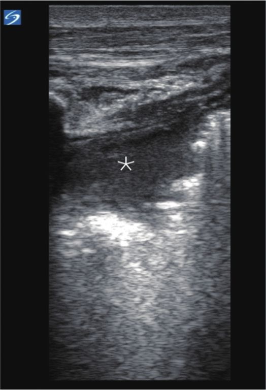
5344                                                                                                Milojevic et al. ICU ultrasound for IP

interpreting the imaging (8). Although the typical role of             Equipment
interventional pulmonologist (IP) in the ICU is to perform
                                                                       Ultrasound machine
advanced procedures, rather than day to day management
of the critically ill, it is necessary to discuss the universal        Lung sonography is based on artifacts described by
principles of lung and pleural ultrasound before focusing              original investigators using technology available in the
on sonographic aspects of pleural drainage procedures,                 1990s; examination can therefore be performed using any
transthoracic biopsies, and percutaneous dilatational                  commercially available two-dimensional (2D) scanner
tracheostomy (PDT).                                                    (10-13). The lung and pleural structures are visualized
                                                                       in 2D B-mode (brightness mode, which displays the
                                                                       acoustic impedance of a tissue cross-section), while
Basic principles of ultrasonography
                                                                       pleural movement can be assessed via M-mode (motion
For a more detailed consideration of applied ultrasound                mode, which emits pulses in quick succession, taking an
physics, comprehensive review is available (9). The                    image each time). Image quality is determined by the
fundamental principle of ultrasound imaging is the                     individual machine characteristics, presets, and post-image
reflection of ultrasound waves from tissues in the path of             processing. One caveat is that modern ultrasound machines
the beam. Echogenicity of the tissue refers to its ability             use numerous techniques to minimize the presence of
to reflect and/or transmit ultrasound waves in the context             ultrasound artifacts while lung ultrasonography often
of surrounding tissues. Translated to a visual greyscale,              relies on analysis of such artifacts. Therefore, experienced
echogenic, highly reflective structures appear white, in               operator is expected to adapt machine settings for optimal
contrast to black, non-reflective, anechoic structures;                visualization of said artifacts, unless lung presets are
“hypoechoic”, “isoechoic” and “hyperechoic” are relative               configured by the manufacturer. Typically, lung presets will
descriptors of brightness of the compared structures, in               turn off smoothing and artifact minimization algorithms
shades of gray.                                                        such as compound and harmonic imaging, and will also
   Whenever there is an interface of structures with                   reduce dynamic range. This is particularly helpful during
different echogenic properties as defined by their acoustic            assessment of lung sliding and B-lines. Abdominal presets
impedances, a difference in brightness will be apparent                may be more appropriate when evaluating pleural effusions
on the screen. The difference in acoustic impedances                   or consolidations. Doppler ultrasound is not a part of
between two tissues is directly proportional to the extent of          diagnostic lung sonography paradigm, but may be useful
reflection of ultrasound waves at their interface. Minimal             as a procedural adjunct in order to avoid vascular injury.
acoustic impedance of air determines large impedance                   Preferred ultrasound devices for bedside use are easy to
mismatch with surrounding tissues and results in almost                transport and clean, with short boot-up times and solid data
complete wave reflection at lung-chest wall interface,                 storage and transfer capabilities (10).
lending the pleural line its distinctive echogenic appearance.
Because only a small proportion of waves penetrates air,
                                                                       Transducers
and air is an unfavorable medium for sound transmission,
aerated lung appears as homogenous amorphic grayness                   Ultrasound probes in use today are multiple element
rather than a discrete structural entity. As the air content in        transducers, commonly called arrays. Various combinations
the lung decreases, acoustic mismatch also diminishes and              of array construction (linear, convex), and array functional
the ultrasound beam gets partially reflected, repeatedly, as it        features such as beam scanning and steering (accomplished
reaches the deeper zones. With even further decrease in air            by electronic sequencing or electronic phasing) and beam
content, acoustic window completely opens and lung can be              focusing (accomplished by electronic phasing) define the
directly visualized as solid parenchyma.                               probe footprint and image formats. Aside from technology
   Pleuropulmonary pathologic processes conducive                      used for beam manipulation which defines array type and
to the ultrasonographic examination such as effusions                  consequently probe footprint and image format, other
and peripherally extending consolidations provide real                 important probe feature is the ultrasound frequency range.
images, while the rest of the lung ultrasound is based on              Simplified terminology that partially describes probes
interpretation of artifacts derived by air/tissue interface.           as linear, phased array, or convex is pervasive (14) and

© Journal of Thoracic Disease. All rights reserved.               J Thorac Dis 2021;13(8):5343-5361 | http://dx.doi.org/10.21037/jtd-19-3564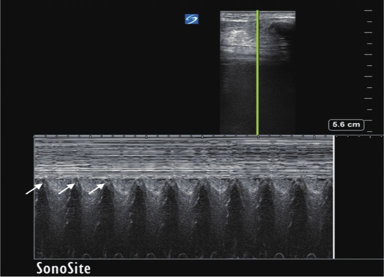
Journal of Thoracic Disease, Vol 13, No 8 August 2021 5345
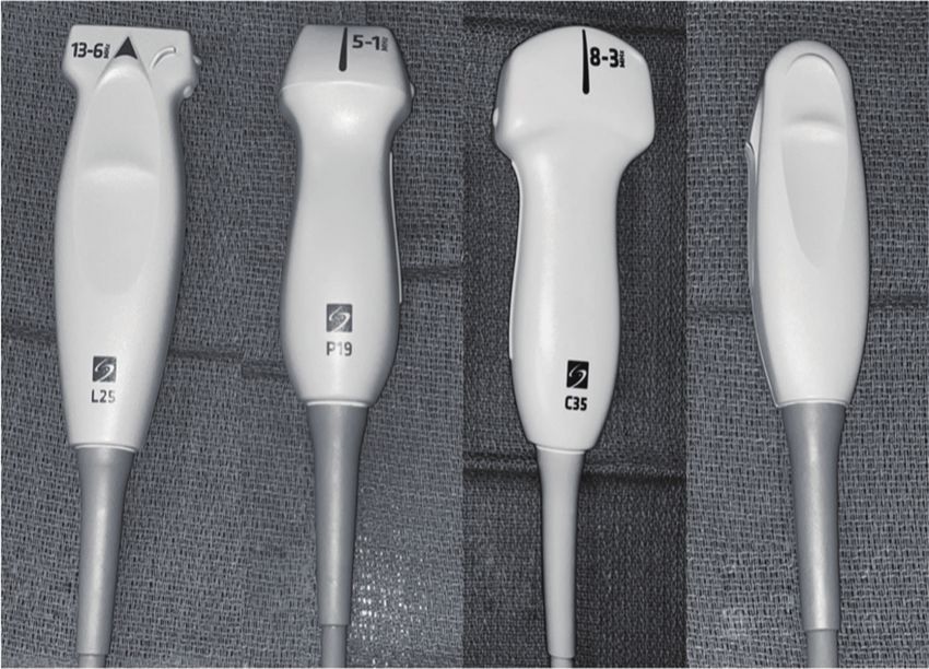
A B C D
Figure 1 Various ultrasound probes. (A) Linear (vascular) probe; (B) phased array (cardiac) probe; (C) convex (abdominal) probe; (D) micro-
convex probe.
used interchangeably with their most prominent imaging particularly important to use configured lung presets.
indication—vascular, cardiac, and abdominal, respectively. P h a s e d - a r r a y ( c a r d i a c ) p r o b e s ( F i g u re 1 B ) a r e
Higher frequency probes are typically used to acquire high characterized by low frequency and small footprint allowing
resolution images of more superficial structures, while for relatively easy image acquisition of deeper chest
lower frequency transducers capture lower resolution structures, albeit with less image resolution and narrow
images of deeper structures. If multiple probes are available, field of view. Therefore, this transducer is a reasonable
a useful rule is to choose the one that will provide the best selection for lung ultrasound if gross and diffuse pathology
resolution for the required depth. Certain probes have wide is suspected.
frequency ranges allowing them to be more versatile and The convex (abdominal) probes (Figure 1C) are
therefore cost-effective. This concept is illustrated by the characterized by intermediate frequency values and large
whole-body critical ultrasound paradigm which was built footprint, allowing decent visualization of the superficial
around a universal wide range microconvex probe (10,11). structures without losing the overview of the chest while
Intercostal acoustic windows are narrow, making it much attempting to search the entire pleural surface for localized
easier to acquire images free of rib artifact using a smaller pathology.
footprint probe, although diverging ultrasound beam Microconvex probes (Figure 1D) have a small footprint in
also means that only a small field of the pleural surface is combination with good resolution for both superficial and
examined (“tunneled vision”) and lateral resolution of the deep structures, making them the probe of choice for lung
image is poor. A larger footprint enables examination of a ultrasonography (10,11).
larger field of the pleural surface and improves resolution,
but it may be more difficult to acquire an image.
Linear (vascular) probes (Figure 1A) are characterized Basic ultrasonography technique
by high frequency (7.5–10 MHz) and large footprint; they
Probe and image orientation
are typically used to visualize the chest wall, pleural line,
and subpleural space in greater detail, and evaluate for Radiologic convention dictates that for imaging in
pneumothorax. Because this evaluation is dependent on longitudinal planes—sagittal and coronal, probe marker
ultrasound artifacts generated by air/tissue interface, it is should be oriented towards the head (cephalad), while for
© Journal of Thoracic Disease. All rights reserved. J Thorac Dis 2021;13(8):5343-5361 | http://dx.doi.org/10.21037/jtd-19-3564
5346                                                                                                  Milojevic et al. ICU ultrasound for IP

imaging in the transverse plane probe marker should be                   chest with its associated pathology. Although organized
oriented towards the patient’s right. Image orientation                  approach is recommended, there is no superior scanning
on the screen is determined by the location of the screen                protocol and flexibility is allowed, to account for clinical
marker which corresponds to the probe marker. Radiologists               context (13,16). Universally, higher patient acuity
keep the screen marker on the left side of the screen and                dictates simpler and faster methodology. Comprehensive
cardiologists traditionally keep it on the right. Critical               28 rib space technique allows for semi-quantification
ultrasound pioneering work was performed using head-to-                  of the interstitial syndrome (16,17), while eight-zone
the-left whole body convention including the heart, while                anterolateral lung examination is often employed during
practices among critical care practitioners are varied (15).             qualitative evaluation for interstitial syndrome (16,18).
                                                                         Bedside Lung Ultrasound in Emergency (BLUE) protocol
                                                                         assumes clinically significant interstitial syndrome and free
Depth
                                                                         pneumothorax should always be detected anteriorly (1),
Prior to beginning an examination, depth should be                       while free pleural fluid can predictably be found posteriorly
considered. Examiner can start by visualizing the deeper                 above the diaphragm regardless of the volume (19). These
structures by selecting the higher numerical depth setting               assumptions allow rapid identification of the cause of
and then zoom in on more superficial structures by                       acute respiratory failure, by examining only three identical
switching to lower depth setting.                                        predetermined points (the upper BLUE-point, lower
                                                                         BLUE-point and the posterolateral alveolar and/or pleural
                                                                         syndrome—or PLAPS—point) in both left and right
Gain
                                                                         hemithorax (20).
The gain function serves to optimize image quality by
compensating for attenuation (sound amplitude reduction).
                                                                         Semiotics of lung and pleural sonography
As sound waves travel deeper into the body the amplitude
of the returning signal diminishes but can be amplified by               Basic consideration of lung and pleural sonography is that
the receiver, therefore creating a brighter and better visible           real images always reflect pathology (pleural fluid and
image on the screen. Gain can be adjusted for the near field,            consolidated lung parenchyma) while artifacts can reflect
far field, or the entire field (overall gain). If the gain is set        both physiological and pathological state.
too low, the image appears dark, while excessive gain makes
the image too bright by adding “noise”.
                                                                         Signs

                                                                         The field of lung ultrasound is built on the following visual
Patient positioning
                                                                         concepts: pleural line (bat-wing sign), lung sliding and
Image acquisition performed with the patient sitting up                  corresponding seashore sign, absence of lung sliding and
allows full access to the chest, but is rarely feasible in a             corresponding stratosphere sign, lung pulse, lung point,
critically ill patient. More often patients are imaged while             A-lines, B-lines, sinusoid sign, shred sign, tissue-like sign,
supine, semi-recumbent, in lateral decubitus, or slightly                and air bronchograms (16,21). Seashore, stratosphere, and
rotated. Because the distribution of lung abnormalities does             sinusoid sign are recorded in M-mode. Lung pulse and
not rapidly change with different body positions, with the               lung point can be sought for in both M- and B-modes.
notable exception of free pleural effusions, it is possible to           Remaining signs pertain to B-mode.
perform an adequate ultrasonographic exam in almost any
position (13). The only true limitation is difficulty assessing          Pleural line (bat-wing sign)
the dorsal chest of a supine patient who is not easily                   The basic image of lung sonography is B-mode longitudinal
mobilized.                                                               view called the bat-wing sign (Figure 2) generated by upper
                                                                         and lower ribs (echogenic wings of the bat) and the pleural
                                                                         line (echogenic back of the bat). Pleural line is visualized
Scanning strategy
                                                                         approximately 0.5 cm below rib lines (Figure 2) and it
By examining various ultrasound planes systematically,                   always represents the parietal pleura, while visceral pleura
expert ultrasonographer can develop a 3D model of the                    may or may not contribute. Utilizing oblique instead of

© Journal of Thoracic Disease. All rights reserved.                 J Thorac Dis 2021;13(8):5343-5361 | http://dx.doi.org/10.21037/jtd-19-3564
Journal of Thoracic Disease, Vol 13, No 8 August 2021                                                                                   5347

                                                                         Figure 4 Barcode (stratosphere) sign (M-mode). Below the pleural
Figure 2 Bat-wing sign. Pleural line is marked by the middle             line marked by the arrow, granular pattern has been replaced by
arrow and corresponds to the “back of the bat”. The shadows of           horizontal lines suggesting the absence of lung sliding.
the upper (left arrow) and lower (right arrow) ribs represent the
echogenic “wings of the bat” (B-mode).
                                                                         lung sliding suggests that parietal and visceral pleura are
                                                                         closely approximated, and regional ventilation is preserved.
                                                                         Presence of anterior lung sliding rules out pneumothorax
                                                                         with 95.3% sensitivity, 91.1% specificity and 100% negative
                                                                         predictive value (22). Normally, lung sliding is more subtle
                                                                         in the upper parts and easier to recognize towards lung
                                                                         bases. Certain pathologic conditions or clinical contexts
                                                                         can significantly reduce lung sliding; for instance, it may be
                                                                         quite difficult to ascertain whether lung sliding is present
                                                                         in a paralyzed patient, protectively ventilated with low tidal
                                                                         volumes (12). For optimal visualization of subtle findings
                                                                         during lung sliding assessment, the harmonic imaging and
Figure 3 Seashore sign (M-mode). Below the pleural line marked
                                                                         compound imaging filters should be disabled and dynamic
by the arrow granular pattern referred to as “sandy beach” is            range (compression) reduced, although one could consider
observed and signifies that lung sliding is present.                     disabling all filters (11).

                                                                         Absence of lung sliding (stratosphere or barcode sign)
longitudinal approach one can evaluate a larger portion of               If lung sliding is absent, pleural line does not move
the pleural line, not interrupted by the rib shadows.                    synchronously with ventilation. Consequently, no
                                                                         shimmering is detected in B-mode (Video 2). In M-mode,
Lung sliding (pleural sliding) and seashore sign                         the granular or sandy beach pattern below the pleural line
In B-mode (Video 1), lung sliding is represented by                      is replaced by horizontal lines, while chest wall appearance
the movement of the pleural line synchronously with                      above the pleural line is unchanged, creating the barcode or
respiration (13). Despite various, even contradictory                    stratosphere sign (Figure 4). Detection of this sonographic
definitions, lung sliding is intuitively recognized as                   pattern in the anterior chest implies that pleural surfaces
homogeneous shimmering starting at the pleural line and                  are separated by air, and/or that regional ventilation is
spreading below (10). Characteristic appearance of lung                  impaired by airway obstruction, hyperinflation, or pleural
sliding in M-mode is named seashore sign (Figure 3).                     adhesions. Further investigation is performed by assessing
Area above the pleural line with a wave-like pattern                     for the presence of B-lines, lung point, and lung pulse (see
represents the chest wall and area below the pleural line                below), while considering the clinical context. In trauma
with a granular/sandy beach-like pattern represents the                  patients, for example, isolated absence of lung sliding has
expanded and ventilated lung. In physiologic terms, evident              98.1% sensitivity and 99.2% specificity for detection of

© Journal of Thoracic Disease. All rights reserved.                 J Thorac Dis 2021;13(8):5343-5361 | http://dx.doi.org/10.21037/jtd-19-3564
5348                                                                                                      Milojevic et al. ICU ultrasound for IP

Figure 5 Lung pulse (M-mode). In the absence of lung sliding, a             Figure 6 Lung point (M-mode). The arrow marks the transition
distinct visual pattern below the pleural line (arrows) can be seen,        from the seashore sign to the stratosphere sign with changes in
and is created by pleural movement synchronously with cardiac               the respiratory cycle. Seashore sign indicates that lung sliding is
pulsations. This pattern is generated by the lack of regional               present during inspiration and stratosphere sign indicates that lung
ventilation and rules out pneumothorax.                                     sliding is absent during expiration.

pneumothorax (23). Absence of lung sliding can be observed
in patients ventilated with high positive end-expiratory
pressure (PEEP) but without pneumothorax (24).

Lung pulse
In the absence of lung sliding, subtle movements of the
pleural line caused by the force of cardiac pulsation and
synchronous with the pulse can be seen in both B-mode
(Video 3) and M-mode (Figure 5). Presence of lung pulse
rules out pneumothorax and confirms absence of regional
ventilation with 93.0% sensitivity, and 100% specificity (25).
                                                                            Figure 7 A-lines (B-mode). Horizontal, hyperechoic lines below
Lung point                                                                  the pleural line, marked by white arrows, and representing
Lung point marks the location on the chest wall where the                   reverberation artifact.
collapsed lung achieves transient contact with the parietal
pleura during inspiration. In B-mode, this phenomenon is
appreciated as inspiratory presence and expiratory absence                  skin and the pleural line. A-lines represent a reverberation
of lung sliding in a particular scanning location. M-mode                   artifact and indicate the presence of air, physiologic or free,
depicts alternating seashore and stratosphere appearances                   below the parietal pleura (28). Significance of A-lines is
in relation to the phase of the respiratory cycle (Figure 6).               further interpreted in the context of lung sliding and lung
Sensitivity of lung point for pneumothorax detection is                     pulse, as well as other signs.
only 66%, while specificity is 100% (26). Notably, complete
lung collapse cannot produce the lung point. Location on                    B-lines
the chest wall where lung point is detected can be used for                 B-lines (Figure 8) are discrete, laser-like, vertical,
pneumothorax semi-quantification (27).                                      hyperechoic reverberation artifacts that arise from the
                                                                            pleural line, extend to the bottom of the screen without
A-lines                                                                     fading, erase A-lines, and move synchronously with lung
A-lines are hyperechoic, horizontal lines seen at regular                   sliding (16). They belong to the comet-tail artifacts family
intervals from the pleural line (Figure 7). These intervals                 and were referred to as lung comets before the nomenclature
are equal to the distance between the transducer on the                     was standardized (16). One theory of B-line genesis is that

© Journal of Thoracic Disease. All rights reserved.                    J Thorac Dis 2021;13(8):5343-5361 | http://dx.doi.org/10.21037/jtd-19-3564
Journal of Thoracic Disease, Vol 13, No 8 August 2021                                                                                   5349

Figure 8 B-lines (B-mode). Vertical, hyperechoic lines arising           Figure 10 Shred sign (B-mode). Subpleural consolidation with
from the pleural line, marked by white arrows. Visualization of          echogenic and irregular borders, marked by arrows, separating it
more than 2 per ultrasound field is considered abnormal.                 from the adjacent aerated lung parenchyma.

                                                                         lung infarct) (31), while diffuse pattern of interstitial
                                                                         syndrome usually reflects cardiogenic pulmonary edema or
                                                                         ARDS.

                                                                         Sinusoid sign
                                                                         Sinusoid sign pertains to the M-mode recording of the
                                                                         lung and visceral pleura respiratory movement within
                                                                         free pleural fluid collection (Figure 9). Sinusoid sign also
                                                                         implies low viscosity of the fluid, which is important during
                                                                         thoracentesis deliberations (19).

Figure 9 Sinusoidal sign. M-Mode recording of the respiratory            Shred (fractal) sign
movement of the lung and visceral pleura represented by the wavy,        Shred sign is seen with smaller subpleural consolidations
hyperechoic line within anechoic, free pleural fluid marked by *.        visualized as hypoechoic areas separated from the rest of the
                                                                         lung by irregular, echogenic borders (Figure 10).
                                                                            These consolidations can be of infectious origin (32), due
ultrasound beam interacts with edematous interlobular septa              to sub-pleural infarcts (33) or diffuse lung disease (34).
originating from the visceral pleura (29). Although anatomic
and physical basis of B-lines is not certain (16), it is widely          Tissue-like sign
accepted that B-lines can only be seen when the visceral                 Tissue-like sign (Figure 11) represents direct visualization of
pleura is opposing the parietal pleura and that their presence           the lung tissue after complete loss of aeration has occurred,
rules out pneumothorax, even if lung sliding is absent (30).             typically in lobar pneumonia pattern (35,36). Term
B-lines represent the densitometer of lung parenchyma,                   sonographic hepatization of the lung effectively describes
and with their increasing number lung density increases, as              echogenicity of the consolidated lung, which is similar to
measured by CT (28). The very presence of B-lines is not                 that of the liver.
abnormal and a maximum of 2 B-lines per scan (longitudinal
plane between two ribs) is observed at the lateral bases in              Air-bronchogram
27% of healthy subjects (29). Interstitial syndrome is defined           Hyperechoic structure recognized within a tissue-like
as diffuse anterolateral presence of 3 or more B-lines per               consolidation, which corresponds to air-filled airway
scan (1). B-line distribution is another cardinal feature to             (Figure 12). If air bronchogram is absent or static,
consider, since focal B-pattern can be correlated with various           potential lack of airway patency is suggested and/or
pathologies (pneumonia, atelectasis, mass, lung contusion,               partial to complete air resorption. Presence of dynamic air

© Journal of Thoracic Disease. All rights reserved.                 J Thorac Dis 2021;13(8):5343-5361 | http://dx.doi.org/10.21037/jtd-19-3564
5350                                                                                                  Milojevic et al. ICU ultrasound for IP

bronchograms (Video 4) supports regional airway patency
and rules out atelectasis (37).

Interpretation

While sensitivity of lung ultrasound is generally robust,
signs discussed above are often not specific for a diagnosis.
Absent lung sliding, multiple B-lines, or lung consolidation
can all be linked to various pathologic conditions and
what influences interpretation the most is patient’s clinical
condition. Directly imaged pathologies, such as effusions
and peripherally extending consolidations are usually                    Figure 11 Tissue-like sign (B-mode). Echogenicity of the
straightforward umbrella diagnoses, while their further                  consolidated lung marked by the arrow, resembles the echogenicity
etiologic classification is nuanced. Artifact interpretation             of the liver, seen below the hyperechoic dome of the diaphragm.
is typically more challenging and is based on sequential
evaluations of the distribution of different signs. The final
result is a sonographic lung profile, which in an appropriate
clinical context is either consistent with a diagnosis, or able
to rule one out. This approach is codified in several focused
assessment protocols, intended for application to a correct
clinical scenario and integration with standard clinical
approach.

Sonographic diagnoses

Normal sonographic appearance of the lung

Presence of lung sliding in combination with A-lines and                 Figure 12 Air-bronchogram (B-mode). Hyperechoic flecks and
maximum of 2 B-lines per view in anterior lung fields                    lines (arrows) within consolidated lung tissue represent air filled
suggests normally aerated lung parenchyma. However,                      airways.
the same profile can be seen with pathologies sparing the
parenchyma, such as thromboembolic or airway disease,
while posterior interstitial changes can be seen in the                  mass, or any combination of these findings (43,44), while
absence of true pathology, as a result of gravity alone (1).             ultrasonography is superior in differentiating pleural fluid
                                                                         from parenchymal processes and hemidiaphragm elevation
                                                                         (39,45). Lung ultrasound yields diagnostic accuracy of
Pleural effusion
                                                                         93% when compared with chest CT for pleural fluid
Ultrasound can easily diagnose the effusion, suggest                     detection (46), while it outperforms CT for complex
etiology, estimate volume, and image-guide fluid removal,                effusion diagnosis due to its superior ability to detect
making evaluation of pleural effusions the most established              septations (47,48).
application of ultrasound in the chest.                                     Obeying gravity, free-flowing fluid in the pleural space
   With regards to pleural fluid detection, ultrasound                   accumulates in the dependent part of the chest. Posterior
is more accurate than clinical examination (38), since it                axillary line above the diaphragm is thus the optimal site
can detect as little as 3–5 mL (39). Better sensitivity and              to evaluate for a non-loculated fluid collection (16). In
reliability of ultrasound in comparison with portable chest              contrast, loculated effusions do not change with body
X-rays is of special interest in critically ill patients (40-42).        position and can occupy non-dependent areas, warranting
Chest radiography often discriminates poorly between                     examination of the entire hemithorax. With probe marker
pleural thickening, effusion, atelectasis, pneumonia,                    oriented towards the head during longitudinal scan,

© Journal of Thoracic Disease. All rights reserved.                 J Thorac Dis 2021;13(8):5343-5361 | http://dx.doi.org/10.21037/jtd-19-3564
Journal of Thoracic Disease, Vol 13, No 8 August 2021                                                                                     5351

Figure 13 Pleural effusion (B-mode) seen above the diaphragm               Figure 15 Complex septated pleural effusion (B-mode). Septations
dome (right arrow) along with atelectatic lung tissue (left arrow).        are indicated by the arrows.
The anechoic pleural fluid is marked by *. This appearance is
compatible with a simple effusion.

                                                                           Figure 16 Homogeneous echogenic pleural effusion (B-mode).
                                                                           Homogeneously echogenic fluid is denoted by *.
Figure 14 Complex non-septated pleural effusion (B-mode)
characterized by non-homogeneous echogenic material without
septations.                                                                hypoechoic top layer and cellular, hyperechoic bottom
                                                                           layer, which constitutes the hematocrit sign. Previously
                                                                           described sinusoid sign in M-mode is characteristic for a
exam starts by identifying the diaphragm as a curvilinear,                 pleural effusion and can help delineate small effusions from
hyperechoic structure, which moves with breathing. Pleural                 pleural thickening, since it represents floating motion of the
fluid is then typically seen as an anechoic or hypoechoic                  lung within low viscosity fluid (19).
collection above the diaphragm (Figure 13, Video 5). In case                  Sonographic appearance of the pleural fluid has value
of a moderate to large effusion with adjacent parenchymal                  in separating transudates from exudates. Four sonographic
consolidation, tissue-like structure is noted flapping in the              effusion patterns have been described: (I) simple, anechoic
fluid collection (flapping lung or jellyfish sign—Video 6).                (Figure 13); (II) complex non-septated, characterized by
Alternatively, aerated lung may be seen moving over the                    non-homogeneous echogenic material and no septations
pleural effusion while covering liver and/or spleen during                 (Figure 14); (III) complex septated, with floating fibrin
inspiration, which is called the curtain sign (Video 7). If                strands or septae in a lattice-like pattern (Figure 15) and
cellular or proteinaceous debris suspended in the pleural                  (IV) homogeneously echogenic (Figure 16). Transudates are
fluid gets agitated by respiratory or cardiac motion, a                    almost always anechoic, while exudates can have any of the
swirling pattern called the plankton sign, is created. The                 four patterns (49,50). Parapneumonic effusions, empyemas,
presence of significant cellular debris is responsible for                 and malignant effusions can all have complex septated
a layering effect caused by gravity, with hypocellular,                    appearance, whereas empyemas and hemorrhagic effusions

© Journal of Thoracic Disease. All rights reserved.                   J Thorac Dis 2021;13(8):5343-5361 | http://dx.doi.org/10.21037/jtd-19-3564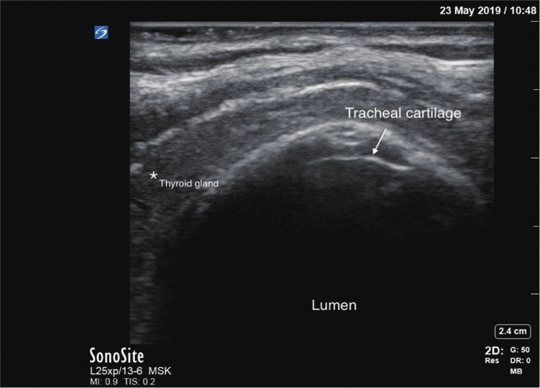
5352                                                                                                  Milojevic et al. ICU ultrasound for IP

may appear homogeneously echogenic (51). Additional                      out pneumothorax due to insufficient sensitivity (66%) (26).
sonographic clues towards etiologic diagnosis of pleural                 Notably, in cases of complete lung collapse lung point
effusion include echogenic swirling pattern and/or presence              will not be seen, although lack of sensitivity can also
of pleural nodules which imply malignancy (52), and                      be explained by suboptimal operator skills, incomplete
adjacent consolidated lung with dynamic air bronchograms                 examination, and unfavorable anatomy. Lung point location
which indicates infectious origin.                                       on the chest wall denotes pneumothorax extension. In one
   Despite the existence of various sonographic methods                  study, 90% of patients with lateral lung-points required
for fluid volume quantification (41,42,53,54) reliable                   drainage versus 8% of patients with anterior lung-point (59).
estimation of the effusion volume remains challenging for
multiple reasons (55). Inspiratory interpleural distance of at
                                                                         Alveolar consolidation
least 15 mm with effusion also identifiable in both adjacent
intercostal spaces has been suggested as a feasibility marker            Alveolar consolidation extends to the pleura in overwhelming
of safe thoracentesis in mechanically ventilated patients (19).          majority of cases, while 90% of consolidations may be
                                                                         imaged from the PLAPS point or intersection of a horizontal
                                                                         line at the level of the lower BLUE-point and posterior
Pneumothorax
                                                                         axillary line (61). Ultrasound has 90% sensitivity and 98%
In mechanically ventilated or trauma patients the                        specificity for the diagnosis of alveolar consolidation, when
importance of rapid pneumothorax recognition cannot be                   compared to CT (61), and may be superior in detection
overstated. Supine portable chest radiographs are widely                 of abscesses and necrotizing areas within the consolidated
available and routinely performed for this indication, albeit            lung (62). Two main consolidation patterns are the smaller,
with a high rate of misdiagnosis (56-58). Chest CT, the                  non-translobar, which yields the shred sign, and the more
gold standard for air detection in pleural space, is highly              extensive, translobar, which produces impressive image
impractical for most critical care settings, not to mention              resembling anatomical lung (tissue-like sign) (61,63). Since
the radiation exposure. The tendency of free air to collect              various pathological processes (compressive or obstructive
in non-dependent anterior chest makes the supine position                atelectasis, pneumonia, ARDS, contusion, infarction or mass)
ideal for ultrasound exploration of pneumothorax. Bedside                can present as consolidation, additional sonographic clues
ultrasound is proven to be an effective tool to rule out                 may help determine the exact etiology. Different pulmonary
pneumothorax (22,25,30,59). Presence of anterior lung                    lesions exhibit characteristic patterns of vascularization on
sliding virtually rules out pneumothorax in a severely                   qualitative color Doppler sonography (64). Lung pulse is
dyspneic patient (22). In other words, in case of severe                 reported to have a 93% sensitivity and 100% specificity for
dyspnea/acute respiratory failure the presence of anterior               the diagnosis of complete atelectasis following right main
lung sliding has a 100% negative predictive value for                    stem intubation in patients without previous respiratory
pneumothorax causing such symptoms. Absence of lung                      disorders (25). Identification of dynamic air bronchograms
sliding, in contrast, is all but specific for pneumothorax               within consolidated lung indicates bronchial patency and
and can also be attributed to a host of other pathologies                argues against resorptive atelectasis, thus yielding 97%
(airway occlusion with mucous plug, tumor or foreign body,               positive predictive value for the diagnosis of pneumonia (37).
ARDS, pneumonia, effusion etc.). In the absence of lung                  Dynamic linear-arborescent air bronchogram seems to
sliding, one can still rule out pneumothorax by detecting                be a specific sign for the diagnosis of ventilator-associated
either lung pulse (25) or B-lines (30). If not ruled out by              pneumonia (32,65).
these sequential evaluations in a critically ill patient with
high pretest probability, it is safe to assume pneumothorax
                                                                         Peripheral lung masses
is present and proceed with urgent management without
confirmatory testing (16,60). Clinical context also weighs               Malignant lesions are hypoechoic or moderately echogenic,
in heavily in post-procedural settings, where if previously              round or oval, inhomogeneous structures with well-defined
present lung sliding disappears on repeated evaluation,                  margins, often serrated or with finger-like projections into
pneumothorax is highly likely. Detection of lung point                   the surrounding lung tissue (Figure 17) (51). Ultrasound
confirms the presence of pneumothorax with 100%                          assessment of the chest wall invasion by the tumor has been
specificity, while inability to identify lung point does not rule        reported to be superior to CT scan (66). Sonographic criteria

© Journal of Thoracic Disease. All rights reserved.                 J Thorac Dis 2021;13(8):5343-5361 | http://dx.doi.org/10.21037/jtd-19-3564
Journal of Thoracic Disease, Vol 13, No 8 August 2021 5353
presence of B-lines in the anterior chest denotes a severe
degree of cardiogenic pulmonary edema, focused anterior
scanning as suggested by the BLUE protocol is highly
accurate in the critically ill, but may be insufficient to
diagnose less symptomatic patients (68), which would
benefit from extended examination to include the lateral
chest (18). Nonhomogeneous distribution of B-lines,
irregular thickened pleura with subpleural and posterior
consolidations favor ARDS over cardiogenic pulmonary
edema in critically ill patients (69). Semi-quantification of
B-lines has found many applications in the critical care arena:
cardiogenic pulmonary edema monitoring (70), assessment
of hemodialysis effectiveness for extravascular lung water
removal (71), fluid management in patients with ARDS
and septic shock (72), predicting ARDS mortality (73),
defining PEEP responders (74), identifying pronation
responders (75,76) and anticipating weaning failure (77).
Figure 17 Peripheral lung mass (B-mode, linear probe) seen as BLUE protocol
hypoechoic lesion marked by the *, with hyperechoic, irregular
BLUE protocol is an algorithm for acute respiratory failure
margins.
evaluation using bedside ultrasound which, when applied
to critically ill patients yields a correct diagnosis in 90% of
cases (1). Protocol combines lung and venous ultrasound in
for chest wall infiltration include rib destruction, pleural line
rapid, sequential assessment for lung sliding, A and B-line
interruption, and limited respiratory excursion of the mass.
artifacts, venous thrombosis, alveolar consolidation and/or
pleural effusion, performed in 3 standardized locations per
Alveolar-interstitial syndrome (AIS) hemithorax. Combination of findings designates a profile
consistent with one of the following: pulmonary edema,
The AIS is a non-specific sonographic manifestation of
pulmonary embolism, pneumonia, asthma/COPD and
different acute and chronic parenchymal conditions, defined
pneumothorax.
as 3 or more B-lines in a single view, diffusely disseminated
over the antero-lateral thorax. In direct comparison to
chest radiography, diffuse sonographic B-pattern identifies Ultrasound procedural guidance
diffuse radiographic AIS with 93% sensitivity and 93%
Pleural drainage procedures
specificity (29). B-line density in a single view correlates
with the severity of AIS documented by chest CT. B-lines Ultrasound reduces complications and increases the yield
that are 7 mm apart at the pleural line imply subpleural of thoracentesis and is integral to the management of
interlobular septal thickening, while B-lines that are only complex pleural disease by allowing targeted placement
3 mm apart at the pleural line correlate with subpleural of chest tubes. It also allows sequential sampling of fluid
ground glass opacities (67). Differentiation among potential pockets, if pH variability is expected (78), while sonographic
causes of AIS, such as cardiogenic and noncardiogenic appearance informs the decision on whether to use tissue
pulmonary edema, ARDS, interstitial pneumonia and plasminogen activator with DNase therapy (79,80) or
lung fibrosis depends on additional sonographic features, recommend surgical intervention.
but even more so on clinical context. Acute cardiogenic Rate of postprocedural pneumothorax for thoracentesis
pulmonary edema is characterized by symmetric spatial without image guidance is unacceptably high (20–39%)
distribution and usually progresses from the dependent and drops significantly when ultrasound is used to identify
zones to the anterior upper chest (13). Considering that the optimal puncture site (81-83). When compared to
© Journal of Thoracic Disease. All rights reserved. J Thorac Dis 2021;13(8):5343-5361 | http://dx.doi.org/10.21037/jtd-19-35645354 Milojevic et al. ICU ultrasound for IP
clinically marked procedural site, ultrasound reduced wall, parietal pleura, lung, and sometimes diaphragm or
complication rate while increasing yield, regardless of mediastinum. The technique can also be applied to the
clinician seniority (38). Superiority of ultrasound-guided nearby extra-thoracic structures such as supraclavicular
thoracentesis has been confirmed by meta-analysis (84) and lymph nodes. US-TTNB of pleural-based lesions performed
a large retrospective cohort study (85) and adopted into by pulmonologists is a safe procedure (95-98), with an
practice guidelines (86). British Thoracic Society Pleural acceptable diagnostic yield (95-97,99-101), and represents a
Disease Guidelines also recommend the assistance of time-saving, cost-effective alternative to CT-guided biopsy
ultrasound when placing any chest drain for fluid, although (98,102). The pneumothorax rate is lower than for CT-
in the absence of robust data, and mostly extrapolating from guided biopsies (98,102), possibly because only peripheral
thoracentesis experience. lesions in direct contact with the pleura are amenable to
The optimal site, angle, and depth of needle insertion US-TTNB. Aside from being more readily available to the
portend procedure success. Lung sliding should be critically ill patient than CT scan, a significant advantage
documented prior to needle insertion, since its absence of ultrasound is real-time guidance that allows dynamic
post procedure implies iatrogenic pneumothorax. Needle evaluation of the target, so vasculature and necrotic areas are
trajectory must replicate the angle and position of the avoided, while biopsy of smaller lesions can be conducted
transducer during image acquisition, while the depth of in favorable respiratory phase with breath holding. The use
insertion is calculated from a frozen image of the fluid of biopsy guide simplifies the diagnostic procedure for the
pocket by using calipers. Two basic techniques are real- operator by moving the needle in the US plane, while free
time guidance and site-marking technique. With real- hand technique should be reserved for experts (103). US-
time guidance the position of the needle is monitored TTNB has a potential role in microbiological diagnosis and/
constantly, allowing true targeted placement in cases of or therapeutic drainage of a pleural-based lung abscess (104),
complex effusions, but is technically more challenging and provided there is evidence of inflammatory pleurodesis at the
not routinely performed. As long as the site marking is planned access site, so that pleural contamination is avoided.
done correctly, and procedure performed immediately after The use of ultrasound guidance during closed pleural
(to avoid fluid redistribution from patient repositioning), biopsy in a challenging population of patients, including
site-marking technique has very low complication rate, cases of failed thoracoscopy, resulted in high diagnostic
even in mechanically ventilated patients (87). Intercostal accuracy (105). Although not a routine bedside procedure in
arteries are not amenable to external compression and the ICU setting, US-TTNB in our opinion should always
inadvertent trauma during chest drain insertion can cause be considered in critically ill patients with suspected, but,
bleeding into a large negative pressure space (88). The risk undiagnosed malignancies where a tumor mass is abutting
of injury is greatest within 6 cm from the spinal column the pleura or an enlarged lymph node is identified with US.
(89,90), justifying guideline recommendation for mid- Under such circumstances US-TTNB can be performed
axillary placement within the “safe triangle” and along easily at the bedside and guide therapeutic options or help
the superior rib border. This approach does not account with patient prognosis.
for anatomical variation (90), the risk still being relatively
high in elderly patients with tortuous arteries (91) or when
Percutaneous dilatational tracheostomy
obesity and subcutaneous edema render the recognition of
surface landmarks difficult (92). Ability of Color Doppler to PDT is the procedure of choice in critically ill patients
identify the intercostal vessels prior to needle insertion was (106-108), although surgical approach may be preferred in
reported (93) and, after testing against the gold standard CT cases of unfavorable anatomical landmarks, unstable cervical
angiogram, recommended prior to thoracentesis, when there spine, malignancy at the site of insertion, and emergency
is an increased risk of bleeding (94). This practice is not yet situations (108,109), Guidelines (108) recommend
widely adapted, since prospective confirmation is lacking. bronchoscopy use during PDT for real-time confirmation
of needle entry in midline position, endotracheal tube
visualization, and prevention of posterior tracheal wall injury.
Ultrasound-guided transthoracic needle biopsies
Reported drawbacks of bronchoscopy in this context are
(US-TTNB)
increase in intracranial pressure and decrease in ventilation
US-TTNB is typically used to obtain tissue from the chest with alveolar de-recruitment (109). Sonography can be
© Journal of Thoracic Disease. All rights reserved. J Thorac Dis 2021;13(8):5343-5361 | http://dx.doi.org/10.21037/jtd-19-3564Journal of Thoracic Disease, Vol 13, No 8 August 2021 5355
Ultrasound-guided PDT differs from bronchoscopic-
guided PDT primarily by visualization of the needle
insertion, laryngoscopic endotracheal tube adjustment,
and the tracheostomy tube confirmation (115). Studies
comparing real-time ultrasound with bronchoscopy as PDT
image guide found ultrasound guidance to be non-inferior to
slightly favorable over bronchoscopy (116-118). Standalone
sonographic guidance has been used in a neurosurgical
population, avoiding the transient increase in intracranial
pressure reported during bronchoscopy (119). However, it
seemed to have an extended learning curve, with at least 50
Figure 18 Trachea in the transverse plane (B-mode, linear probe). procedures required to develop competence (120).
Education and competence in chest ultrasound
Chest sonography for both diagnostic purposes and image-
guidance depends on the operator competence in image
acquisition and interpretation at the point-of-care. However,
there are barely any validated tools to evaluate demonstrated
skills (121), no universally accepted training methodology
exists, and accreditation programs are typically geared
toward local clinical environments (122). Paucity of relevant
literature justifies, to some extent, extrapolation from 2011
guidelines on training methodology for general critical care
ultrasound (123), which were developed around American
Figure 19 Trachea in the longitudinal plane with endotracheal
College of Chest Physicians/La Société de Reanimation
tube in the lumen (B-mode, linear probe).
de Langue Française competence statement (124).
Certification programs in critical care sonography generally
acknowledge the necessity of longitudinal experience
used during PDT as a neck screen, to identify patients
needed to master ultrasound techniques (8). In 2017
unsuitable for the procedure and assist with landmark
Interventional Pulmonology Special Interest Group of the
recognition if anatomy is difficult (110,111), estimate the
Thoracic Society of Australia and New Zealand developed
distance from the surface of the skin to the trachea (112),
an expert-consensus program for recognition of thoracic
and prevent puncture of aberrant vessels. In addition, real-
ultrasound competence, where competence was defined as
time ultrasound-guidance may be employed for visualization
“the ability to accurately and safely use bedside ultrasound
of the needle and dilator entering the trachea. Bleeding is
to evaluate a pleural effusion, determine a safe site for
a significant concern with PDT (113). Ultrasound allows
thoracentesis or chest tube insertion, to assess pleural
preoperative identification of aberrant neck vessels, which pathology or parenchymal lung disease and rule out a
in one case series resulted in changing the intended tracheal pneumothorax” (122). From IP’s perspective competency
puncture site in every fourth patient (114). In our practice requirements should be expanded to include proficiency
we routinely use US preoperatively to estimate the distance in performing transthoracic biopsies, vascular and anterior
from the surface of the skin to the trachea, and to identify neck imaging, with the ultimate goal being an integrated
any aberrant vessels in the field. In real-time ultrasound- training methodology for both the use of ultrasound and
guided PDT technique, a high-frequency linear transducer the performance of ultrasound-guided procedures.
is used to image proximal trachea, cricoid cartilage,
cricothyroid membrane, thyroid gland, and thyroid
Ultrasound limitations
cartilage, allowing accurate puncture of the target tracheal
interspace at midline (Figures 18,19). Since air is a poor acoustic medium, subcutaneous
© Journal of Thoracic Disease. All rights reserved. J Thorac Dis 2021;13(8):5343-5361 | http://dx.doi.org/10.21037/jtd-19-35645356 Milojevic et al. ICU ultrasound for IP
emphysema, pneumothorax and aerated lung all degrade sponsorship. The authors have no other conflicts of interest
ultrasound images and block visualization of nearby to declare.
structures. Image quality may also be poor in morbidly
obese or anasarcic patients. Critically ill patients tend to be Ethical Statement: The authors are accountable for all
immobile and can be sub-optimally positioned during exam, aspects of the work in ensuring that questions related
while the presence of catheters and dressings additionally to the accuracy or integrity of any part of the work are
interferes with image acquisition. appropriately investigated and resolved.
Open Access Statement: This is an Open Access article
Conclusions
distributed in accordance with the Creative Commons
Past several decades have witnessed bedside ultrasonography Attribution-NonCommercial-NoDerivs 4.0 International
evolve into a cornerstone of critical care medicine, while License (CC BY-NC-ND 4.0), which permits the non-
lung ultrasound gained momentum mainly in the last commercial replication and distribution of the article with
decade, as an integral part of rapid evaluation algorithms the strict proviso that no changes or edits are made and the
for acute respiratory failure, shock and cardiac arrest. Wide original work is properly cited (including links to both the
application of semiquantitative sonographic techniques formal publication through the relevant DOI and the license).
for assessment of lung aeration in the modern ICU keeps See: https://creativecommons.org/licenses/by-nc-nd/4.0/.
the momentum going. Lack of radiation exposure, cost-
effectiveness, high diagnostic accuracy for actionable life-
References
threatening conditions, and point-of-care procedural
image-guidance are difficult to match by other imaging 1. Lichtenstein DA, Mezière GA. Relevance of lung
technologies. In the critically ill, less invasive advanced ultrasound in the diagnosis of acute respiratory failure: the
diagnostic and therapeutic procedures involving the BLUE protocol. Chest 2008;134:117-25.
chest and airways are preferred over the standard surgical 2. Lichtenstein D. FALLS-protocol: lung ultrasound in
approach. This trend dictates increased bedside presence of hemodynamic assessment of shock. Heart Lung Vessels
the IP who must be proficient in pleural, lung, and vascular 2013;5:142-7.
ultrasound. Ultrasound competence for the IP’s scope of 3. Lichtenstein D. Fluid administration limited by lung
practice would be best enhanced by a validated training sonography: the place of lung ultrasound in assessment of
curriculum. acute circulatory failure (the FALLS-protocol). Expert Rev
Respir Med 2012;6:155-62.
4. Volpicelli G, Lamorte A, Tullio M, et al. Point-of-
Acknowledgments
care multiorgan ultrasonography for the evaluation
Funding: None. of undifferentiated hypotension in the emergency
department. Intensive Care Med 2013;39:1290-8.
5. Lichtenstein DA. How can the use of lung ultrasound
Footnote
in cardiac arrest make ultrasound a holistic discipline.
Provenance and Peer Review: This article was commissioned The example of the SESAME-protocol. Med Ultrason
by the Guest Editors (Jonathan S. Kurman, Ashutosh 2014;16:252-5.
Sachdeva and Rahul Nanchal) for the series “Interventional 6. Lichtenstein D, Malbrain ML. Critical care ultrasound in
Pulmonology in the Intensive Care Unit Environment” cardiac arrest. Technological requirements for performing
published in Journal of Thoracic Disease. The article has the SESAME-protocol--a holistic approach. Anaesthesiol
undergone external peer review. Intensive Ther 2015;47:471-81.
7. Lien WC, Liu YP, Chong KM, et al. A novel US-
Conflicts of Interest: All authors have completed the ICMJE CAB protocol for ultrasonographic evaluation
uniform disclosure form (available at http://dx.doi. during cardiopulmonary resuscitation. Resuscitation
org/10.21037/jtd-19-3564). The series “Interventional 2017;115:e1-2.
Pulmonology in the Intensive Care Unit Environment” was 8. Lyn-Kew KE, Koenig SJ. Bedside ultrasound for
commissioned by the editorial office without any funding the interventional pulmonologist. Clin Chest Med
© Journal of Thoracic Disease. All rights reserved. J Thorac Dis 2021;13(8):5343-5361 | http://dx.doi.org/10.21037/jtd-19-3564Journal of Thoracic Disease, Vol 13, No 8 August 2021 5357
2013;34:473-85. Intensive Care Med 2003;29:2187-92.
9. Shriki J. Ultrasound physics. Crit Care Clin 2014;30:1-24, v. 26. Lichtenstein D, Mezière G, Biderman P, et al. The “lung
10. Lichtenstein D. Novel approaches to ultrasonography of point”: an ultrasound sign specific to pneumothorax.
the lung and pleural space: where are we now? Breathe Intensive Care Med 2000;26:1434-40.
(Sheff) 2017;13:100-11. 27. Volpicelli G, Boero E, Sverzellati N, et al. Semi-
11. Lichtenstein DA. Lung ultrasound in the critically ill. Ann quantification of pneumothorax volume by lung
Intensive Care 2014;4:1. ultrasound. Intensive Care Med 2014;40:1460-7.
12. Lichtenstein DA. Lung ultrasound (in the critically ill) 28. Chiumello D, Mongodi S, Algieri I, et al. Assessment of
superior to CT: the example of lung sliding. Korean J Crit lung aeration and recruitment by CT scan and ultrasound
Care Med 2017;32:1-8. in acute respiratory distress syndrome patients. Crit Care
13. Gargani L, Volpicelli G. How I do it: lung ultrasound. Med 2018;46:1761-8.
Cardiovasc Ultrasound 2014;12:25. 29. Lichtenstein D, Mézière G, Biderman P, et al. The comet-
14. Kremkau FW. Multiple-element transducers. tail artifact. An ultrasound sign of alveolar-interstitial
Radiographics 1993;13:1163-76. syndrome. Am J Respir Crit Care Med 1997;156:1640-6.
15. Lichtenstein DA. Current misconceptions in lung 30. Lichtenstein D, Mezière G, Biderman P, et al. The comet-
ultrasound: a short guide for experts. Chest 2019;156:21-5. tail artifact: an ultrasound sign ruling out pneumothorax.
16. Volpicelli G, Elbarbary M, Blaivas M, et al. International Intensive Care Med 1999;25:383-8.
evidence-based recommendations for point-of-care lung 31. Volpicelli G, Caramello V, Cardinale L, et al. Detection
ultrasound. Intensive Care Med 2012;38:577-91. of sonographic B-lines in patients with normal lung or
17. Jambrik Z, Monti S, Coppola V, et al. Usefulness of radiographic alveolar consolidation. Med Sci Monit
ultrasound lung comets as a nonradiologic sign of 2008;14:CR122-8.
extravascular lung water. Am J Cardiol 2004;93:1265-70. 32. Mongodi S, Via G, Girard M, et al. Lung ultrasound for
18. Volpicelli G, Mussa A, Garofalo G, et al. Bedside lung early diagnosis of ventilator-associated pneumonia. Chest
ultrasound in the assessment of alveolar-interstitial 2016;149:969-80.
syndrome. Am J Emerg Med 2006;24:689-96. 33. Nazerian P, Vanni S, Volpicelli G, et al. Accuracy of point-
19. Lichtenstein D, Hulot JS, Rabiller A, et al. Feasibility and of-care multiorgan ultrasonography for the diagnosis of
safety of ultrasound-aided thoracentesis in mechanically pulmonary embolism. Chest 2014;145:950-7.
ventilated patients. Intensive Care Med 1999;25:955-8. 34. Reissig A, Kroegel C. Transthoracic sonography of diffuse
20. Lichtenstein DA, Mezière GA. The BLUE-points: three parenchymal lung disease: the role of comet tail artifacts. J
standardized points used in the BLUE-protocol for Ultrasound Med 2003;22:173-80.
ultrasound assessment of the lung in acute respiratory 35. Cortellaro F, Colombo S, Coen D, et al. Lung ultrasound
failure. Crit Ultrasound J 2011;3:109-10. is an accurate diagnostic tool for the diagnosis of
21. Mojoli F, Bouhemad B, Mongodi S, et al. Lung ultrasound pneumonia in the emergency department. Emerg Med J
for critically ill patients. Am J Respir Crit Care Med 2012;29:19-23.
2019;199:701-14. 36. Reissig A, Copetti R, Mathis G, et al. Lung ultrasound
22. Lichtenstein DA, Menu Y. A bedside ultrasound sign in the diagnosis and follow-up of community-acquired
ruling out pneumothorax in the critically ill. Lung sliding. pneumonia: a prospective, multicenter, diagnostic accuracy
Chest 1995;108:1345-8. study. Chest 2012;142:965-72.
23. Blaivas M, Lyon M, Duggal S. A prospective comparison 37. Lichtenstein D, Mezière G, Seitz J. The dynamic
of supine chest radiography and bedside ultrasound for the air bronchogram. A lung ultrasound sign of alveolar
diagnosis of traumatic pneumothorax. Acad Emerg Med consolidation ruling out atelectasis. Chest 2009;135:1421-5.
2005;12:844-9. 38. Diacon AH, Brutsche MH, Solèr M. Accuracy of pleural
24. Markota A, Golub J, Stožer A, et al. Absence of lung puncture sites: a prospective comparison of clinical
sliding is not a reliable sign of pneumothorax in patients examination with ultrasound. Chest 2003;123:436-41.
with high positive end-expiratory pressure. Am J Emerg 39. Gryminski J, Krakówka P, Lypacewicz G. The diagnosis
Med 2016;34:2034-6. of pleural effusion by ultrasonic and radiologic techniques.
25. Lichtenstein DA, Lascols N, Prin S, et al. The “lung Chest 1976;70:33-7.
pulse”: an early ultrasound sign of complete atelectasis. 40. Eibenberger KL, Dock WI, Ammann ME, et al.
© Journal of Thoracic Disease. All rights reserved. J Thorac Dis 2021;13(8):5343-5361 | http://dx.doi.org/10.21037/jtd-19-3564You can also read