2014 Eye Special Tropical Ophthalmology - Working Group Tropical Ophthalmology The Netherlands - Tropische Oogheelkunde
←
→
Page content transcription
If your browser does not render page correctly, please read the page content below
2014 Eye Special
Tropical Ophthalmology

Working Group Tropical Ophthalmology
          The Netherlands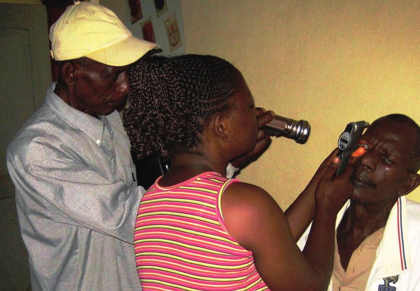
Table of content
AIDS EYE DISEASES IN HIV/AIDS 2
IMMUNE RECOVERY UVEITIS 25
BLINDNESS CHILDHOOD BLINDNESS AND VISION LOSS IN AFRICA 8
CATARACT AGE RELATED CATARACT 5
CONJUNCTIVA CONJUNCTIVITIS OF THE NEWBORN 13
TUMOURS OF THE BULBAR CONJUNCTIVA 67
DIABETES DIABETES 20
EQUIPMENT MAINTENANCE OF INSTRUMENTS 31
TRANSFORMING A PAPERCLIP INTO AN EYE-OPENER 40
EYE EXAMINATION RED REFLEX AND MORE 47
WHAT TO DO WITH A PATIENT WHO HAS EYE 73
PROBLEMS?
EYELID STYE AND OTHER AFFECTIONS OF THE EYELIDS 52
GENERAL THE SHIFT IN POTENTIALLY BLINDING EYE DIS- 59
EASE BEFORE AND DURING THE AIDS PANDEMIC IN
CAMEROON
VISION 2020, THE RIGHT TO SIGHT 69
GLAUCOMA GLAUCOMA 21
ONCHOCERCIASIS ONCHOCERCIASIS (RIVER BLINDNESS) 33
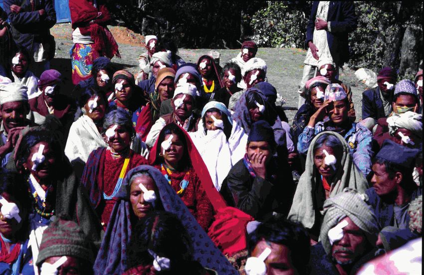
PREVENTION PREVENTION: COMMUNITY-BASED PREVENTION OF 10
CORNEAL BLINDNESS
HOW TO IMPROVE EYE SERVICES IN YOUR AREA 27
RED EYE RED EYE: ACUTE RED EYES 41
CORNEAL ULCERS 16
REFRACTION HOW TO HELP PROVIDE LOW VISION CARE, ESPECIALLY 28
FOR CHILDREN
REFRACTION: THE NEED FOR REFRACTIVE SERVICES IN 49
LOW‐INCOME COUNTRIES
TRACHOMA TRACHOMA 61
TRAUMA TRAUMATIC EYE LESIONS 65
XEROPHTHALMIA XEROPHTHALMIA, MEASLES AND MALNUTRITION 74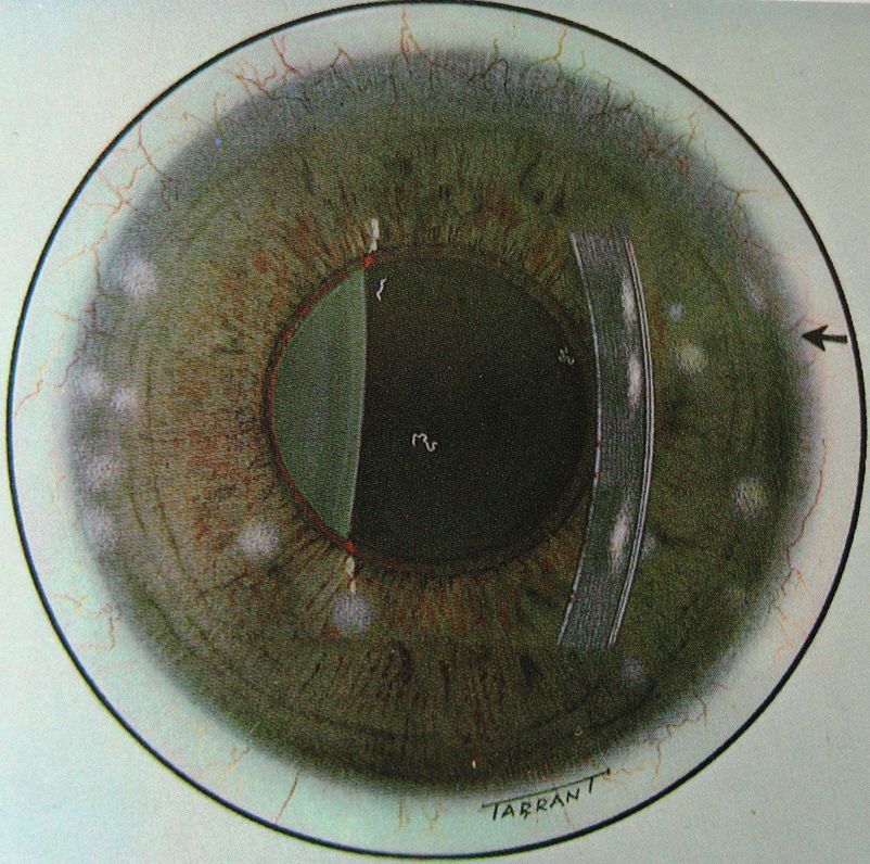
Editorial Eye Special

In 2002 a Memisa edition was dedicated to ophthalmology. It was used in the ‘Tropical Course’, the
preparation for doctors going to low income countries. Now this training is an official specializa-
tion: Arts Internationale Gezondheidszorg en Tropengeneeskunde (AIGT). Though most of the
content was still relevant, it was felt that a revision was needed.

The first MTb issue in 2014 gives an overview where eye care in low income countries stands now.
MTb, Medicus Tropicusi is the official bulletin of the Netherlands Society for Tropical Medicine
and International Health. The content of this issue is added to the updated Memisa special.

It is outlined how the ophthalmological world thinks to improve the quality of sight (Vision 2020)
There is a special article about the care for children. The last years there has been a shift in eye pa-
thology. This shift is described and 2 articles highlight an important cause, the aids epidemic. There
is an example how a simple action in the field can reduce blindness. The important (low income
countries) eye diseases are discussed.
All articles put emphasis on what workers in the frontline can do.

We hope that you enjoy this new Eye Special and find it useful. You can also find it on the site of
Werkgroep Tropische Oogheelkunde (Working Group Tropical Ophthalmology). http://www.
tropischeoogheelkunde.nl You can find also some more information there.

The Werkgroep can be reached via e-mail mailto:secretariaat@tropischeoogheelkunde.nl

Jan Geert Bollemeijer, Peter Hardus, Ype Henry, Margreet Hogeweg, Coen Koppert

On the front you see the cover of the Memisa Eye special 2002 combined with measuring the ocular tension
with the modern Icare tonometer in a rural setting.

                                                                                                           1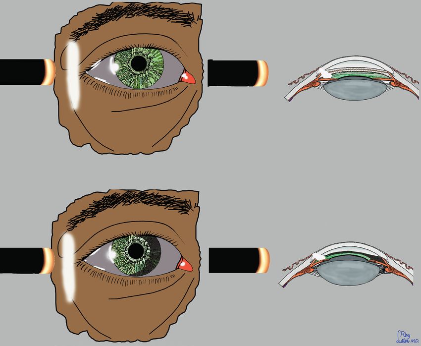
Eye diseases in HIV/AIDS –
some practical tools for diagnosis and
treatment
Jan Geert Bollemeijer MD, ophthalmologist,
Rotterdam Eye Hospital, formerly Zimbabwe
e-mail j.bollemeijer@oogziekenhuis.nl

                                                      Fig. 3 Kaposi (Photo C. Meenken and G.J. van
                                                      den Horn)

                                                      Imagine, you are a general practitioner in a
                                                      middle-sized city somewhere in Sub-Sahara
                                                      Africa. On Monday morning your first patient
                                                      in the OPD is a 33 year old male with since two
                                                      weeks the symptoms of herpes zoster ophthal-
                                                      micus on the left side. He can’t close his left eye
                                                      anymore due to scar formation of the upper
                                                      eyelid, his eye is painful, red and filled with
                                                      puss, the vision in this eye is markedly reduced.
                                                      (fig 1)

                                                      The second patient is a 40 year old woman with
                                                      since several months a conjunctival growth on
Fig. 1 Herpes zoster ophthalmicus. If the tip, side   the right eye. (fig 2) The third patient is a 29
and root of the nose is involved (sign of Hutchin-    year old male with since several weeks a strange
son) the n. nasociliaris is involved and more risk    thickened dark red upper eyelid of the left eye.
of ocular problems                                    The eyelid is not painful. (fig 3) He has dark red
                                                      patches in his oropharynx. The fourth patient,
                                                      a 36 year old woman, has been put on HAART
                                                      recently as she was found to be HIV positive.
                                                      She is complaining of loss of vision.

                                                      30 years ago it was very rare to start the week in
                                                      the OPD like this. However, with the arrival of
                                                      HIV opportunistic infections like herpes zoster
                                                      ophthalmicus (patient 1) and neoplasmata like
                                                      squamous cell carcinoma of the conjunctiva
                                                      (patient 2) and Kaposi sarcoma (patient 3) are
Fig. 2 Squamous cell carcinoma                        nowadays common in relatively young patients.

2
And since the introduction of Highly Active          Table 1 Ophthalmic manifestations of HIV infec-
Antiretroviral Treatment (HAART) in 1996             tion by CD4 T cell count
new phenomena like Immune Recovery Uveitis            CD 4 < 500 Herpes zoster ophthalmicus
(patient 4) have appeared (see article Immune                    Kaposi sarcoma
Recovery Uveitis, C. Meenken and others. pp.                     Lymphoma
25.).                                                            Squamous cell carcinoma conjunctiva

Worldwide more than 35,3 million people are           CD4 < 200   Tuberculosis
                                                                  Toxoplasmosis
infected with HIV, the greater part in Sub-                       Fungal infections like Coccidioidomycosis,
Saharan Africa (25 million), Asia (4,8 million)                   Cryptococcosis, Histoplasmosis, Pneumocystis
and North America/Western Europe (2,1 mil-                        jirovecii
lion). In lower income countries it is the second
cause of death after lower respiratory infections.    CD4 < 100   Cytomegalovirus retinitis, Herpes zoster virus
                                                                  retinitis
Adnexal and orbital complications affect more                     Mycobacterium avium complex infection
than 25% of untreated HIV-positive patients                       Microsporidiosis
and could be the presenting sign of the disease.                  Progressive multifocal leucoencephalopathy
Keratoconjunctivitis sicca occurs in 10 to 20%                    Retinal/conjunctival microvasculopathy
of patients and in more advanced stages of the
disease posterior segment manifestations like
retinal microvasculopathy and CMV retinitis          In diagnosing an opportunistic infection a
are seen in some areas in 40-50% of patients.        handy tool is the hand ophthalmoscope. (see
                                                     article Red Reflex and More, P.Hardus and oth-
Eye diseases diminish the quality of living of       ers. pp. 47.)
patients suffering from AIDS. For the general
practitioner it is important to recognise that an    Therapy depends on the availability of drugs. In
infection can be opportunistic, due to AIDS. As      individuals with advanced immunodeficiency
many general practitioners are the first doctors     more aggressive therapy is mandatory as they
patients visit they are the ones who diagnose the    have an increased risk of permanent vision loss.
disease and who decide on therapy and referral.
In this paper some tools are presented to rec-       A patient with herpes zoster ophthalmicus
ognise HIV related eye diseases and to facilitate    could benefit from aciclovir (800 mg five
treatment and/or referral.                           times daily) or valaciclovir (1000 mg three
                                                     times daily), topical calamine lotion or emol-
Two important skills are necessary to fulfil this    lient, potassium permanganate soaks, systemic
task: knowledge and examination skills. The          antibiotics in case of secondary infection and
general practitioner should have knowledge           analgesics like indomethacin (50 mg three times
about which opportunistic infection fits into the    daily), while amitryptillin (75-150 mg at night)
picture of a certain level of immunodeficiency.      and carbamazepine (100 mg once or twice
And the general practitioner should be capable       daily) can reduce post herpetic neuralgia. Local
to perform a basic eye examination.                  therapy for cornea exposure due to a retracted
                                                     upper eyelid could consist of eye ointment and
HIV is acting by reducing the number of CD4          a tarsorraphy.
cells eventually leading to deep immune incom-
petence, which paves the way for opportunistic       In case of a Kaposi sarcoma of the eyelid and
infections and neoplasmata. Table 1 shows an         conjunctiva there are more Kaposi’s in the
overview.                                            mouth. Median survival in Sub-Saharan Africa
                                                     is 3.5 months. Treatment is in the first place to
                                                     start HAART as soon as possible as it greatly
                                                     improves the survival. Surgical excision is
                                                     sometimes possible, but as Kaposi sarcoma is
                                                     a heavily vascularized tumor can be difficult.
                                                     Radiotherapy and intralesional vinblastine
                                                     chemotherapy are other options.

                                                                                                                   3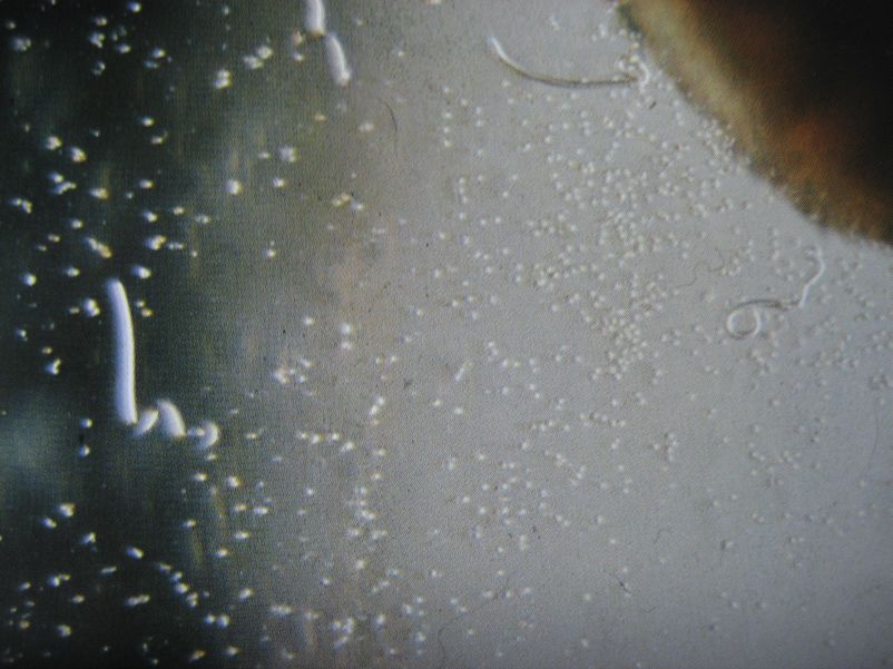
The differential diagnosis of a conjunctival          References:
growth is (apart from a few other rare tumors         1. Cunningham ET Jr, Margolis TP. Ocular manifestations of
like non-Hodgkin lymphoma, pyogenic granu-               HIV infection. N Engl J Med 1998; 339: 236-244
loma after trauma and papilloma) pinguecula           2. http://www.unaids.com
(harmless hyalin degeneration), pterygium
(from the conjunctiva spreading “wing” over
the cornea with parallel blood vessels giving rise
to astigmatism and eventually covering of the
pupil) and conjunctival intraepithelial neopla-
sia leading to squamous cell carcinoma of the
conjunctiva. Squamous cell carcinoma of the
conjunctiva must be excised completely with a
free zone around the process of at least 2 mm as
recurrences can be very aggressive. If possible
the whole area is treated with double freeze-
thaw cryotherapy. An alternative is application
of 5-fluoro-uracil 1% eye drops four times daily
during four days followed by ten days rest and
reinstallation of 5-fluoro-uracil 1% four times
daily. In case of metastasis the first stations are
the submandibular- and pre-auricular lym-
phnodes.

If the CD4 count is below 200 patients are more
likely to suffer from intra-ocular infections.
Those infections reduce the visual acuity and
can be diagnosed by funduscopy. Look for local
and systemic signs of the infections of table 1.

AIDS is still a deadly disease. In general prompt
diagnosis of HIV and timely start of treatment
with HAART is essential to improve the condi-
tion of the patient.

Many thanks to Hans van den Horn and Ina
Meenken, ophthalmologists, for reviewing the
manuscript.

4
Cataract
Margreet Hogeweg, ophthalmologist, CBM medical advisor CE Asia
a huge backlog of cataract patients in need of
surgery.
Patients with age related cataract are elderly,
and present with a history of gradual and pain-
less loss of vision, due to progressive clouding
of the lens. It is essentially a bilateral condition.
The most important risk factor is age. In low
income countries, age related cataract seems to
develop about 10 years earlier compared to the
West. Many patients are only in their early six-
ties. Cataract may also be secondary to trauma,
intra-ocular inflammation and long-term use of
steroids. Cataract can also occur in young chil-
dren, as congenital or developmental cataract.
Cataract requires surgery as soon as possible, to
prevent amblyopia (lazy eyes).
Examination and diagnosis
Assessment of Visual Acuity (VA): to qualify
for cataract surgery in remote rural areas, VA
should usually be less than 6/60 but of course
depends on local guidelines. Light perception
and light projection must be intact: patients
should be able to indicate whether a torch light
is ‘’on’’ or ‘’off ’’, and to indicate from which
direction the light comes, even in ‘mature cata-
ract’. The pupil must react briskly to light.
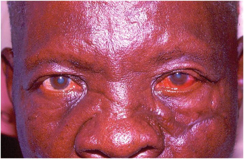
Fig 1. Bilateral mature cataract In mature cataracts, the opaque lens is visible
immediately behind the iris, as a grey-white
The prevalence of blindness* (*WHO definition opacity (fig. 1) (not to be confused with a
of presenting Visual Acuity (VA) of less than corneal scar: an irregular white grey opacity in
3/60 (< 0.05, cannot count fingers at 3 meter) front of the iris and partly or completely cover-
in the better eye) is usually between 2-4% in ing it). Immature cataract (in particular central
people over the age of 50 years. Of all blindness, posterior sub-capsular cataract) can be visually
40-50% is due to age related cataract, making very disturbing but cannot be recognised as eas-
this the single most important cause of blind- ily during an external ocular examination. The
ness. WHO advises a ‘Cataract Surgical Rate’ colour of the ‘’pupil’’/ lens in elderly people is
(CSR: number of cataract surgeries per year per always slightly greyish: this does not necessarily
million population) of 2000 for Africa and 3000 indicate the presence of cataract.
for Asia. In many African countries the CSR is
still estimated as < 500/million/year, indicating The best method to estimate the amount of
5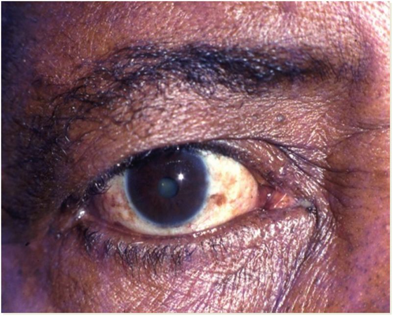
cataract is by examining the ‘red reflex’ with        gaining more and more popularity in major eye
a direct ophthalmoscope in a semi-darkened            departments, but the machine is very expensive
room. A clear lens and clear vitreous will show       and even the cheapest foldable IOL’s are 10 x
a bright red reflex, emerging from the well           more expensive than standard PMMA IOL’s.
vascularized choroid. An advanced immature            This is usually not affordable for poor rural pop-
cataract will show partial obscuration of the red     ulations. In addition: advanced cataracts are less
reflex. The red reflex cannot be seen in mature       suitable for this technique. Final results can be
cataract. The level of VA should be in balance        almost equally good in all three techniques, but
with the obscuration of the red reflex. Note: in      much depends on the right selection of patients,
rare cases, obscuration of the red reflex may be      the quality of surgery and the postoperative
caused by a vitreous haemorrhage, not cataract.       care. All techniques require a good microscope.
Corneal scars will of course also obscure the         The choice of technique will depend on local
red reflex. (see article Red Reflex and More,         circumstances and the preference and training
P.Hardus and others. pp. 47.)                         of the surgeon. Unless the patient is a child,
                                                      surgery is done under local anaesthesia.
It should always be realised that there are other     An A-scan and keratometer can calculate the
causes for gradual loss of vision in the elderly,     best IOL power before surgery so that no glasses
apart from cataract: for instance gradual change      will be necessary postoperatively. If not avail-
in refraction, glaucoma (chronic open angle),         able, a standard 21 or 22 D IOL is implanted
diabetic retinopathy, age related macular degen-      and any residual refractive errors can be cor-
eration, various other chorio-retinal conditions      rected with glasses, if needed. A late complica-
and optic atrophy. Therefore assessment of            tion of the extracapsular cataract extraction
visual acuity, in combination with assessment         is secondary posterior capsule opacification,
of the red reflex, is essential before making the     causing visual impairment. If severe, this
diagnosis ‘cataract’ and selecting patients for       requires Nd:YAG laser treatment or opening of
cataract surgery.                                     the capsule with a sharp needle. In case an IOL
                                                      cannot be implanted (aphakia), the patient will
Treatment                                             need S+10 glasses after surgery.
‘Anti-cataract’ medicines are useless and a
waste of money. The only effective treatment          Couching is still common in parts of Sub
is surgery, with removal of the lens material.        Saharan Africa. Traditional healers perforate
Intraocular lens (IOL) implantation (fig. 2)          the eye with a sharp ‘instrument’ and push the
has become standard (pseudophakia) in most            lens away from the pupillary area, deep into the
countries. Cheap IOL’s are available for $ 5-10.-     vitreous. The complication rate is very high,
or even less.                                         but the intervention can be successful. A well
    The surgical technique will usually be either     couched eye has an uncorrected VA of 2-3/60
ECCE (extra capsular cataract extraction) or          (count fingers at 2-3 metres), which can im-
‘’SICS’’, (small incision cataract surgery). Phako-   prove to 6/18 or even better with aphakic S+10
emulsification is another good technique,             spectacles.

                                                      Best indications for surgery:
                                                      • patient with bilateral VA< 6/60, due to cata-
                                                         ract (depending on policies and available
                                                         resources).
                                                      • one-eyed patient with a VA of< 3/60, due to
                                                         cataract.
                                                      • bilateral dense congenital cataract in chil-
                                                         dren (no red reflex).
                                                      • eyes with lens-induced glaucoma (prognosis
                                                         guarded).

                                                      Relative indications for surgery:
Fig 2. Intraocular lens                               • unilateral cataract (second eye has no/imma-

6
ture cataract, patient still can see).           hospital once or twice a year. For this, take into
•   second eyes, in unilateral pseudophakia (vi-     account the farming season and local festivities.
    sion already restored by surgery in the first    Particular attention should be paid to acces-
    eye).                                            sibility for women, as they suffer from cataract
•   unilateral traumatic cataract (often a poor      blindness more often than men. Good publicity
    prognosis, second eye good).                     is essential. It may well be possible to obtain
                                                     financial support through local churches and
No indications for surgery:                          service clubs or through the various (I)NGO’s
• no/doubtful perception/projection of light         that focus on eye care. Rural health insurance
   (no improvement to be expected).                  systems may include refund for cataract surgery,
• cataract extraction in eyes with poor vision,      thus removing the barrier of costs. For follow
   but good red reflex (other eye disease likely).   up patient will usually come back to the local
• unilateral congenital cataract (eye deeply         hospital. It is important to monitor the outcome
   amblyopic).                                       of cataract surgery. In the ideal situation, < 5%
                                                     of the patients should have an uncorrected VA
‘Referral’ for cataract surgery is not sufficient.   of < 0.1, six weeks after surgery. In reality the
Only 10% of the ’’referred patients’’ will finally   final VA is often < 0.1. in 10-15%, but this per-
present for surgery; the reasons for this include    centage should not be higher than that.
costs and lack of transport.
    It is very worthwhile to arrange for cataract    Cataract blindness is unnecessary. Vision can be
surgery. If there is no eye department in the        restored by a comparatively simple operation. It
vicinity, it may be useful to liase with an eye      is one of the most rewarding and cost effective
team. The team could visit regularly as part of      interventions in medicine!
their outreach work and take patients with them
for surgery at their base and back. An alterna-
tive is to invite an eye team for surgery at the

                                                                                                     7
Childhood blindness and vision loss in
Africa
Paul Courtright M.D. Ph.D , ophthalmologist
Kilimanjaro Centre for Community Ophthalmology, Moshi, Tanzania.
Kilimanjaro Centre for Community Ophthalmology, Division of Ophthalmology, University of
Cape Town, Cape Town, South Africa.
Vision loss and blindness in children is rare, The aetiology of congenital or developmental
even in Africa. While accurate estimates are not cataract in many of these children remains
available, is it likely that, in most of Africa, less poorly understood. While rubella, a treatable
than one child in 5,000 is blind. (1) That said condition, does contribute to some cases of
the impact of vision loss and blindness in child- congenital cataract, evidence suggests that its
hood can be significant; for the children, their contribution is not more than 20%.
families, and societies at large the impact can
endure for decades. Managing the present causes of blindness and
vision loss in children requires sophisticated
Due to successful vitamin A supplementation services to provide good quality surgical,
and measles immunization programmes in medical, and optical interventions. Also, these
many countries, corneal blindness has reduced children require comprehensive care through-
significantly. As a result most incident blindness out their childhood in order for them to achieve
and vision loss in children is no longer prevent- their full visual potential. Unlike cataract
able; instead, it is a mix of treatable and untreat- surgery in adults, managing cataract in children
able causes. The major treatable causes include is a lifelong undertaking. The links between the
congenital or developmental cataract (fig. 1), health care services and educational services
glaucoma, and refractive error. need to be strengthened to ensure that these
children achieve their educational potential.
One aspect of dealing with childhood vision
loss is still true: children with serious eye
disease need to be seen by a qualified eye care
provider as soon as possible because of amblyo-
pia prevention. In many countries, particularly
in eastern Africa, “Child Eye Health Tertiary
Facilities” (CEHTF) have been established at
key tertiary hospitals, each striving to serve
a population of approximately 10 million.
(2) These facilities need to be staffed by well-
trained paediatric ophthalmologists, paediatric
anaesthetists, optometrists, low vision special-
ists, and Childhood Blindness & Low Vision
Coordinators. Ideally they will have strong
links to the communities they serve in order to
identify children in need of services as well as to
ensure that children receive adequate health and
educational follow up.
Experience gained suggests that:
1. Key community members (key informants)
Fig. 1 Congenital bilateral cataract (Photo Prof. can be very effective in identifying and
Khumbo Kalua, Malawi) referring children in need of eye care ser-
8vices. Studies in a number of countries have 4. In collaboration with the CEHTF consider
demonstrated the impact of this approach conducting key informant programmes in
and training manuals have been developed the area.
and disseminated.(3-5) 5. Do not forget vitamin A or measles related
2. Less success has been demonstrated in using blindness; if corneal opacity secondary to
general health workers to identify and refer vitamin A/measles is detected, this should
children, whether through routine clinic be a trigger to report to health authorities.
activities such as immunization, or through Corneal opacification is the “tip of the ice-
community campaigns. (4,5) Currently, berg” and indicates a serious public health
the knowledge and skills of general health problem.
workers regarding childhood vision loss is Continuing to reduce the burden of vision loss
generally weak (6). Every district hospital in in children in Africa requires good planning,
Africa should have at least one trained clini- a comprehensive approach, good partnership,
cal person dedicated to eye care; Their role is a strong link between all sectors of the health
crucial to ensure that a sufficient diagnosis is care services, a viable system for follow up, and
made and proper referral done. They should engagement with the educational sector. Only
have a strong relationship with the CEHTF by including all these aspects will children be
both for referral and for follow up. able to achieve their best visual and educational
3. Where possible children need to be referred potential.
to a CEHTF for proper assessment and
treatment. Since children require long term References:
follow up, which may be difficult to always 1. Gogate P, Kalua K, Courtright P. Blindness in childhood in
carry out at the CEHTF, a plan of action, developing countries: time for a reassessment? PLoS Med.
tailored to each child and the clinical and 2009 Dec;6(12):e1000177.
educational environment needs to be ad- 2. Agarwal PK, Bowman R, Courtright P. Child eye health
tertiary facilities in Africa. JAAPOS 2010;14:263-266
opted. This is one of the tasks of the Child- 3. Shija F, Kalua K, Shirima S, Lewallen M, Courtright P. Using
hood Blindness & Low Vision Coordinator, key informants to identify and refer children who need
who works alongside clinical personnel. eye care services: A manual for Africa. AED. 2010
4. Shija F, Shirima S, Lewallen S, Courtright P. Comparing key
For general clinicians working in Africa, some informants to health workers in identifying children in
recommendations include: need of surgical eye services. International Health 2012;4:
1. Find out where the nearest CEHTF is 1-3
located, visit the facility, and establish a 5. Kalua K, Ng’ongola RT, Mbewe F, Gilbert C. Using primary
relationship with the relevant personnel. health care (PHC) workers and key informants for commu-
2. Assess the current knowledge and skills nity based detection of blindness in children in Southern
Malawi. Hum Resourc Health 2012; Sept 27; 10(1):37
of eye care personnel in the area related to 6. Kishiki E, Dieleman M, Hogeweg M, Lewallen S, Courtright
child eye health and provide upgrade train- P. Is the existing knowledge and skills of health workers
ing, as needed. regarding eye care in children sufficient to meet needs?
3. Insert short educational messages in train- International Health 2012; 20: 260-266
ing of general health workers, particularly
on the need to refer children, regardless of
age, with serious eye problems to the rel-
evant eye care providers as emergencies.
9Community-based prevention of corneal blindness, a successful programme in Takeo province, Cambodia Manfred Mörchen Dr.med., FEBO OphthalmologistCARITAS Takeo Eye Hospital, CBM Te Serey BonnMPH, MA Project Director CARITAS Takeo Eye Hospital Margreet Hogeweg, M.D,ophthalmologist CBM CEARO Medical AdvisorCBM CEARO regional office, Bangkok. Introduction treatment with antibiotic e.o. also prevents the Corneal ulceration as a result of untreated development of fungal corneal ulcers, that are traumatic corneal abrasion is one of the leading otherwise very hard to treat. causes of ocular morbidity and blindness world- wide.[1] In developing countries the main cause Corneal scarring unrelated to trachoma was of corneal ulcer is a minor agricultural injury identified as the second main cause of bilateral sustained during farming, e.g. during plantation blindness in a Rapid Assessment of Avoidable and harvest. Patients usually prefer treatment Blindness (RAAB) in Cambodia in 2007. [6] nearby (such as by unlicensed pharmacies, In a hospital-based study (2005) at the CARI- traditional healers, private doctors, or apply eye TAS Takeo Eye Hospital (CTEH) 130 patients drops, already used by others). They therefore had been admitted within a period of only 6 present late at the hospital with severe bacterial- months because of a severe corneal ulcer: 50 or fungal ulcers, that are resistant to treatment. % was due to trauma, 75 out of 99 eyes were The widespread availability of steroid-contain- blind (VA
corneal abrasion in 1,004 cases (87,5%). The
                                                    main results of these 1,004 cases are presented
                                                    in table 1.

                                                    Table 1: Outcome in 1,004 patients with corneal
                                                    abrasions, as diagnosed and treated by VHWs
                                                     Corneal abrasion                      1,004    100%
                                                     Healed                                949      94,5%
                                                     Referred because of corneal ulcer     34       3,3%
                                                     despite treatment
                                                     Dropped out                           14       1,4%
                                                     Missing results                       7        0,7%

                                                    In total 713 (71.3%) patients reported an injury
                                                    of organic nature, of whom 392 (39.2%) had
                                                    an injury with rice. Table 2 demonstrates the
                                                    seasonal correlation between location and agent
                                                    of ocular injuries. In December 2008 and 2009
                                                    (main harvest season in Cambodia), around
Fig. 2 Corneal abrasion                             70% of all ocular traumas were reported to have
                                                    happened during work in the paddy fields, with
VHWs were also taught to record visual acuity       rice grains as major agent. A second peak with
(VA) using an E-chart, identifying corneal ulcer    a similar pattern could be observed in April
and other common eye diseases, and how to re-       and May (minor harvest and early plantation
fer to CTEH. They were advised to treat a) only     season).
residents of their intervention area, b) patients
presenting within 48 hours of the injury with       Visual acuity was less than 6/60 in 26.4% of all
confirmed corneal abrasions and c) patients         patients before treatment. After treatment, only
aged 5 years and older. Every month the VHWs        1.1% could see less than 6/60.
were called to CTEH for reporting and follow-
up.                                                 Of the 34 patients referred because of corneal
                                                    ulcer, 9 (26.5%) were lost to follow-up. Of the
Results                                             remaining 25 patients, 7 (28%) corneal ulcers
During 13 months, 1,147 individuals (female         could be confirmed at CTEH. None of these
56.9%, male 43.1%) reported to the VHWs.            eyes had to be removed. In 18 patients (72%)
783 (78.2%) were farmers. VHWs diagnosed            corneal ulcer could not be confirmed. Addition-

                                                                                    Table 2: Reported loca-
                                                                                    tion of ocular injury
                                                                                    and agent from Decem-
                                                                                    ber 2008 until Decem-
                                                                                    ber 2009 by VHWs.

                                                                                                       11
ally, 46 sight threatening (cataract, pterygium mass radio messages at the start of the har-
etc.) and 28 conjunctivitis cases were referred vest season in order to create awareness of the
by the VHWs. importance of early treatment after sustained
corneal injury.
discussion
This intervention project aimed to prevent Advocacy efforts by CTEH resulted in signifi-
traumatic corneal ulcer in a region dominated cant support by the local government institu-
by agriculture, with a hot and humid climate tions, especially the Provincial Health Depart-
and known high prevalence of corneal blind- ment (PHD) of Takeo Province. The project
ness.[6,7] Hospital-based data indicate that at continued during 2010 and 2011 with support
CTEH the overall number of patients that had from CTEH and was handed over to the PHD
to be treated because of corneal ulcer decreased in February 2012. In these 3 years, all together
from 745 in 2007 to 442 in 2013, a decrease of 1,985 patients with corneal abrasions were iden-
41% while yearly more patients attended ! This tified (healing rate 98.9%). 24 Patients with sus-
study therefore shows that early application pected corneal ulcer and 246 patients with other
of chloramphenicol e.o. probably prevented a eye diseases, like cataract, pterygium etc., were
considerable number of corneal ulcers. referred to CTEH. We hope, that the Cambo-
dian Ministry of Health, will adapt community
Only 28% of the patients referred with corneal prevention of corneal ulceration as a national
ulcer could be confirmed. As the VHWs had strategy in the next multi-year plan.
been trained only for one week, such misdiag-
noses had been expected. There was confusion References
with a variety of other causes of red eyes, - not 1) Whitcher JP et al: Corneal blindness: a global perspective.
ulcers -, but yet in need of treatment. We con- Bull World Health Organ 2001;79:214-21
sider this therefore as a positive outcome. 2) Upadhyay MP et al: The Bhaktapur eye study: ocular
trauma and antibiotic prophylaxis for the prevention of
corneal ulceration in Nepal. Br J Ophthalmol 2001;85:388-
VHWs had to be selected from the communes 392
in collaboration with the local authorities. 3) Maung N et al: Corneal ulceration in South East Asia. II: A
Therefore, this Cambodian experience may strategy for the prevention of fungal keratitis at the village
reflect the ground reality and may serve as a level in Burma. Br J Ophthalmol 2006;90:968-970
feasible model of intervention despite some 4) Gethsen K et al: Corneal ulceration in South East Asia. I: A
limitations. model for the prevention of bacterial ulcers at the village
level in rural Bhutan. Br J Ophthalmol 2006;90:276-278
The strong correlation between the harvest 5) Srinivasan M et al: Corneal ulceration in south-east Asia
season, location of ocular trauma and reported III: prevention of fungal keratitis at the village level in
agent is important: massive awareness cam- south India using topical antibiotics. Br J Ophthalmol
2006;90:1472-1475
paigns before the harvest season and basic 6) Rapid assessment of avoidable blindness (RAAB). National
training of primary health care workers for Program for Eye Health, Ministry of Health Cambodia,
a short period may be able to prevent many 2007
corneal ulcers in communities with a large agri- 7) Hall T, Lion F: Corneal ulcer in a Cambodian eye hospital.
cultural sector and hot and humid climates. As Community Eye Health Journal Vol 18 No.53 2005 p81
a result of our study, we have indeed initiated
12Conjunctivitis of the new-born
N. Buisman, ophthalmologist Ex-Zimbabwe, Cameroon and Tsjaad
P. Hardus, ophthalmologist, previously Dept. ophthalmology Groningen
University ,Nigeria, Angola
This article was originally written by Nico Buisman who was much involved in ophthalmology in low
resource countries ; he worked as general medical doctor and ophthalmologist for many years in dif-
ferent countries, especially in Africa. Unfortunately he died much too young due to a fatal disease. The
content of his article is still very actual. Where necessary some updating is done.
Introduction melt the cornea. This can happen extremely
Every case of conjunctivitis with an onset in the quickly and may cause a corneal perforation.
first 28 days after birth is called conjunctivitis Both gonococci and chlamydia cause systemic
of the newborn or ophthalmia neonatorum. (1) infections and this will be evident physically.
The contamination usually takes place dur- It is impossible to make a clinical distinction
ing the passage through the birth canal. lt is, between gonococci and chlamydia infections.
therefore, a sexually transmitted infection (STI). Gonococcal conjunctivitis may start a bit earlier
The incidence of STI’s is high in Africa, 10-30%’. than chlamydia conjunctivitis. Gonococci and
Common causes are Neisseria gonorrhoea and chlamydia often cause a more severe conjuncti-
Chlamydia trachomatis. Chlamydia occurs more vitis than other microorganisms. In Cameroon
often than gonorrhoea. In addition, other less a sexually transferred micro-organism was
harmful microorganisms occur. Prompt diag- detected seven times more often in severe con-
nosis and treatment of the conjunctivitis of the junctivitis than was detected in the lighter cases.
new born is important as an untreated gonococ- (2) The severity of the inflammation is a strong
cal conjunctivitis can lead to blindness within indicator of the presence of an STI in mother
a short time. Furthermore, a genital infection and child. A dual infection of both gonorrhoea
in the mother that remains undetected can lead and chlamydia is common.
to sterility. Despite the effort in the struggle
against AIDS to reduce the number of STI’s, the
prevalence of these infections is still increasing.
An additional problem is the high prevalence
of Penicillinase-Producing Neisseria gonorrhoea
(PPNG) in Africa.
Clinical picture
A conjunctivitis caused by gonococci and chla-
mydia generally leads to a severe inflammation
with much swelling and redness of the eyelids
and conjunctiva (Fig.1). Often great quantities
of half-liquid green-yellow discharge are pres-
ent. When the eye is opened by the examiner Fig. 1 Neonatal conjunctivitis
this discharge may flow out of the eye. This
picture, however, may also be much milder. Diagnosis
A simple method of diagnosing gonococci
lt may start on one side and remain unilateral and chlamydia as a cause of conjunctivitis in a
for the first days. Gonococci produce toxins that newborn is needed in order to treat mother and
13child adequately for STI’s. Gonococci can be Treating Gonococcal Infections
diagnosed reliably with Gram stain microscopy. The best treatment is single dose, systemic and
Following the removal of the bulk of secre- on an outpatient basis. WHO and CDC propose
tion, a smear of the conjunctiva can be taken several drugs. It is important to first rinse the
and Gram stained. The diagnosis is positive eye thoroughly with water or saline (every 5
for gonococci when Gram negative diplococci minutes) in order to remove the toxins that are
are seen in the leucocytes. A culture does not dangerous for the cornea.
give much more information than does careful
microscopy. Laboratory diagnosis of chlamydia The following antibiotics are useful:
is practically impossible in a general hospital in • Kanamycin 25 mg/kg IM 1x + Tetracycline
a low resource country. The distinction between eye ointment, 1week (recommended)
a severe and a light conjunctivitis can be very • Kanamycin 25 mg/kg IM 1x +Gentamycin
helpful. The treatment scheme makes use of this eye ointment, 1week
distinction.(Table 1) • Ceftriaxone 20-25 mg/kg IM 1x; do not
exceed 125 mg
The clinical picture of newborn conjunctivitis • Cefotaxim 25 mg/kg IM 1x
may be confused if a child is given traditional
eye medicines (TEM) by a local healer. Herpetic Ceftriaxon and cefotaxim PM are practical but
conjunctivitis is very uncommon. Most times expensive. Kanamycin combined with topical
there are herpetic skin abnormalities around the gentamycin or tetracycline is very useful and
eye as well . Also nasal lacrimal duct obstruc- less expensive.
tion can give rise to discharge but will be at a bit Gonococcal strains tend to have become
later age and is usually unilateral. Pressure on susceptible to antimicrobials that are not fre-
the lacrimal sac will give discharge. quently used, such as kanamycin, gentamicin
and spectinomycin. Spectinomycin, especially,
Treatment scheme would seem an appropriate second-line treat-
A Gram stained smear should be made when- ment for gonorrhoea, although it is expected
ever a newborn has conjunctivitis. If gonococci that widespread use of these antibiotics will
are present mother and child must be treated. quickly result in development of resistant N.
In all cases of severe conjunctivitis mother and gonorrhoea. (3,4 )
child also need to be treated for chlamydia The best will be to get information which
as the chance that chlamydia is an underly- treatment is locally most effective to treat the
ing cause of the infection is very high. A dual gonococ.
treatment is recommended in all severe cases
of gonorrhoea. The remaining cases of light Treating chlamydia infections
conjunctivitis can be treated as a nonspecific Systemic treatment for the child consists of
infection. If the conjunctivitis persists following erythromycin orally 12.5 mg/kg q.i.d for two
two to three days’ of antibiotics, treatment for weeks. (1) Obviously both the mother and her
chlamydia should be given. partner should also be treated for the STI’s. It
is worth noting that the Chlamydia trachomatis
Tabel 1 Treatment of ophthalmia neonatorum that causes the conjunctivitis of the newborn is
Gonococci Kanamycin 25 mg/kg IM 1x + of a different serotype (D-K) than the chlamydia
Tetracycline (alternative: Genta- that causes trachoma (A-C). The clinical picture
mycin) eye ointment 1 week + of the conjunctivitis is also different.
Erythromycin orally 12.5mg/kg 4x a
day for 2 weeks Prophylaxis
The most important activity is the thorough
No gonococci
cleansing of the eyelids as soon as the child’s
Severe conjunctivitis Erythromycin orally 12.5mg/kg 4x a head is born in order to prevent cervical secre-
day for 2 weeks tions from reaching the conjunctiva. Topical
Light conjunctivitis Tetracycline eye ointment 0.5% erythromycin and 1% tetracycline are con-
sidered equally effective for prophylaxis of ocu-
lar gonorrhoea infection in newborn infants. (1)
14Where available (not in USA), povidone-iodine
2.5% seems at least as effective. A second ap-
plication has no additional effect.(5)
References
1. Neonatal Conjunctivitis Medscape update march 2013
McCourt E.A
http://emedicine.medscape.com/article/1192190-over-
view
2. Buisman NJF, Abong Mwemba T, Garrigue Getal.
Chlamydia Ophthalmia Neonatorum in Cameroon. Doc.
Ophthalmol. 1988; 70: 257-64
3. Guidelines for the management of sexually transmitted
infections. WHO, 2001
4. Report of the expert consultation and review of the latest
evidence to update guidelines for the management of
sexually transmitted infections WHO/RHR/11.37 World
Health Organization 2011.
http://apps.who.int/iris/bitstream/10665/75194/1/
WHO_RHR_11.37_eng.pdf
5. S J Isenberg, L Apt, M Del Signore, S Gichuhi, N G Berman
A double application approach to ophthalmia neonatorum
prophylaxis Br J Ophthalmol 2003;87:1449–1452
http://www.ncbi.nlm.nih.gov/pmc/articles/
PMC1920568/
15Corneal Ulcers (Infective Keratitis):
The silent epidemic
Jan van der Hoek, Consultant Ophthalmologist Scarborough Hospital
Woodlands Drive
Scarborough YO12 6QL UK. Formerly Nepal
Introduction caused by fungus, particularly in a community
Unilateral blindness resulting from microbial engaged in agricultural activity. Fusarium or
keratitis has gone under-reported in the large Aspergillus are the most commonly implicated
national blindness surveys, although it is now filamentary fungal species. Bacterial infec-
recognised that superficial corneal trauma, tion make up most of the remaining 50%,
often sustained during agricultural work and both gram positive and gram negative bacteria
leading to microbial keratitis, is a major world- such as Staphylococcus aureus, Streptococci and
wide cause of unilateral ocular morbidity and Pseudomonas are commonly found in infec-
blindness. (see article Community-based preven- tive keratitis. Acanthamoeba is less frequently
tion of corneal blindness Manfred Mörchen a.o. encountered but may present as a non-contact
p. 10) The incidence of ulcerative keratitis in the lens related cause of keratitis. More unusual
US has been reported at 11 per 100,000 popula- organisms such as Nocardia and some species of
tion. In southern India the incidence was found mycobacterium are occasionally seen as causes
to be closer to 11 per 10,000 population or 10 of keratitis.
times the US rate. Extrapolation of the results of Another features of infective keratitis in the
studies in India suggests that annually world- developing world is the often marked delay in
wide one and a half million people become uni- seeking medical help; the delayed patients may
laterally blind as a result of corneal ulceration. have tried traditional eye medicines and or
In Western countries microbial keratitis is steroid eyedrops prior to presentation. Children
often caused by contact lens wear (30-40% as may have poor vitamin A status, particularly if
reported in literature) or seen in severe ocular associated with measles although some reports
surface disease and/or in debilitated elderly in the literature appear to suggest that this is on
patients. In the developing world the most the decline.
common cause is trauma, which in rural areas is Patients may also have pre-existing trichia-
often sustained during agricultural work (up to sis from trachoma (particularly common in
50% in reported series). The incidence usually women), poor eyelid closure and/or reduced
follows a seasonal pattern with peak incidences corneal sensation from leprosy.
occurring at times of increased agricultural
activity, particularly during harvest and plant- History
ing seasons. Children and young adults may be The farmer with keratitis is unlikely to present
heavily represented in the patient group with to an ophthalmic trained person within hours of
infective keratitis thought to be the result of the original injury. There may be a considerable
unsupervised playing and the general lack of delay in seeking advice as small eye injuries are
awareness of the dangers of eye injury. Corneal probably common and many may heal without
ulcers may account for 10% of all red eyes seen sequelae. Treatment with topical steroids (which
in eye clinics in developing countries. may be available at the local pharmacy shop
Factors that make the presentation of infec- and may help to mask the symptoms of inflam-
tive keratitis different from that in the more mation) or harmful traditional eye medicines
temperate parts of Europe include the different (TEM) can significantly worsen the situation.
etiology: fungal keratitis is very much more TEM may be available from local healers or
common in hotter climates. Up to an estimated simply bought from local market stalls. Not all
50% of infective keratitis in hot climates may be TEM will be harmful for the eye, but patients
16may be persuaded to try unsuitable and ag- tis requires an early diagnosis and it should be
gressive treatments some of which were not realised that in developing countries acantham-
intended as eye treatments (tiger balm) or con- oeba is only rarely associated with contact lens
tain ingredients unsuitable as medicine (human wear. The latter may be considered rare outside
urine). The patient may be reluctant to make urban centres.
a long and arduous journey to an unfamiliar It is important to bear in mind that no ap-
eye clinic, often having to spend considerable pearance is pathognomonic of a particular or-
length of time away from home resulting in loss ganism and polymicrobial infections do occur.
of income and unanticipated costs. Viral keratitis may occasionally masquerade
as a bacterial infection although the typical
Diagnosis staining pattern usually provides clues as to
Infective keratitis is generally not difficult to the correct diagnosis. Ulceration secondary to
diagnose as the history of trauma, the loss of Herpes zoster should raise suspicion of an un-
vision, watering and the typical clinical features derlying HIV infection in endemic areas. Sterile
of corneal infiltration and hypopyon are usu- ulcers and in particular Mooren’s ulcers are not
ally self-evident. A history revealing the type uncommon, especially in sub-Saharan Africa.
of matter or material that entered the eye and The underlying process is autoimmune related
caused the keratitis may be helpful. and gives rise to mostly peripheral corneal melt-
Always look if there is a foreign body left. ing which can be relentlessly progressive. Males
(rotate eyelid) tend to be more affected than females.
A baseline record of epithelial defect,
amount of infiltration and hypopyon should be Treatment Protocol
obtained. Deep corneal scrapings can obtained The treatment of microbial keratitis requires the
for direct microscopy with Gram stain; KOH intensive topical application of broad spectrum
or Lactophenol Cotton Blue may be used for antibiotics. Oral or intravenous antibiotics
the detection of fungi. However, it takes con- are not thought to be helpful. In cases where
siderable skill and experience to confidently endopthalmitis is suspected oral ciprofloxacin
diagnose a particular infection on the basis of (750 mg b.d.) provides good penetration into
direct microscopy. If microbiology facilities are the eye and broadspectrum activity.
available a scrape for culture and sensitivity may Topical ciprofloxacin (ofloxacin or levo-
lead to identification of the offending organism, floxacin may be alternatives) has good broad
although cultures may only be positive in about spectrum activity which includes activity
half of all cases. against Pseudomonas, but more restricted
Infections should initially be assumed to be activity against gram positive organisms. It is
bacterial unless there are features strongly sug- easily available in Asia and Africa and can be
gestive of other organisms. Trauma involving kept outside a refrigerator. In some parts of
vegetative matter should lead to a high index of the world quinilone resistance may be on the
suspicion of fungal keratitis, although it obvi- increase.
ously does not rule out a bacterial cause. The The combination of topical gentamycin
presence of a dry, raised plaque on the cornea, (which may be fortified) and a cephalosporin
particularly if associated with pigmentation, also has excellent broad spectrum range and
a feathery infiltrate and a sticky non-moving does cover gram positive organisms. Pseudomo-
hypopyon make a diagnosis of fungal kerati- nas resistance may occur and tobramycin has
tis much more likely. A history of steroid use better activity against that organism. Cephalo-
(abuse) is contributory to the diagnosis. Pain sporin drops are not commercially available and
may be a late features of fungal keratitis. have to be manufactured; these can therefore
Acanthamoeba may often be mistaken for not be kept for long and will require refrigera-
a herpetic infection in the early stages with a tion. Resistence to cephalosporins may be on
waxing waning course. Pain is a prominent and the increase in some parts of the world. Genta-
early feature and often out of proportion with mycin causes corneal toxicity and prolonged use
the initially scanty clinical signs. The classic ring may delay healing of corneal defects.
ulcer often appears later in the disease process. Tobramycin, gentamycin and cephalospo-
Successful treatment of acanthamoebal kerati- rins are widely available, although the latter may
17have to be made locally. Other antibiotics, such anti-mycotic which has been used successfully
as penicillins can be manufactured into eye in individual cases. Most anti-fungals contain
drops. large molecules that penetrate the cornea poorly
The initial intensive treatment regime and debridement and scraping of epithelium
requires hourly medication round the clock at may help. Oral and intravenous anti-fungals
least for the first 48 hours and during wak- have been tried in the treatment of corneal ulcer
ing hours thereafter for 5-7 days (sterilization as well as subconjunctival injections and even
phase). intrastromal injections.
Additional measures are important and Perforations can be glued with a histoacrylic
may include cutting of the eye lashes to aid lid glue where available, but this may be difficult or
hygiene and adequate instruction to the patient impossible without an operating microscope.
and his helpers regarding hand washing and If the infection is under control perforations
other hygienic measures. The eye should not be can do quite well following tarsorraphy . Leav-
padded. ing adequate space for inspection of the eye is
Mucoceles, trichiasis and entropion should helpful and in successful cases the AC deepens
be corrected in the early treatment phase. within 1-2 days. Maintain good antibiotic cover
Children should always receive vitamin A and be aware that panophthalmitis is a real risk
supplementation in those areas where marginal if the original infection festers under the tarsor-
vitamin A status can be expected. The same ap- rhapy.
plies to pregnant and lactating mothers.
Five days of intensive treatment clears the Prognosis
cornea of susceptible bacteria and in eyes that In all cases it is important to be realistic in
show definite signs of improvement the treat- terms of what can be achieved for a particular
ment can be tailed off with the emphasis shift- patient. Often vision cannot be saved and keep-
ing towards adequate lubrication and epithelial ing the eye and making the eye symptom free-
healing. becomes the main goal.
Indolent and worsening ulcers should Keratoplasty for corneal scarring or ac-
initially be treated with alternative regimes and tive keratitis is rarely an option outside large
in tropical countries polymicrobial ulcers and/ ophthalmic centres and not available at all
or fungal keratitis should be suspected. Delayed in some African countries. The possibility of
healing of defects may benefit from a tarsorrha- evisceration in advanced and difficult cases
phy once the initial infection is under control. should be considered as a means to make the
patient symptom free. Heroic attempts at treat-
Fungal & Acanthamoeba ing unsalvageable eyes may be very costly to the
Chlorhexidine drops are active against acan- patient even in clinics where the treatment is
thamoeba and possibly have antifungal proper- free. Every day away from home is expensive for
ties. Neomycin –in combination with chlorhexi- the patient and family members who may have
dine- can also be used against acanthamoeba. accompanied the patient. Earnings may be lost
The treatment is often long and difficult. Poly- and the need to pay for transport and food may
hexamethyl biguanide (PHMB) in combination be a considerable burden. Evisceration/ con-
with dipropamidine (Brolene) is the standard junctival flaps and tarsorrhapies may have to be
treatment in most European countries. considered in earlier phases of the disease than
Both Fusarium and Aspergillus are thought is customary outside less developed and poorer
be sensitive to natamycin and often to ampho- parts of the world.
tericine B. Natamycin is the usual choice for fil-
amentous fungal infections and is commercially Prevention
available. Amphotericine B may be used as an Primary prevention of eye trauma is difficult
alternative but is not readily available as an eye and require education programmes, e.g. at
treatment; floconazole and/or miconazole may schools and the availability of eye protection.
be less effective in ocular mycosis according to Simple antibiotic ointment (e.g. chlorampheni-
published data. None of these drugs are com- col and clotrimazole ointment) applied soon
mercially available as eye drops and will have after an eye injury has been shown to be of use
to be manufactured. Voriconazole is a newer in the prevention of corneal infections. BAK
18and chloramphenicol have invitro anti-fungal engaging pharmacies in medical education pro-
properties. Povidon Iodine drops may also be grammes may yield benefits. The same applies
useful. The training of local health workers to to the use of traditional eye medicines if these
recognize corneal trauma and institute appro- are found to be harmful and the engagement of
priate treatment is important. The use of topical local healers in medical education.
steroids should be strongly discouraged –and
Summary of Treatment Protocol
Topical Ofloxacin/ Ciprofloxacin hourly for 5 days
Or
Topical fortified Gentamycin AND Cephazoline
alternating hourly for 5 days
Plus
Cut eye lashes, treat trichiasis and mucocele
Vitamin A supplementation in children
AND
Gt Natamycin if fungal cause is suspected
Fortified Gentamycin (1.5%)
2 ml of parenteral 40 mg/ml (intravenous) Gentamycin is added to normal concentration Genta-
mycin eyedrops (0.3%)
Note: risk of corneal toxicity in prolonged use
Fortified Cephalosporin (5%)
500 mg of parenteral antibiotic is diluted with 2.5 ml sterile water and added to 7.5 ml of preser-
vative free artificial tears
or
Dilute 750 mg of antibiotic with 5 ml of sterile water and add the solution to 10 ml of sterile wa-
ter; the resulting solution is placed in sterile drop bottles. These solutions require refrigeration
For information on the manufacture of (fortified) eye drops :
Pharmacy Department of Moorfields Eye Hospital may be able to offer advice.
Address: City Road , London EC1V 2PD , UK
Telephone +4420 76849090
Fax +4420 7253 4696
Email: pharmaceuticals@moorfields.nhs.uk
19Diabetes mellitus
P.Hardus, ophthalmologist previously Department of ophthalmology
Groningen University, Nigeria, Angola
The globalisation makes diabetes and so dia- anti-VEGF treatment and preferably also vitreo-
betic retinopathy a global problem. Four out of retinal surgery are present.
every five people with diabetes now live in low What to do for the physician who does not
income countries, with most affected men and have these facilities? Screen your patients for
women being of working age. (2010 international eye disease by using funduscopy. Though direct
diabetes federation) Factors are the change in ophthalmoscopy does not show the peripheral
lifestyle with less movement, obesity and change retina, screening the central retina gives most
of food.(1) of the times sufficient information. Fundus
Certain ethnic groups are more susceptible than camera’s, mounted on smart phones may soon
others. become available and will greatly facilitate
Prevalence figures especially in developing diagnosis of diabetic retinopathy. A grading and
countries are not very reliable. referral scheme is available. (5)
A recent review included a total of 72 Try also to find out where advanced treat-
articles from 33 countries. There were only 26 ment can be given so that you can advise your
population-based studies using fundus photog- patient.
raphy (12 in developing countries), of which Good blood sugar regulation, weight reduc-
only 16 (eight in developing countries) were tion in case of overweight, sufficient exercise,
published since 2000. healthy food and hypertension control delay the
Prevalence estimates of diabetic retinopathy progression of diabetic retinopathy and other
varied from as low as 10% to as high as 61% in diabetes related complications.
persons with known diabetes and from 1.5 to
31% in newly diagnosed diabetes. Across all References
the studies, the median (interquartile range) 1. Diabetes Action Now: An initiative of the World Health
prevalence of any diabetic retinopathy in known Organisation and the International Diabetes Federation
diabetes was 27.9% (22-37%) and 10.5% (6-16%) 2004 ISBN 92 4 159151 http://www.who.int/diabetes/
in newly diagnosed diabetics. Prevalence of actionnow/en/DANbooklet.pdf
2. Ruta LM. Magliano DJ, Lemesurier R, Taylor HR. Zimmet
diabetic retinopathy was higher in low income PZ. Shaw JE. Prevalence of diabetic retinopathy in Type 2
countries.(2) diabetes in developing and developed countries. Diabetic
An excellent review about modern treat- Medicine. 30(4):387-98, 2013 Apr.
ment is written by Hall for ICEH.(3)The presen- 3. Hall.A Recognising and managing diabetic retinopathy.
tation seems basically not different in the first Comm Eye Health Vol. 24 No. 75 2011 pp 05 – 09.
and third world.(4) http://www.cehjournal.org/wp-content/uploads/down-
In type 1 diabetes, neovascularisation is load/ceh_24_75_005.pdf
more prominent. In type 2 there is more back- 4. Padmaja Kumari Rani, Rajiv Raman, Vikranth Sharma,
ground retinopathy. Severe retinopathy can be Sachin Vasant Mahuli, Arokiasamy Tarigopala, RR Sudhir,
present while at the same time visual acuity can Govindasamy Kumaramanickavel, and Tarun Sharma.
Analysis of a comprehensive diabetic retinopathy screen-
be fine: so screening only on visual acuity gives ing model for rural and urban diabetics in developing
a false feeling of comfort and is therefore insuf- countries. Br J Ophthalmol. 2007 November; 91(11):
ficient. 1425–1429. http://www.ncbi.nlm.nih.gov/pmc/articles/
Diagnostic means techniques such as fundus PMC2095459/
photography, fluorescence angiography and 5. Diabetic retinopathy (DR): management and referral 2011
ocular coherence tomography are only limitedly http://www.cehjournal.org/wp-content/uploads/down-
available. They are only useful if laser treatment, load/ceh_24_75_005.pdf
20You can also read