A rare cause of chronic elbow pain in an adolescent baseball player: a case report
←
→
Page content transcription
If your browser does not render page correctly, please read the page content below
ISSN 0008-3194 (p)/ISSN 1715-6181 (e)/2016/226–232/$2.00/©JCCA 2016

A rare cause of chronic elbow pain in an
adolescent baseball player: a case report
David Wasylynko, BSc, MS, ND, DC1

Objective: To present a case of chronic elbow pain as a                       Objectif : Présenter un cas de douleur chronique du
result of a hidden underlying osteochondral defect.                           coude résultant d’une anomalie ostéo-cartilagineuse
   Clinical Features: A 17-year old baseball player                           sous-jacente cachée.
presented with chronic lateral elbow pain. Examination                           Caractéristiques cliniques : Un joueur de baseball de
revealed swelling of the elbow with signs of possible                         17 ans souffrait d’une douleur chronique latérale du
ligament, muscle, and tendon injury.                                          coude. L’examen a révélé un gonflement du coude avec
   Diagnosis and Treatment: Although there was                                des signes de blessures possibles au ligament, au muscle
apparent soft-tissue injury, the elbow swelling created                       et au tendon.
immediate suspicion of a more serious underlying                                 Diagnostic et traitement : Bien qu’il y ait une blessure
condition. Examination revealed a swollen and tender                          évidente des tissus mous, le gonflement du coude a
elbow, with plain x-ray confirming a subchondral bone                         immédiatement indiqué une affection sous-jacente plus
disorder (osteochondral defect) of the capitellum.                            grave. L’examen a révélé un coude enflé et douloureux,
Surgical repair was performed by an orthopedic                                une radiographie simple confirmant un trouble de l’os
surgeon using DeNovo NT Natural Tissue Grafts:                                sous-chondral (anomalie ostéo-cartilagineuse) du
the implantation of small pieces of juvenile joint                            capitellum de l’humérus. Une chirurgie réparatrice
cartilage into the affected area, using glue-like fibrin.                     a été réalisée par un chirurgien orthopédiste qui a
Rehabilitation of the elbow began immediately following                       eu recours à des greffes de tissus naturels DeNovo
surgery.                                                                      NT : l’implantation de petits morceaux de cartilage
   Summary: Examination and imaging indicated that                            articulaire juvénile dans la zone touchée, avec de la
elbow pain in an adolescent baseball player could be                          colle de fibrine. La réhabilitation du coude a commencé
from multiple sources, however, the chronic swelling                          immédiatement après l’intervention chirurgicale.
                                                                                 Résumé : L’examen et l’imagerie indiquent que la
                                                                              douleur du coude chez un joueur adolescent de baseball
                                                                              pourrait provenir de sources multiples; cependant, le

1
 	Private practice, Surrey, BC

Corresponding author: David Wasylynko
102 10366 136A St., Surrey, BC V3T 5R3
e-mail: nscclnc@telus.net
Tel: 604 585-1588

The author reports no conflicts of interest in the preparation of this manuscript. Patient consent to submit this case for publication was obtained.
© JCCA 2016

226                                                                                                              J Can Chiropr Assoc 2016; 60(3)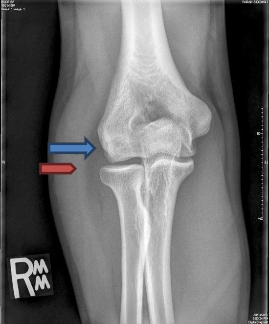
D Wasylynko

raised suspicion of a condition requiring immediate and          gonflement chronique a éveillé des soupçons d’une
further investigation.                                           condition exigeant un examen plus poussé.

(JCCA. 2016;60(3):226-232)                                       (JCCA. 2016;60(3):226-232)

k e y w o r d s : chiropractic, elbow pain,                      m o t s c l é s : chiropratique, douleur du coude,
chondroblastoma, osteochondral bone defect, joint                chondroblastome, anomalie ostéo-cartilagineuse,
swelling                                                         gonflement articulaire

Introduction                                                        OCD in the capitellum of the elbow has been described
Elbow pain in an adolescent baseball player is not an un-        in the literature by numerous authors.1-9 Although it is
usual occurrence. The reasons for the elbow pain may at          considered a rare condition when present, it is most com-
times be due to unusual circumstances such as tumours            monly found in the capitellum. The exact incidence of the
and undiagnosed fractures. Although most elbow com-              condition is speculative, with current estimates of 1.3% to
plaints in young baseball players are the result of either       1.6% amongst little league players.4 It is not uncommon
traumatic injury or repetitive stress, it is important to rec-   for this injury to be confused with osteonecrosis, Panner’s
ognize that elbow pain may signify the presence of an            disease, Little Leaguers elbow, and hereditary epiphyseal
injury not restricted to the soft-tissues. This is especial-     dysplasia.5
ly true when a red flag such as painful joint swelling is           This case is of particular interest to chiropractors who
present.                                                         treat athletes in general, and most relevant to those who
   One possible reason for such joint swelling is an osteo-      treat baseball players. Elbow injuries from various mech-
chondral defect. Such a defect is a progressive stage of         anisms are common in adolescent baseball players. It is
osteochondrosis/osteochondritis dissecans. This condi-           important for clinicians to realize that at times muscu-
tion is relatively rare, necessitates different treatment op-    loskeletal conditions requiring a more thorough investi-
tions, and is associated with variable recovery dependent        gation may masquerade as sprains and strains and could
on the stage of the condition’s development. Osteochon-          potentially be missed. The misdiagnosis may result in
dritis dissecans (OCD) is a commonly used term. How-             damaging sequelae such as disruption of the normal endo-
ever, due to the paucity of inflammatory cells found on          chondral ossification process (disruption of the growth
the osteochondral articular surface of the bone, a more          plate).6 Chronic joint swelling is one such red flag that
appropriate term would be osteochondrosis, which does            should not be overlooked.
not assume the presence of an inflammatory process.1,2
   A similar condition commonly confused with OCD                Case Presentation
is Panner’s disease (an osteonecrosis of the capitellum).        A 17-year-old baseball player presented with right elbow
Although the two conditions appear similar, there are sig-       pain of approximately one year in duration. The pain
nificant differences between them. The most important            was diffuse throughout the elbow, and had progressive-
difference is the age of onset. Panner’s disease occurs in       ly worsened. The elbow was aggravated with throwing,
children below the age of 12, with some authors stating          particularly on the follow-through and the player did
the upper range to be at 10 years of age. Panner’s disease       not recall any specific trauma or injury to the elbow. He
tends to be self-limiting and presents as a focal osteone-       had been previously diagnosed and treated for a sus-
crosis of the entire capitellum. OCD on the other hand           pected chronic lateral epicondylitis. Regrettably for the
commonly occurs in adolescence, is laterally located, and        patient there had been no relief from any of the treat-
is variable in terms of prognosis. Some authors feel that        ment received. He reported that the treatment consisted
there may be a progressive connection between the two            of muscle stimulation, Graston technique, massage, and
disorders.1,3                                                    “strong” stretching.

J Can Chiropr Assoc 2016; 60(3)                                                                                         227
A rare cause of chronic elbow pain in an adolescent baseball player: a case report
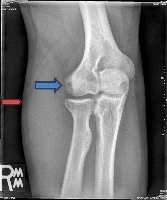
Figure 1.
Plain radiographs of the elbow prior to surgical intervention.
Blue arrow: Osteochondral defect; red arrow: Soft tissue swelling. Left: AP view; Right: oblique view.
Clinical Findings decrease in external rotation. All other ranges, strength,
Inspection of the elbow revealed significant swelling on upper limb reflexes, and sensation appeared to be normal.
the lateral aspect of the joint. Range of motion was lim-
ited in extension to approximately 150° with discomfort. Differential Diagnosis and Follow-up
Flexion of the elbow was minimally limited. Varus and The physical examination indicated that this could have
valgus stress on the medial and lateral holding elements been an atypical or complicated case of soft-tissue dam-
respectively did not induce any excessive movement due age. Soft-tissue differentials included a mild sprain of
to ligamentous laxity. The ulnar collateral ligament, al- the ulnar collateral and annular ligaments, and strains of
though tender, appeared intact. Digital percussion of the the pronator teres, biceps, brachialis, and elbow extensor
epicondyles produced pain on the lateral epicondyle. group.
There was also soft-tissue swelling and tenderness to pal- The presence of swelling with progressively worsening
pation over the lateral aspect of the elbow. pain was potentially ominous and indicated a need to refer
Strength testing of the elbow resulted in pain-related for plain x-ray imaging and subsequent medical evalua-
weakness in flexion, extension, pronation, and supination. tion. In the initial stages of the evaluation, benign tumours
Palpation revealed multiple tender areas around the elbow such as osteoid osteoma, giant cell tumour, or chond-
joint, particularly over the distal aspect of the brachialis roblastoma were considered. Of these, chondroblastoma
and lateral head of the biceps muscles. Similar tenderness was most consistent with both the age of the patient and
was found on the lateral forearm extensors, common ex- presence of swelling. Malignant tumours that required
tensor tendon, and pronator teres muscle. Notwithstand- consideration in order of incidence were osteosarcoma,
ing an absence of ligamentous laxity, pain was elicited Ewing sarcoma, and synovial sarcoma.7
during palpation of the ulnar collateral ligament and an- The radiographs revealed an osteochondral defect in
nular ligament. The shoulder joint revealed a significant the right lateral capitellum of the elbow (Figure 1). Fol-
228 J Can Chiropr Assoc 2016; 60(3)D Wasylynko
Figure 2.
Plain radiographs of the elbow 7 months post-surgical intervention.
Left: AP view, 8 months post-surgery; Right: Oblique view, 8 months post-surgery.
lowing confirmation of the diagnosis, the patient con- During the late cocking and early acceleration phases
sulted an orthopedic surgeon where a De Novo NT allog- of throwing a significant valgus stress occurs at the elbow,
raft procedure was performed. At approximately 7 months producing abnormal compressive forces across the radio-
post-surgery and rehabilitation, the patient was able to capitellar joint. This is especially significant in a develop-
begin light throwing. Post-surgical radiographs were re- mentally immature elbow. The continued stresses result
ported as normal with no focal or suspicious abnormality in a localized injury to the subchondral bone in the form
noted. In addition, there was no evidence of joint effusion of fatigue fractures and diminished support for the over-
identified (Figure 2). lying articular cartilage. Consequently, this combination
of events results in a breakdown and potential fragmenta-
Discussion tion of the cartilage and bone. It is believed that 60% of
axial compression forces across the elbow are transmitted
Etiology to the radiocapitellar joint.5,9
It is generally accepted that the etiology of OCD of the According to Yadao et al.5 the vascular supply to the
elbow is largely unknown. A review of the literature distal humerus may be considered marginal, which sug-
indicates that there appears to be some common ground gests that the lack of blood (ischemia) may play a part
amongst most authors as to the most likely origins of the in the development of OCD. Further to this, the small
condition.1-9 The most obvious suspicions for the develop- blood vessels enter posteriorly to the capitellar epiphysis
ment of an osteochondral defect are repetitive micro- and extend over the epiphyseal cartilage, which is a site
trauma, and compromised blood flow to the elbow. OCD of significant contact and compression. It is therefore
is found primarily in throwing athletes and gymnasts due conceivable that repetitive stress in this area may lead to
to the excessive compressive and shearing forces on the the development of OCD. If this injury is left untreated,
lateral compartment of the elbow. symptomatic degenerative change appears to be inevit-
J Can Chiropr Assoc 2016; 60(3) 229A rare cause of chronic elbow pain in an adolescent baseball player: a case report
able, leading to damage and fragmentation of the articular 14.9 months to heal, while the stage II patients resolved
cartilage.5 There is some belief that a genetic component in 12.3 months. According to that study, treatment con-
to the disease may exist based on reports of OCD occur- sisted of refraining from heavy use of the elbow for six
ring in generations of families.5 months.13 In contrast, Takahara et al.14 found that there
OCD may initially be asymptomatic in the early stages was only a slight chance of the capitellum healing with
but with continued overuse, will progress to a symptom- conservative care, regardless of stage.
atic state that can potentially end an athlete’s career.10 In this particular case the condition had been pro-
Takahara et al.9 described the early development and de- gressing for 12 to 18 months and required surgical re-
tection of OCD utilizing MRI. They observed capitellar pair. Although different surgical techniques have been
abnormalities while screening adolescent baseball pitch- developed to repair chondral and osteochondral lesions,
ers and noted that those individuals developed OCD over the literature demonstrates only fair to good results when
time. Early changes in the capitellum were identified performed, even in the appropriate circumstances. Addi-
by low signal intensity on T1-weighted images, while tionally, there is significant controversy in regards to the
T2-weighted images displayed no abnormalities. They indications and outcomes of these various procedures.13
also found that diagnostic ultrasound revealed a localized The specific procedure utilized in this case was a DeNovo
flattening of the subchondral bone with a normal outline NT allograft. This particular technique has not been used
of the articular cartilage. Plain radiographs with the elbow often in the elbow but has been administered with empir-
at 45° of flexion displayed a slight flattening and sclerosis ically good success in the knee. Therefore, its application
of the superficial aspect of the capitellum.9 for the elbow was largely untested. Additionally, rehabili-
As noted by Krych et al.10, many bone and soft-tissue tation of the elbow following this specific surgery was
tumours present disproportionately in young and active done without the benefit of previous cases, but was guid-
patients who are often involved in athletic activities. In ed simply by general basic principles of rehabilitation for
such instances the clinician may misdiagnose these rare other post-surgical orthopedic conditions. According to
tumours as more common sports injuries.10 Walker et al.11 Tompkins et al.15 “most surgeons develop personal treat-
emphasize that both joint-related tumours and sports in- ment preferences guided by training, published literature,
juries often afflict young, active patients, and the symp- outcome data, education conferences, expert opinion, and
toms may overlap significantly. In these cases, lack of ad- personal experience. As a recently developed cartilage-re-
equate imaging studies may result in either a significant pair technique, the role of DeNovo NT has not been clear-
delay in diagnosis or an inappropriate and unnecessary ly defined”.15 This is especially true with regards to the
arthroscopic procedure.11 capitellum.
The DeNovo NT (natural tissue) repair process inserts
Therapeutic Intervention juvenile articular cartilage into the damaged bone. The
Many authors have discussed the treatment options avail- particulated allograft chondral fragments are attached to
able for OCD. Although conservative treatment may be the bone defect and held in place with adhesive fibrin.
effective in certain circumstances, there is general agree- The fibrin has been shown in vitro to support chondrocyte
ment that a variety of surgical options tend to provide bet- overgrowth, thereby assisting in the healing process. The
ter outcomes.12-17 juvenile chondrocytes are used due to their greater poten-
The therapeutic protocols for OCD are somewhat de- tial for cell division and matrix production.13
pendent on the stage of the condition. If caught early in Rehabilitation of the elbow by the chiropractor in this
the process, conservative management may be effective. case began immediately following surgery and was guided
Matsuura et al.13 produced a retrospective paper on 176 by general principles of rehabilitation for other post-sur-
individuals with osteochondrosis of the humeral capitel- gical orthopedic conditions. The protocol initially involved
lum. Of the 101 patients that received conservative care, pain-free passive movement, which continued for 10
healing occurred in 90.5% of stage I lesions (radiolucent weeks, producing approximately 175° of extension. Fol-
areas on plain radiograph) and 52.9% of stage II lesions lowing the passive movement stage, light resisted motion
(non-displaced fragments). On average, stage I required was introduced. Minimal dumbbell weight (2-3 lbs) and
230 J Can Chiropr Assoc 2016; 60(3)D Wasylynko
stretch tubing was utilized to strengthen flexion, extension, played may also be considerations.4 Generally in regard
pronation, and supination. The volume of work consisted to the latter, one would expect a higher incidence of OCD
of one to two sets of 20 repetitions initially, progressively in pitchers and catchers, due to the high number of throws
increasing the number of sets and weight as tolerance to executed. In this particular case, the patient was a first
the exercise increased. The patient also developed gener- baseman, which simply exemplifies the fact that playing
al fitness, utilizing an exercise bike, and maintained core position may not be a reliable proxy measure of volume
strength and upper and lower limb strength where possible of throwing activity.
without aggravating the repaired elbow. The general fit-
ness program was supervised by an athletic therapist who Summary
included left arm goblet squats, left side back rows, plyo- Chronic swelling within a joint should always be con-
metric boxes, weighted sit-ups and agility drills. At 16 to sidered suspect, particularly in young athletes, and not
20 weeks weighted wrist pronation was performed with 5 dismissed as simply an atypical soft-tissue sprain or
lbs, progressing by 24 to 28 weeks to 10 lbs. The volume strain. Serious conditions are rare but when present, may
remained consistent at 10 reps for 3 sets. Dumbbell biceps masquerade as a common athletic injury. Conditions that
curls and triceps extension were performed with 5 to 15 lb are unresponsive to initial therapy should be investigated
weights (15 reps/3 sets) progressing from 16 to 28 weeks. further. OCD is an example of a potentially serious con-
At week 24, light push-ups were introduced. dition that can mimic a common athletic soft-tissue in-
By 28 weeks post-surgery, the patient began light jury, yet if diagnosed early, may resolve simply with rest.
throwing in the form of wrist flicks and light hitting off a Conversely, if misdiagnosed and mismanaged, OCD can
tee at 40-50% of maximum. He continued to strengthen potentially become a career-ending injury.
the arm with his normal workout routine, while increasing
general fitness under the supervision of the team trainer. Acknowledgments
By 32 to 36 weeks, the goal of rehabilitation was achieved The author would like to thank Dr. Jeffrey Quon, Dr. John
with a return to non-competitive play. Taylor and Ms. Stacey Kumar for their assistance in pre-
paring this manuscript.
Clinical Significance
OCD of the capitellum is rare, but is most often found in References:
adolescent overhead athletes, and was therefore a condi- 1. Gonzalez-Lomas G, EllAttrache N. Elbow osteochondritis
tion requiring a high degree of suspicion in the current dissecans and olecranon stress fractures in young athletes.
case.6 More importantly to clinicians is the awareness J Am Acad Orthop Surg. 2010:18: 87-105.
of the multiple implications of chronic joint swelling. In 2. Kobayashi K, Burton K, Rodner C, Smith B, Caputo A.
Lateral compression injuries in the pediatric elbow:
this case the patient claimed to have been assessed and Panner’s disease and osteochondritis dissecans of the
treated by numerous different practitioners for the afore- capitellum. J Am Acad Orthop Surg. 2004;12: 246-254.
mentioned signs and symptoms. That so many different 3. Wroblewski R, Urban M, Michalik D, Zakrzewski P,
individuals failed to recognize a condition more signifi- Langner M, Pomianowski S. Osteochondrosis of
cant than a soft-tissue injury highlights the need to under- the capitellum of the humerus (Panner’s disease,
stand the many reasons for chronic joint swelling. osteochondritis dissecans). Case study. Ortopedia
Traumatologia Rehabilitacja. 2014;1(6); 79-90.
The treatment for OCD, particularly when chronic, is 4. Kida Y, Morihara T, Kotoura Y, Hojo T, Taciiri H,
surgical intervention. If the condition is recognized early, Sukenari T, et al. Prevalence and clinical characteristics of
prior to frank symptoms, but in the presence of the typical osteochondritis dissecans of the humeral capitellum among
mechanism of injury and age of the athlete, development adolescent baseball players. Am J Sports Med. 2014;42(8):
of more serious sequelae may be prevented. According to 1963-1971.
5. Yadao M, Field L, Savoie F. Osteochondritis dissecans of
Kida et al.4 introduction to the game at an early age may the elbow. AAOS Instructional Course Lectures. 2004;53;
be a risk factor for OCD of the humeral capitellum. Addi- 599-605.
tionally, the duration of competitive play, a history of cur- 6. Laor T, Zbojniewicz A, Eismann E, Wall E. Juvenile
rent and prior elbow pain when throwing, and position osteochondritis dissecans: is it a growth disturbance of
J Can Chiropr Assoc 2016; 60(3) 231A rare cause of chronic elbow pain in an adolescent baseball player: a case report
the secondary physis of the epiphysis? American Journal 12. Ruchelsman D, Hall M, Youm T. Osteochondritis
Rheum. 2012;199: 1121-1128. dissecans of the capitellum: current concepts. J Am Acad
7. Mihata T, Quigley R, Robicheaux G, McGarry M, Neo M, Orthop Surg. 2010;18: 557-567.
Lee T. Biomechanical characteristics of osteochondral 13. Matsuura T, Kashiwaguchi S, Iwase T, Takeda Y, Yasui N.
defects of the humeral capitellum. Am J Sports Med. Conservative treatment for osteochondrosis of the humeral
2013;41(8); 1909-1914. capitellum. Am J Sports Med. 2008;36(5): 868-873.
8. Bradley J, Petrie R. Osteochondritis dissecans of the 14. Takahara M, Ogino T, Fukushima S, Tsuchida H,
humeral capitellum. Clin Sports Med. 2001;20(3): 565-590. Kaneda K. Nonoperative treatment of osteochondritis
9. Takahara M, Shundo M, Kondo M, Suzuki K, Nambu T, dissecans of the humeral capitellum. Am J Sports Med.
Ogino T. Early detection of osteochondritis dissecans of 1999;27(6):728-733.
the capitellum in young baseball players. J Bone and Joint 15. Tompkins M, Adkisson D, Bonner K. DeNovo NT
Surg. 1998;80-A(6): 892-897. Allograft. Oper Tech Sports Med. 2013;21: 82-89.
10. Krych A, Odland A, Rose P, Dahm D, Levy B, Wenger D 16. Cain E, Dugas J, Wolf R, Andrews J. Elbow injuries in
et al. Oncologic conditions simulate common sports throwing athletes: a current concepts review. Am J Sports
injuries. J Am Acad Orthop Surg. 2014;22: 223-234. Med. 2003;31(4): 621-634.
11. Walker E, Brian P, Longo V, Fox E, Frauenhoffer E, 17. Baumgarten T, Andrews J, Satterwhite Y. The arthroscopic
Murphey M. Dilemmas in distinguishing between tumor classification and treatment of osteochondritis dissecans of
and that posttraumatic lesion with surgical or pathologic the capitellum. Am J Sports Med. 1998;26(4): 521-525.
correlation. Clin Sports Med. 2013;32: 559-576.
232 J Can Chiropr Assoc 2016; 60(3)You can also read