A Sarcina bacterium linked to lethal disease in sanctuary chimpanzees in Sierra Leone - Robert Koch-Institut
←
→
Page content transcription
If your browser does not render page correctly, please read the page content below
ARTICLE
                  https://doi.org/10.1038/s41467-021-21012-x                 OPEN

                  A Sarcina bacterium linked to lethal disease in
                  sanctuary chimpanzees in Sierra Leone
                  Leah A. Owens 1, Barbara Colitti2, Ismail Hirji 3, Andrea Pizarro3, Jenny E. Jaffe 4,5, Sophie Moittié                                                 6,7,

                  Kimberly A. Bishop-Lilly8, Luis A. Estrella8, Logan J. Voegtly 8,9, Jens H. Kuhn 10, Garret Suen 11,
                  Courtney L. Deblois11, Christopher D. Dunn1, Carles Juan-Sallés12 & Tony L. Goldberg 1 ✉
1234567890():,;

                  Human and animal infections with bacteria of the genus Sarcina (family Clostridiaceae) are
                  associated with gastric dilation and emphysematous gastritis. However, the potential roles of
                  sarcinae as commensals or pathogens remain unclear. Here, we investigate a lethal disease of
                  unknown etiology that affects sanctuary chimpanzees (Pan troglodytes verus) in Sierra Leone.
                  The disease, which we have named “epizootic neurologic and gastroenteric syndrome”
                  (ENGS), is characterized by neurologic and gastrointestinal signs and results in death of the
                  animals, even after medical treatment. Using a case-control study design, we show that ENGS
                  is strongly associated with Sarcina infection. The microorganism is distinct from Sarcina
                  ventriculi and other known members of its genus, based on bacterial morphology and growth
                  characteristics. Whole-genome sequencing confirms this distinction and reveals the presence
                  of genetic features that may account for the unusual virulence of the bacterium. Therefore,
                  we propose that this organism be considered the representative of a new species, named
                  “Candidatus Sarcina troglodytae”. Our results suggest that a heretofore unrecognized complex
                  of related sarcinae likely exists, some of which may be highly virulent. However, the potential
                  role of “Ca. S. troglodytae” in the etiology of ENGS, alone or in combination with other factors,
                  remains a topic for future research.

                  1 Department of Pathobiological Sciences, School of Veterinary Medicine, University of Wisconsin-Madison, Madison, WI, USA. 2 Department of Veterinary

                  Science, University of Torino, Torino, Italy. 3 Tacugama Chimpanzee Sanctuary, Freetown, Sierra Leone. 4 Tai Chimpanzee Project, Max Planck Institute for
                  Evolutionary Anthropology, Leipzig, Germany. 5 Epidemiology of Highly Pathogenic Microorganisms, Robert Koch Institute, Berlin, Germany. 6 School of
                  Veterinary Medicine and Sciences, University of Nottingham Sutton Bonington Campus, Sutton Bonington, Leicestershire, UK. 7 Twycross Zoo,
                  Atherstone, UK. 8 Genomics and Bioinformatics Department, Biological Defense Research Directorate, Naval Medical Research Center, Fort Detrick, MD,
                  USA. 9 Leidos, Reston, VI, USA. 10 Integrated Research Facility at Fort Detrick, Division of Clinical Research, National Institute of Allergy and Infectious
                  Diseases, National Institutes of Health, Fort Detrick, MD, USA. 11 Department of Bacteriology, University of Wisconsin-Madison, Madison, WI, USA. 12 Noah’s
                  Path, Elche, Spain. ✉email: tony.goldberg@wisc.edu

                  NATURE COMMUNICATIONS | (2021)12:763 | https://doi.org/10.1038/s41467-021-21012-x | www.nature.com/naturecommunications                                   1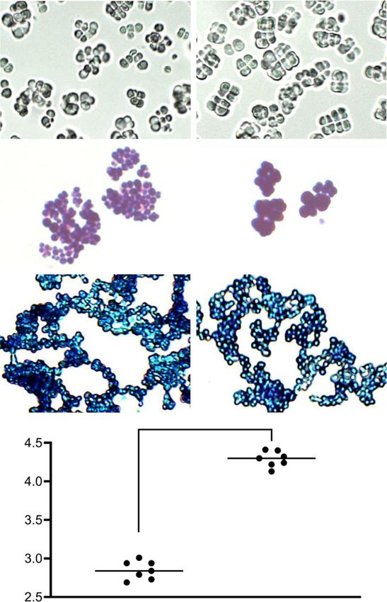
ARTICLE                                                                  NATURE COMMUNICATIONS | https://doi.org/10.1038/s41467-021-21012-x

E
      merging pathogens pose a substantial risk to animal and                biochemical pathways potentially contributing to enhanced
      human health1,2. Pathogens can emerge due to the acqui-                virulence, including an encoded urea degradation biochemical
      sition of virulence factors through genetic mutation and               pathway, consistent with the clinical signs observed in chim-
horizontal gene transfer3–5 and due to ecological processes that             panzees. We conclude that the genus Sarcina likely contains an
alter their epidemiology/epizootiology and host range6–8. These              overlooked complex of species ranging from benign commensals
processes are accelerating due to factors such as agricultural               to frank pathogens. In light of these findings, the importance of
intensification9, demographic shifts10, ecosystem perturbations11,            sarcinae in human and animal clinical disease should be re-
geophysical processes12, and global environmental changes13.                 evaluated.
Furthermore, improved diagnostic methods facilitate detection
and characterization of new pathogens and the genetic features
                                                                             Results
that contribute to their emergent phenotypes14–16.
                                                                             Epizootiology, clinical signs, and pathology. From 2005 to 2018,
   Emerging pathogens of non-human primates are especially
                                                                             56 resident chimpanzees of TCS died of ENGS. In 32 of 56 cases,
salient examples of this phenomenon because of the high
                                                                             affected individuals displayed signs including anorexia, neuro-
potential for such pathogens to infect humans, who are geneti-
                                                                             muscular weakness, ataxia, seizures, vomiting, and abdominal
cally similar hosts17,18. For example, one of the main causative
                                                                             distension (Fig. 1; “Clinical signs” group). Signs persisted for a
agents of human malaria, Plasmodium falciparum, once thought
                                                                             median of 6 days (range: 1–90 days) prior to recovery or death
to have co-evolved with humans, actually arose from a recent
                                                                             (Supplementary Table 1). In all recovered cases, clinical disease
zoonotic transmission from a western gorilla (Gorilla gorilla
                                                                             subsequently recurred and resulted in death. In the remaining 24
(Savage, 1847))19–21. Nowhere are such risks more evident than
                                                                             cases, individuals were discovered post-mortem with no pre-
in zoological and sanctuary settings, where captive and semi-
                                                                             monitory signs noted by care staff or developed signs which
captive primates come into frequent close contact with
                                                                             progressed to death in 12 h or less (Fig. 1; “Sudden death” group).
people22,23. For example, contact with New World primates led to
                                                                             Despite these disparate manifestations, all clinical presentations
simian foamy virus transmission to primate workers in Brazil24
                                                                             were associated and clearly recognizable as the same “mystery
and monkeypox virus transmission occurred in staff at a primate
                                                                             disease” (described as “unmistakable” by veterinarians). We
sanctuary following a monkeypox outbreak in sanctuary chim-
                                                                             therefore chose the term “syndrome” to reflect the heterogenous
panzees (Pan troglodytes (Blumenbach, 1775)) in Cameroon25.
                                                                             nature of the clinical presentations and the suspicion of a com-
   Since 2005, western chimpanzees (Pan troglodytes verus
                                                                             mon etiopathogenesis. ENGS represented the highest cause of
Schwarz, 1934; “chimpanzees” hereafter) in Sierra Leone’s
                                                                             mortality in this population, affecting 33.7% of chimpanzees and
Tacugama Chimpanzee Sanctuary (TCS, in Western Area
                                                                             accounting for 63.6% of deaths during this time period (Fig. 2a),
Peninsula National Park) have suffered from a lethal disease of
                                                                             with a case fatality rate of 100% and a seasonal distribution
unknown etiology. Characteristic signs are neurologic (weakness,
                                                                             peaking in March (Fig. 2b). The etiological agent of ENGS does
ataxia, seizures) and gastrointestinal (abdominal distension,
                                                                             not appear to be transmitted directly, as there were few instances
anorexia, vomiting), resulting in death even after aggressive
                                                                             of cases clustering in time and space (Supplementary Table 1).
medical treatment by staff veterinarians. To date, a total of 56
                                                                                Frequent lesions included acute shock (congestion involving
individuals at this facility have died of this condition, which we
                                                                             multiple organs), neutrophilic margination in the microcircula-
have named “epizootic neurologic and gastroenteric syndrome”
                                                                             tion, moderate to marked gastric dilation, pulmonary edema,
(ENGS), constituting a medical emergency in this population,
                                                                             acute aspiration of digestive contents, and acute hemorrhage in
which averages 93 chimpanzees at any given time. TCS is the
                                                                             the thymus, pancreas, or both. In total, post-mortem evaluation
largest repository of Sierra Leone’s chimpanzee genetic diversity,
                                                                             documentation was available for 28 chimpanzees, but in only 17
a training site for conservationists throughout western Africa, an
                                                                             of these was the gastrointestinal tract assessed. Of these 17
educational/ecotourism destination important for the local
                                                                             patients, 14 had gross evidence of acute gastric dilation, 1 did not
economy, and the only home for displaced or orphaned chim-
                                                                             show such evidence, and 2 were inconclusive (Fig. 1). One of the
panzees in the country.
                                                                             chimpanzees with acute gastric dilation also had massive
   Despite ongoing efforts by veterinary staff and international
                                                                             hemorrhagic diathesis (Fig. 3a) and multiple gas-filled lesions in
collaborators, the etiology of ENGS has remained elusive. Ence-
                                                                             the cecal wall (emphysematous typhlocolitis; Fig. 3b). Micro-
phalomyocarditis virus (EMCV; Picornaviridae: Cardiovirus)
                                                                             scopically, these gas-filled lesions were surrounded by infiltrates
infection and toxicity from certain plants (Dichapetalum tox-
                                                                             of macrophages, eosinophils, and multinucleate giant cells
icarium (G. Don) Baill./D. heudelotii (Planch. ex Oliv.) Baill.)
                                                                             (Fig. 3c).
were both suspected but, after investigation, deemed unlikely to
be causal. In such circumstances where infection has been sus-
pected but known agents have not been identified, diagnostic                  Samples. We selected 95 archived samples from 32 chimpanzees
approaches based on metagenomics have proven useful26–28. We                 for analysis (Supplementary Data 1). These samples comprised 19
therefore undertook a case-control epizootiological investigation            individuals (7 males and 12 females) that had died from ENGS
to identify potential pathogens of all major types (viruses, bac-            (cases), representing a subset of the 56 total cases that occurred
teria, and eukaryotes) using metagenomics and traditional                    since the epizootic began in 2005 (Fig. 2), and 14 healthy indi-
methods in the TCS chimpanzees, to detect associations between               viduals (7 males and 7 females) sampled during routine veter-
particular microbes and ENGS.                                                inary health checks or, in 2 instances, sampled post-mortem
   Here we report the finding of a Sarcina genus bacterium in 13              when cause of death was known and clearly unrelated to ENGS
of 19 ENGS cases but no controls. We also report the occurrence              (e.g., from trauma; controls). In one instance, matched samples
of gross and histopathological lesions in affected chimpanzees               were available from a healthy chimpanzee who subsequently
consistent with the most severe forms of Sarcina infection                   became ill and died from ENGS (hence this individual was first a
reported in humans and animals. By studying the morphology                   control and then a case). The chimpanzees in this study ranged in
and growth characteristics of the bacterium, and by sequencing               age from 5 to 27 years (median age = 12 years) and were sampled
the complete genome of an isolate, we identify features that dis-            between 14 March 2013 and 11 July 2016, although not all cases
tinguish it from all previously described members of its genus. In           within this time period were sampled or available for study.
particular, we show that the bacterium possesses genes encoding              Among ENGS cases included, clinical signs were similar to those

2                NATURE COMMUNICATIONS | (2021)12:763 | https://doi.org/10.1038/s41467-021-21012-x | www.nature.com/naturecommunications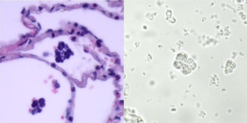
NATURE COMMUNICATIONS | https://doi.org/10.1038/s41467-021-21012-x                                                                                   ARTICLE

                                                                                                              ENGS case, female                Gastric dilation
                                                                                                              ENGS case, male                  No gastric dilation
                                                                                                              Control, female                  Inconclusive for
                                                                                                              Control, male                    gastric dilation
                    Sudden death                                       Clinical signs
                                                                                                                                Positive by diagnostic PCR
                                                                                                                                Negative by diagnostic PCR
                                                                                                                               * Same individual

             Not analyzed                                   Not analyzed                                                       Prevalence of clinical signs

                                                                                                              Prevalence (%)
                                                                                                                                                 Not analyzed
                                                                                                                                                 Analyzed

    *      Analyzed                  Analyzed                                      *      Analyzed

                                     6 positive
          0 positive                   (n=6)                                             9 positive
           (n=14)                                                                         (n=13)

Fig. 1 Graphic representation of epizootic neurologic and gastroenteric syndrome (ENGS) cases and study samples. Individual chimpanzees are
represented by rectangles and colors denote sex and case/control status (red, female ENGS case, n = 30; blue, male ENGS case, n = 26; pink, female ENGS
control, n = 7; light blue, male ENGS control, n = 7). Post-mortem findings regarding gastric dilation are shown for those cases where documentation was
available (n = 17; black circle, gastric dilation, n = 14; open circle, no gastric dilation, n = 1; filled gray circle, inconclusive for gastric dilation, n = 2).
“Analyzed” indicates an individual from whom we obtained at least one sample used in this study (n = 32) and the asterisk indicates the single individual
from whom we obtained samples pre- and post-ENGS. Pie charts are scaled to sample size with green representing individuals positive by diagnostic PCR
and white representing individuals negative by diagnostic PCR.

of cases that were not available for inclusion (Fig. 1), the most                 quality (~21% of reads removed during quality filtering;
common of which were ataxia (n = 12), seizures (n = 12),                          Supplementary Table 2). Due to the pan-eukaryotic nature of
vomiting (n = 10), and abdominal distention (n = 9).                              the primers, and despite the use of a mammal-blocking primer,
                                                                                  the majority of reads were identified as host (~83%; Supplemen-
Parasitology. Microscopic examinations of fecal samples were                      tary Table 2). This identification was not surprising due to the
performed on site for 30 chimpanzees (17 ENGS cases and 13                        sample types (host tissues) and because of previously published
controls) from 2005 to 2018 using standard direct and sedi-                       similar findings using the same primers and protocols35. We
mentation methods29, comprising 155 analyses with a median 5                      processed data from all samples through all filtering steps, after
analyses per chimpanzee. Nine records indicated no detectable                     which we excluded those samples that represented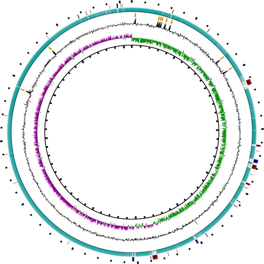
ARTICLE                                                                     NATURE COMMUNICATIONS | https://doi.org/10.1038/s41467-021-21012-x

                                                                                hereafter; mean per sample 39.7, standard deviation 45.5; average
                                                                                length per contig 969.5, standard deviation 690.8) at an average
                                                                                sequence depth of 63.4 (standard deviation 172.4, minimum 3.5,
                                                                                maximum 851). Overall, 21.0% of reads assembled into contigs
                                                                                and 79.0% did not. Analyses of sequence data at the individual
                                                                                read level confirmed the results of the analysis of contigs (i.e.
                                                                                identified the same viruses) and did not identify any additional
                                                                                viruses. Eleven viruses were thus identified, each of which was
                                                                                identical to or closely related to a known virus (Supplementary
                                                                                Table 3), and no viruses were case-associated (Table 1). One
                                                                                control animal was infected with a rhinovirus C (Picornaviridae:
                                                                                Enterovirus) subsequent to an outbreak of respiratory illness in
                                                                                the population. This pathogen was previously documented as a
                                                                                cause of epizootic respiratory disease in wild chimpanzees37, but
                                                                                the clinical features of this rhinovirus C infection (characterized
                                                                                by upper respiratory signs) are not consistent with ENGS.

                                                                                Bacterial metabarcoding. PCR amplification of the 16S rDNA
                                                                                V4 region was attempted on all 96 samples (19 cases and 14
                                                                                controls; Supplementary Data 1) which yielded amplicons in 10
                                                                                of 19 ENGS cases (35 total samples) but none of the controls
                                                                                (0 samples). In total, 1,131,561 raw sequences were generated for
                                                                                all 35 samples of which 787,263 were high quality after filtering in
Fig. 2 Deaths attributed to epizootic neurologic and gastroenteric              mothur. Good’s coverage estimation was calculated for all sam-
syndrome (ENGS) at Tacugama Chimpanzee Sanctuary from 2005                      ples and only those samples that had a value >0.99 were retained
through 2018. a Annual chimpanzee deaths from ENGS (black, overall              for downstream analysis. As a result, 23 samples were considered
total = 56) and other causes (gray, overall total = 32). Numbers above the      for this analysis (Supplementary Data 2), which totaled 774,339
bars indicate the yearly total chimpanzee population and the horizontal         high-quality sequences with an average of 33,667 ± 17,122 stan-
bracket below the x-axis denotes the period during which we obtained            dard deviation per sample. These sequences were binned at 97%
samples for analysis. b Summed totals of ENGS fatalities by month over          similarity into 2592 OTUs. The OTU counts were normalized to
2005‒2018 (n = 56). Source data are provided as a Source Data file.              5900 sequences per sample, and these normalized sequences were
                                                                                used for all further analyses.

                         a.                                                   c.

                         b.

Fig. 3 Gross and histopathologic images of a chimpanzee that died of epizootic neurologic and gastroenteric syndrome (ENGS). Photographic images
from an adult male chimpanzee who died of ENGS showing moderate to severe gastric dilation (a), hemorrhagic diathesis (b), and emphysematous
typhlocolitis (c; arrows point to gas-filled pockets within the cecum wall, which has reddened areas [in inset, white arrowheads point to gas bubbles in the
cut surfaces of the formalin-fixed cecum]; scale bars = 1 cm). On histology (c), gas-filled space (*) in the cecal submucosa, surrounded by inflammatory
infiltrates (arrows) and hemorrhage (h) were visualized by hematoxylin and eosin staining (inset depicts inflammatory infiltrates, which include eosinophils
[arrow] and multinucleate giant cells [arrowheads]; scale bars = 300 µm [main image] and 30 µm [inset]; micrograph is a representative image of three
similar sections obtained from the same individual).

4                   NATURE COMMUNICATIONS | (2021)12:763 | https://doi.org/10.1038/s41467-021-21012-x | www.nature.com/naturecommunications
NATURE COMMUNICATIONS | https://doi.org/10.1038/s41467-021-21012-x ARTICLE
Table 1 Case/Control statistical summary.
Organism ID Organism type Diagnostic Prevalence in Prevalence in Pa Odds ratio 95% CI low 95% CI high
cases (%) controls (%)
Blastocystis Parasite 18S MB 50.00 33.33 0.633 2.000 0.244 16.363
Oesophagostomum Parasite 18S MB 0.00 16.67 0.375 0.175 0.006 5.041
Troglodytella Parasite 18S MB 30.00 33.33 >0.999 0.857 0.098 7.510
GB virus C Virus Virus Seq 70.59 100.00 0.273 0.175 0.008 3.678
Rhinovirus C Virus Virus Seq 0.00 16.67 0.261 0.105 0.004 2.959
Gemykibivirus 2 Virus Virus Seq 41.18 83.33 0.155 0.140 0.013 1.474
Chimpanzee Virus Virus Seq 5.88 0.00 >0.999 1.182 0.042 32.915
parvovirus
Human Virus Virus Seq 23.53 0.00 0.309 4.333 0.202 93.159
picobirnavirus 4
Macaque Virus Virus Seq 11.76 16.67 >0.999 0.667 0.049 9.022
picobirnavirus 24
Chimpanzee Virus Virus Seq 11.76 50.00 0.089 0.133 0.015 1.176
anellovirus
Torque teno virus 4 Virus Virus Seq 23.53 16.67 >0.999 1.538 0.137 17.335
Torque teno virus 23 Virus Virus Seq 35.29 33.33 >0.999 1.091 0.153 7.802
Torque teno virus 14 Virus Virus Seq 23.53 33.33 >0.999 0.615 0.081 4.704
Torque teno virus 16 Virus Virus Seq 17.65 33.33 0.576 0.429 0.052 3.522
“Ca. S. troglodytae” Bacterium PCR 68.42 0.00 0.0001 56.077 2.866 1097.182
Virus Seq Virome shotgun sequencing, 18S MB 18S metabarcoding, CI Confidence interval around odds ratio.
aFisher’s Exact test, 2-tailed.
Analysis of these samples showed that 9 of 23 samples contained from ENGS; the pre-illness blood sample (collected in February
a large proportion of sequences (>5% of total reads and up to 97.4% 2016) was PCR-negative whereas the post-mortem blood sample
in one sample) belonging to a single OTU belonging to an unknown (collected in July 2016) was PCR-positive. Sanger sequences of all
member of the bacterial family Clostridiaceae (Clostridia: Clostri- amplicons were identical to each other and to the representative
diales) (Fig. 4a). This OTU most closely resembled Clostridium sequence generated by metabarcoding, except for one sample with
perfringens in the Greengenes database39,40. However, C. perfringens a single nucleotide polymorphism (C → T transition) at position
diagnostic PCR using published protocols41–43 failed to yield 51 of the diagnostic fragment. Sequences of the diagnostic frag-
amplicons in any instances, including in tissues found positive by ment were also identical to published 16S rDNA sequences in
16S sequencing. GenBank for S. ventriculi (AF110272) and Clostridium ventriculi
Re-examination of the representative sequence from this OTU DSM286 (NR026146), with the exception of the one variant
against the National Center for Biotechnology Information’s sequence (1 nucleotide mismatch to the aforementioned pub-
(NCBI’s) GenBank (GenBank hereafter) non-redundant database lished sequences).
excluding uncultured organisms identified a putative match
(97.2% nucleotide identity) to Clostridium (Sarcina) ventriculi
from feces of Japanese macaques (Macaca fuscata Blyth, 1875; Bacterial isolation and characterization. We attempted to cul-
GenBank accession numbers LC101491 and LC101492). Re- ture the bacterium using 44 combinations of cell preparations and
examination of all samples by including the Clostridium (Sarcina) culture conditions (Supplementary Table 4), 2 of which resulted
ventriculi sequence from GenBank in the Greengenes database in growth of colonies that resembled sarcinae. Specifically, wet
demonstrated this organism to be present in all 23 ENGS case mounts of colonies grown on egg yolk agar plates and Sarcina
samples (Supplementary Data 2). Notably, the organism was ventriculi growth medium (SVGM) plates revealed refractile,
present not only in gastrointestinal contents but also in internal cuboid cells in packets, a morphology that is distinctive of
organs such as brain, liver, and spleen, sometimes at high members of the genus Sarcina (Fig. 5a, right panel). These
abundance (Fig. 4b). The nomenclature of the genus Sarcina is colonies were derived from the liver of one individual (1 colony
contested (and sometimes the genus name Clostridium is on egg yolk agar plates) and the brain of another (many colonies
substituted) because Sarcina is phylogenetically situated within on SVGM plates; Supplementary Data 2). We confirmed the
the “cluster I” group of Clostridia44, considered the “true” identity of every colony using diagnostic PCR and Sanger
Clostridia, although these organisms are polyphyletic. A proposal sequencing (see above).
was made to change the name Sarcina to Clostridium, but was not We were repeatedly able to isolate the organism by plating
approved because the name Sarcina predates the name Clos- brain tissue onto SVGM plates, but the organism ceased to
tridium and therefore has priority45. remain viable after 2‒3 passages and did not grow in any of the
seven liquid media tested (Supplementary Table 4). These results
are consistent with previous studies reporting great difficulty in
Diagnostic PCR. Oligonucleotide primers specific to the 16S isolating and propagating sarcinae46,47. Furthermore, we were
rDNA gene of the unknown organism were successfully devel- unable to recover live organisms after freezing colonies placed in
oped. PCR with these primers yielded amplicons of the predicted 10% or 20% glycerol under various conditions. In contrast, we
length (289 base pairs [bp]) in 13 of 19 ENGS cases (68.4%) but 0 successfully grew the type strain S. ventriculi “Goodsir” (Amer-
of 13 controls (Supplementary Fig. 3) which was statistically ican Type Culture Collection [ATCC] 29068) under the same
significant (odds ratio = 56.1; 95% CI 2.87–1097.2; Fisher’s exact conditions with ease, including propagating the strain in solid
P = 0.0001, two-tailed; Table 1). For one individual, blood sam- and liquid media, freezing the strain in 10% glycerol, and
ples were available both before and after clinical illness and death subsequently recovering the bacterium. On SVGM media, our
NATURE COMMUNICATIONS | (2021)12:763 | https://doi.org/10.1038/s41467-021-21012-x | www.nature.com/naturecommunications 5ARTICLE NATURE COMMUNICATIONS | https://doi.org/10.1038/s41467-021-21012-x
a.
97.4 91.3 66.7 29.7 18.1 9.6 7.5 6.8 5.2
100 Clostridiaceae Blautia
Lactobacillus Collinsella
Acinetobacter Faecalibaculum
% abundance
Prevotella Atopobium
50 Weissella Alloprevotella
Ileibacterium Treponema 2
Lachnospiraceae Anaerovibrio
Prevotellaceae Allobaculum
0 Bifidobacterium Subdoligranulum
) nt nt nt nt ol er it
en m Erysipelotrichaceae Phoenicibacter
ple rte nte nte nte nte - sto - liv vom
i
- s mo co co co co ca eb a - Ruminococcaceae Micrococcales
ita t- ch ch ch ch c B ecc
N pos ma ma ma ma ebe Streptococcus Other
( to to to to R eb
R
o od - s i - s - s - s
l e b a lé
- b afo Be Nit O
ita K
N
b.
Stomach Vomit Colon Spleen Kidney Heart Brain
content Stool Blood Liver Lung Muscle
100 97.4
91.4
% abundance
66.7
50
29.7
18.1
9.6 7.5 6.8
5.2 2.31.3 1.61.3
1.5 1.3 0.9 0.1 1.0 0.7 0.4 0.5 0.1 0.1
0
bi
bi
N i
bi
ya i
bi
bi
Be t
bi
Be e
Be y
ita
lé
Bu ca
bu
Bu ca
bu
lé
ita
G ita
ita
a
Be a
b
N b
n
c
fo
it
w
Be
Be
Be
Be
O
O
ra
Lu
ec
ec
N
N
N
N
Ka
eb
eb
y
am
R
R
M
Sarcina
Other
Fig. 4 Bacterial 16S rDNA microbiome analysis of epizootic neurologic and gastroenteric syndrome (ENGS) case samples. a The percentage of total
reads (n = 5900 per sample) from 9 ENGS case samples at genus-level OTU with % reads mapped to the Clostridiacea OTU shown on top of each bar.
b Percent abundance of reads from 23 ENGS case samples classified to genus-level OTUs Sarcina (% above each bar) and other, arranged by tissue type.
isolate (JB1) grew more slowly than S. ventriculi “Goodsir” (~3‒ 16S rDNA phylogeny. Alignment of 16S rDNA sequences from
4 days until colonies were visible for JB1, versus 24 h for S. the “cluster I” group of Clostridia44, including the new organism
ventriculi “Goodsir”). The JB1 isolate displayed morphology (isolate JB1), yielded a final alignment length of 1585 positions. A
(Fig. 5a), Gram’s staining characteristics (Fig. 5b), and methylene maximum-likelihood phylogeny built from this alignment shows
blue staining characteristics (Fig. 5c) similar to S. ventriculi isolate JB1 to represent a sister taxon to S. ventriculi, forming a
“Goodsir”, as both were Gram-positive with a darkly staining clade with S. maxima, Eubacterium tarantellae, and C. perfringens
outer layer. However, JB1 cells were statistically significantly (Fig. 7). Bacteria of 13 other recognized species pairs included in
larger than those of S. ventriculi “Goodsir” (mean diameters of the analysis had a lower phylogenetic distance between them than
4.29 µm versus 2.83 µm, respectively, Mann–Whitney U P = the distance between isolate JB1 and S. ventriculi (Supplementary
0.0006, two-tailed; Fig. 5d). The cellular diameter of JB1 falls Table 5), lending support to the designation of the bacterium as a
within the published range for S. maxima (4–4.5 µm)48, but representative of a distinct species. To reflect the discovery of this
methylene blue staining showed a cellulose-containing cell wall bacterium in chimpanzees (Pan troglodytes spp.), we designated it
for isolate JB1 which is not characteristic of S. maxima (Fig. 5c). “Candidatus S. troglodytae”. We propose the Candidatus desig-
In addition, the flattened cellular morphology and large packet nation in this instance because we were unable to generate a
size of JB1 cells resemble S. ventriculi and not S. maxima49–51. culture suitable for deposition in the requisite two publicly
Archived histologic preparations of tissues collected from accessible culture repositories in two different countries52.
ENGS cases during postmortem examination and stained with
hematoxylin and eosin clearly revealed sarcinae, visible as packets
of darkly staining basophilic cells in gastric contents of the Whole-genome sequencing, assembly, and annotation. To
chimpanzee with hemorrhagic diathesis, gastric dilation, and generate sufficient material for whole-genome sequencing, we
emphysematous gastritis, and in the pulmonary alveoli of another repeated bacterial isolation from the brain tissue described above
chimpanzee (Fig. 6a). A wet mount direct smear of homogenized using identical methods and allowed colonies to grow to a large
brain tissue from the aforementioned ENGS case also demon- size on SVGM plates. We then harvested a single, large colony,
strated the presence of packets of sarcinae (Fig. 6b). confirmed its identity using microscopy and PCR/sequencing,
6 NATURE COMMUNICATIONS | (2021)12:763 | https://doi.org/10.1038/s41467-021-21012-x | www.nature.com/naturecommunicationsNATURE COMMUNICATIONS | https://doi.org/10.1038/s41467-021-21012-x ARTICLE
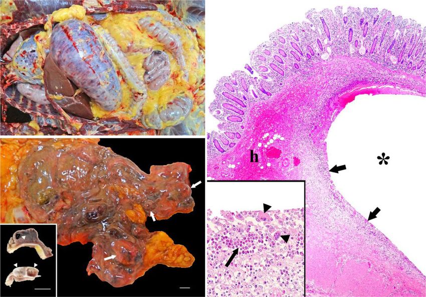
S. ventriculi “Ca. S. troglodytae” enzymes, including the urease sub-units alpha, beta, and gamma,
“Goodsir” isolate JB1 and ORFs whose products are predicted to be urease accessory
a. proteins UreE, UreF, and UreG, which are needed for urease
maturation. Ureases are nickel-containing enzymes, found in a
Live
variety of bacteria, which catalyze the breakdown of urea (a
cells ubiquitous metabolic byproduct in most animals) to ammonia
and carbon dioxide57. Bacterial and fungal ureases play a key role
in gastrointestinal tract colonization and in chronic human
diseases such as gastritis and peptic ulcers58. Additionally, in the
b. extrachromosomal sequences, we found a 34 kb plasmidial
prophage containing ORFs for an ATP-binding cassette (ABC)
Gram transporter. ABC transporters use ATP to move specific
stain substrates across a cellular membrane and can function either
as an importer (e.g., for uptake of nutrients) or an exporter (e.g.,
to efflux toxic molecules, including xenobiotic compounds such
as drugs)59. ABC importers have been associated with increased
c. bacterial survival during colonization of hosts60 and exporters
with bacterial drug-resistance61, both of which may enhance
Methylene pathogenesis of an organism.
blue stain Sarcinae are not known to produce toxins62. However, because
of the unusual neurologic disease associated with ENGS, we
scanned the genome sequence of “Ca. S. troglodytae” for ORFs
with sequence homology to known virulence genes using
d. ShortBRED63 and a customized version of the Virulence Factor
*
Database (VFDB)64, but found no evidence of such genes. Using
Cell diameter (µm)
the Comprehensive Antibiotic Resistance Database (CARD)65, we
*P=.0006
identified two antibiotic resistance ORFs, OXA-241 on the
chromosome and salA on plasmid 1, which confer resistance to
carbapenems (OXA-241) and lincosamides and streptogramins
(salA)66,67.
S. ventriculi “Ca. S. troglodytae” Summary description of the provisional species. “Candidatus
“Goodsir” isolate JB1 Sarcina troglodytae” is a proposed member of the established genus
Fig. 5 Comparative morphology of type strain Sarcina ventriculi
Sarcina, most closely related to S. ventriculi, as determined by full-
“Goodsir” ATCC 29068 and “Ca. S. troglodytae” isolate JB1. a–c Cells
length 16S rDNA phylogenetic analysis. It is an uncultivated, Gram-
were imaged live (a) or heat-fixed and stained with Gram stain (b) or
positive coccus with a tetrad structure and slightly flattened cell
methylene blue stain (c). Micrographs are from a single experiment and are
morphology and may be identified using the PCR primers: Tacu-
representative of three independent experiments with similar results. Scale
Sarc_Diag_F: 5′-TGAAAGGCATCTTTTAACAATCAAAG-3′ and
bars = 10 µm. d Live cell diameters were compared, with lines representing
TacuSarc_Diag_R: 5′-TACCGTCATTATCGTCCCTAAA-3′ or the
median diameters among seven distinct bacterial colonies. Source data are
full genome sequence (accessions CP051754–CP051764). We iso-
provided as a Source Data file. *Calculated using a Mann–Whitney U test,
lated “Ca. S. troglodytae” in an anerobic environment and at
two-tailed.
mesophilic temperature (37 °C) but were unable to maintain a
viable culture for deposition in at least two publicly accessible
culture collections, hence the Candidatus status. Samples described
and extracted DNA from this colony (isolate JB2). We performed here are derived from the brain, liver, and lung tissues of sanctuary
whole-genome sequencing using a hybrid approach of mate-pair western chimpanzees (Pan troglodytes verus) diagnosed with ENGS.
and shotgun sequencing (Supplementary Table 6) followed by de
novo genomic assembly using SPAdes53 and in silico genome
closure. The resulting full, high-quality “Ca. S. troglodytae” Discussion
assembly consists of a circular chromosome of 2,435,860 base The genus Sarcina within the Clostridiaceae is poorly studied in
pairs resolved into one single contig and 10 plasmids (totaling comparison to the highly studied toxigenic clostridia. In 1842,
205,993 base pairs, range: 4.6–78.9 kb; Supplementary Table 7). Goodsir described the type species, S. ventriculi, in the stomach
We annotated the genome with PATRIC54 and confirmed that contents of a human patient with recurrent vomiting68. Subsequent
the organism is closely related, but not identical, to S. ventriculi studies have provided evidence that bacteria morphologically con-
(98.5% nucleotide similarity; Supplementary Table 8)55,56. Total sistent with S. ventriculi cause abdominal pain, nausea, anorexia,
GC content in our organism was 27.6%, which is similar to that of vomiting, hematemesis, dysphagia, diarrhea, and generalized
S. ventriculi (27.7%). The organism shares 96.5% of its open- weakness in people69, with esophagitis70 and duodenitis71 noted
reading frames (ORFs) with S. ventriculi, with notable differences surgically or as a post-mortem finding72. Morphologically indis-
in sugar pathways and capsule biosynthesis (Fig. 8). The genome tinguishable bacteria assumed to be S. ventriculi have also been
of the JB2 strain contains DNA elements encoding metabolic associated with abomasal bloat in young pre-ruminant animals73–75,
pathways with the potential for formation of bacterial endospores characterized by sudden onset of anorexia, abdominal discomfort,
in addition to anaerobic fermentation pathways, including lethargy, dehydration, and shock culminating in high lethality
alcohol fermentation, sulfur reduction, and nitrogen reduction. (75–100%) despite treatment76. Gastric dilation in monogastric
Interestingly, the “Ca. S. troglodytae” genome, but not S. animals (horses, dogs, and cats) has also been linked to putative
ventriculi, possesses ORFs encoding for urea degradation S. ventriculi infection77,78.
NATURE COMMUNICATIONS | (2021)12:763 | https://doi.org/10.1038/s41467-021-21012-x | www.nature.com/naturecommunications 7ARTICLE NATURE COMMUNICATIONS | https://doi.org/10.1038/s41467-021-21012-x
a. b.
Fig. 6 Characteristic cuboidal packets of Sarcina-like organisms in tissues of ENGS-affected chimpanzees. a Basophilic packets of cells in a tetrad
formation can be seen amongst and within alveoli of hematoxylin and eosin-stained lung tissue of one individual (“Jumu”). b Unstained brain tissue
homogenate from another individual (“Joko”) contains highly refractile, cuboid packets of cells. Scale bars = 10 µm. Micrographs are representative images
of one of at least three sections (a) or smears (b) obtained from the same individual with similar results.
Our results demonstrate a statistically significant association ORFs similar to those present in toxigenic clostridia84. The
between a bacterium, “Ca. S. troglodytae”, and ENGS, a protracted pathogenic effects of “Ca. S. troglodytae” on chimpanzees may
lethal epizootic syndrome in sanctuary chimpanzees in Sierra therefore be caused by mechanisms other than toxicity. For
Leone. We designed a case-control epizootiological study using a example sarcinae have an unusual yeast-like metabolism85 that is
case definition that encompassed the range of clinical presentations active over a wide pH range86, allowing bacteria to produce
associated with the syndrome, which included both sudden death carbon dioxide gas and ethanol prolifically, both of which can
and gastrointestinal and neurologic signs prior to death. This case cause disease in the gastrointestinal tract and the central nervous
definition will likely become more refined as the syndrome is stu- system87,88.
died further. By applying metabarcoding and metagenomics, we did We also found that the genome sequence of “Ca. S. troglody-
not find differences between case and control groups with respect to tae” contains ORFs encoding for a predicted urease. Although
infection with any parasite or virus and therefore deemed these urease expression is associated with normal microbial flora in
types of organisms unlikely to be causes of ENGS. However, bac- some instances, ureases are better known as a key virulence fac-
terial metabarcoding and a subsequent PCR revealed infection with tors in pathogenic bacteria such as C. perfringens, Helicobacter
“Ca. S. troglodytae” in 68.4% of ENGS cases but no controls. In one pylori, and Klebsiella pneumoniae89 and are associated with dis-
instance, a chimpanzee was PCR-negative for “Ca. S. troglodytae” eases including ammonia encephalopathy, hepatic encephalo-
when healthy but subsequently became PCR-positive after suc- pathy, hepatic coma, and gastroduodenal infections57. Because, in
cumbing to ENGS. other bacteria, ureases have established roles in infection and
Sarcinae are notoriously difficult to culture, particularly from persistence in the host90, stimulation of host inflammatory
non-environmental sources such as animal tissues46,47. Despite reactions91, cytotoxic effects on host cells92, and damage to
being studied since the 1800s, sarcinae have been isolated suc- extracellular matrix93 and tight junctions94, the presence of a
cessfully from only a handful of animal or human sources75,79,80 urease biochemical pathway in “Ca. S. troglodytae” could help
(see Supplementary Data 3 for review). Prior to this study, only explain the bacterium’s pathogenesis and dissemination outside
one photomicrograph of unfixed sarcinae cells in their native of the gastrointestinal tract. For example, urease activity in the
morphology was published81. Although we were able to isolate yeast Cryptococcus neoformans is responsible for central nervous
JB1, it did not survive repeated passages or freezing, distin- system invasion; unlike the wild-type organism, mutants lacking
guishing it from its closest relative, S. ventriculi, as does its larger this enzyme do not disseminate to the brain and cause menin-
cell size in culture and slower growth. Flattened cell morphology goencephalitis95. Moreover, the major product of urea degrada-
and a cellulose-containing cell wall distinguish “Ca. S. troglody- tion, ammonia, could enhance the ability of “Ca. S. troglodytae”
tae” from S. maxima51, its next closest relative, despite over- to cause neurologic signs, because ammonia is highly neurocy-
lapping cell size. Phylogenetic analysis based on 16S rDNA totoxic in vivo96.
demonstrates the difference between “Ca. S. troglodytae” and S. In some cases of Sarcina infection in humans, symptoms are
ventriculi to be greater than the difference between bacteria of 13 preceded by evidence of delayed gastric emptying69,70,97–99. With
other recognized species pairs within the clostridial rDNA group ENGS, however, affected chimpanzees appeared healthy prior to
I82. Whole-genome sequencing and genetic characterization the onset of signs. It is therefore noteworthy that several studies
revealed 69 ORFs that were not found in the genome of S. ven- have shown colonization of Sarcina and lesions in the absence of
triculi “Goodsir.” For these reasons, we propose that this organ- delayed gastric emptying71,100,101. For example, a recent pub-
ism be considered the representative of a new species within the lication concerning a lethal case of human emphysematous gas-
genus Sarcina. tritis highlights several similarities to ENGS, including lack of
Bacteria within the family Clostridiaceae include organisms gastroparesis, afebrile and normotensive presentation, gastro-
linked to life-threatening diseases, as well as benign commensals intestinal and neurologic signs, and rapid death102. The occur-
and environmental bacteria62. Patterns of virulence/toxigenicity rence of acute gastric dilation and emphysematous lesions in the
do not correspond to phylogeny, and pathogenicity cannot be digestive tract of one ENGS case included in our study recalls
predicted based on 16S rDNA sequence grouping alone83. Whole cases of Sarcina infection in humans and other animal species,
genome sequencing of “Ca. S. troglodytae” revealed no toxin which include acute gastric dilation and emphysematous gastritis.
8 NATURE COMMUNICATIONS | (2021)12:763 | https://doi.org/10.1038/s41467-021-21012-x | www.nature.com/naturecommunicationsNATURE COMMUNICATIONS | https://doi.org/10.1038/s41467-021-21012-x ARTICLE
98 C. saccharobutylicum NR121710, NR122061, NR122051
C. beijerinckii NR113388
C. beijerinckii NR029230
C. diolis NR025542
100 C. saccharoperbutylacetonicum NR102516
82.9 C. diolis NR113244, NR112170, NR042144
C. puniceum NR026105
82.1 C. chromiireducens NR122090
100 C. botulinum B X68173
C. botulinum E X68170 C. botulinum group II
99.6 C. botulinum F X68171
C. paraputrificum NR113021, NR119032, NR026135
96.4 C. auranibutyricum NR044841
C. chartatabidum NR029239
C. vincentii NR026336
98.8 C. baratii NR029229
C. budayi NR024682
C. absonum X77842
C. sardiniense NR112226
85.7 C. sardiniense NR041006
Eubacterium multiforme NR024683
99.9 Eubacterium moniliforme NR113037
Eubacterium moniliforme NR104892
94.3 C. colicanis NR028964
C. tertium NR037086, NR113325
C. sartagoforme NR026490
99.8 C. chauvoei NR026013
C. septicum NR026020
C. carnis NR044716
C. gasigenes NR024945
99.1 C. disporicum NR026491
98.2 C. celatum NR026167
93.4 C. saudiense NR144696
C. quinii NR026149
93.6 “Ca. Sarcina troglodytae” isolate JB1
100 Sarcina ventriculi NR026146
92.4 Sarcina maxima NR026147
Eubacterium tarantellae NR104741
C. perfringens NR112169
100 C. perfringens NR113204
C. perfringens NR121697
100 C. putrefaciens NR119282, NR024995
C. algidicarnis NR041746
99.9 C. amylolyticum NR044386
C. polynesiense NR144690
C. intestinale NR029263
C. polyendosporum NR026496
80.5 C. fallax NR044714
93.3 C. frigidicarnis NR024919
C. hydrogeniformans NR115712
Desnuesienlla massiliensis NR144724
100 C. cadaveris NR104695
C. cellulovorans NR102875, NR027589
C. drakei NR044942
100 C. carboxidovorans NR104768
C. scatologenes NR118727
C. magnum NR119084
95.1 C. kluyveri NR074447
100 C. ljungdahlii NR074161
C. tyrobutyricum NR044718
100 C. acidisoli NR028898
C. pasteurianum NR104822
C. botulinum A NR029157
86.7 C. botulinum F L37593
C. botulinum B X68186 C. botulinum group I
99.6 C. sporogenes NR029231
C. oceanicum NR117132
C. acetireducens NR026179
100 C. cochlearium NR044717
82.9 C. tetani NR029260
C. malenominatum NR104780
C. frigoris NR036822
100 C. estertheticum NR042153
C. lacusfryxellense NR025558 C. botulinum group IV
100 C. argentinense NR029232
C. botulinum G M59087
C. subterminale NR041795
C. pascui NR026322
100 C. acetobutylicum NR074511
93.9 C. roseum NR153749
82.3 C. collagenovorans NR029246
C. botulinum C FN552458
99.3 C. haemolyticum NR024749
C. botulinum D X68187 C. botulinum group III
C. novyi NR040855
86.9 100 C. thermobutyricum NR044849
92.8 C. thermopalmarium NR026112
C. homopropionicum NR026148
C. grantii NR026131
Hathewaya histolytica NR104889
Fig. 7 Maximum-likelihood 16S rDNA gene phylogeny of the Clostridiaceae. The phylogeny is based on the complete 16S rDNA sequence of “Ca. Sarcina
troglodytae” isolate JB1 (arrow and silhouette) and 98 other Clostridia sensu stricto, with Hathewaya histolytica as the outgroup. Gray boxes indicate
Clostridium botulinum groups62. Numbers above the branches are bootstrap values (%) based on 1000 bootstrap replicates (only values ≥75% are shown).
Scale bar indicates nucleotide substitutions per site.
A more consistent gross and histopathologic evaluation of autolysis or overlooked grossly. For example, a chimpanzee in this
affected chimpanzees in the future may reveal a higher propor- population who died of ENGS subsequent to the analyses pre-
tion of affected chimpanzees because acute gastric dilation and sented here clearly showed emphysematous lesions throughout
emphysematous gastrointestinal lesions may be misinterpreted as the gastrointestinal tract. Although each of the clinical
NATURE COMMUNICATIONS | (2021)12:763 | https://doi.org/10.1038/s41467-021-21012-x | www.nature.com/naturecommunications 9ARTICLE NATURE COMMUNICATIONS | https://doi.org/10.1038/s41467-021-21012-x
sau3AIR, HP, HP, hhaIM tRNA (6) GC Skew(-)
rbr3B
HP GC Skew(+)
ADP-ribose hydrolase HP
scgC Urease Accessory Proteins GC Content
HP
rRNA
tRNA (4)
Strain Goodsir
Regions unique to JB2
2400 kbp
Variable regions in JB2 vs Goodsir
200 kbp
2200 kbp
Capsule Biosynthesis Proteins
400 kbp
2000 kbp
MSMEI_2347 Ca. Sarcina troglodytae JB2
2435860 bp 600 kbp
1800 kbp
Lichenan Biosynthesis Proteins
tyICV
HP
Mannitol Biosynthesis Proteins
800 kbp Rhamnose Biosynthesis Proteins
1600 kbp
HP
phoR Spore Coat Proteins
1000 kbp ndhl
1400 kbp
1200 kbp sir
treP
HP, HP
CRISPR cas2 Region
HP HP
calB
HP, fldZ, HP, HP oleD CRISPR cas3 Region
Fig. 8 Whole chromosome comparison of “Ca. Sarcina troglodytae” isolate JB2 compared to the type strain S. ventriculi “Goodsir”. Red: regions which
are unique to JB2. Blue: different ORF sets in the same relative region in JB2 and Goodsir. Teal: 90–100% identity gradient between JB2 and Goodsir
(Goodsir chromosome sequence is based on a manually scaffolded genome).
characteristics of ENGS (abdominal distention, nausea, vomiting, contribute to germination of spores and overgrowth. Seasonal
anorexia, diarrhea, and neurologic deficits) are common to changes known to be important in chimpanzees, such as habitat
diverse diseases, they are all consistent with emphysematous use, diet (including exposure to plant or arthropod toxins), and
gastroenteritis, as is acute lethality, which likely results from physiological condition108, may increase infection risk at certain
irreversible hemodynamic instability and resulting systemic shock times of the year. Alternatively, “Ca. S. troglodytae” may be re-
in emphysematous gastritis cases103. introduced seasonally, for instance by migratory animals109, per-
Notably, we found “Ca. S. troglodytae” not only in the gas- haps with seasonal weather patterns facilitating its establish-
trointestinal tracts of affected individuals, but also in internal ment110. The potential role of “Ca. S. troglodytae” in the etiology
organs, including the brain. For DNA extraction and culture, all of ENGS, alone or in combination with other factors, remains a
tissues were maintained on dry ice, carefully sectioned using topic for future research.
sterile technique, and subsampled from the innermost area while Due to lack of infrastructure at TCS and limitations on sample
still frozen, leading us to conclude that our findings are not likely shipments to the U.S., our analysis could only include samples
due to environmental contamination and instead reflect true obtained between 2013 and 2016, even though ENGS was first
infection. In human cases of S. ventriculi infection, there is pre- noted in 2005. Ideally we would have obtained the same tissues,
cedent for bacteremia, likely originating from gastrointestinal especially from the gastrointestinal tract, from all cases, in addi-
translocation104,105, but to our knowledge presence of viable tion to complete medical records and post-mortem examination
sarcinae in the central nervous system has not been previously notes including gross and histological findings. Unfortunately,
reported. Central nervous system colonization may therefore be this was not possible due to the resource-limited setting and
an overlooked clinical feature of severe Sarcina infection, or “Ca. resulting opportunistic sampling. Fortunately, the veterinary staff
S. troglodytae” may be a particularly virulent bacterium within at TCS have begun standardizing sample and record collection for
the genus Sarcina. We advocate that prior and future human and future cases.
animal cases of severe disease associated with sarcinae, particu- Practical and ethical considerations preclude collecting invasive
larly those cases without clear predisposing factors, be revisited, samples from sanctuary chimpanzees, so control samples were
as they could represent other heretofore unrecognized presenta- limited to serum and feces collected at annual health checks and
tions of infection with Sarcina bacteria. We also speculate that samples collected post-mortem from individuals determined to
many documented cases of infection that were assumed to be have died from causes other that ENGS (e.g., accidental death),
caused by S. ventriculi based on morphology alone may actually which are rare at TCS. Furthermore, the evidence presented here
have been caused by infection with taxonomically distinct sarci- supports an association between “Ca. S. troglodytae” and ENGS
nae. If so, the genus Sarcina may contain a complex of mor- which, due to ethical considerations, cannot be further investi-
phologically cryptic species varying from benign environmental gated by experimentation on chimpanzees. Likewise, infection
bacteria to lethal pathogens. trials in an animal model (e.g., laboratory mice) would require a
Many questions regarding ENGS and “Ca. S. troglodytae” pure culture and, as of yet, we are unable to maintain a culture of
remain unexplained. For example, epizootiologically, ENGS inci- this bacterium, hence the Candidatus designation52.
dence peaks in March each year. As with other disease-associated We also note that clinically similar cases have not been
clostridia, sarcinae form environmentally stable spores106 and may reported in other captive or wild populations of chimpanzees or
be ubiquitous in soil79,99,107, but environmental factors may other primates. Moreover, despite over 10 years of illness among
10 NATURE COMMUNICATIONS | (2021)12:763 | https://doi.org/10.1038/s41467-021-21012-x | www.nature.com/naturecommunicationsNATURE COMMUNICATIONS | https://doi.org/10.1038/s41467-021-21012-x ARTICLE
the TCS chimpanzees, human cases have not been reported, even morphology and/or Gram staining alone with no other diag-
among personnel with close daily contact with affected indivi- nostics for confirmation (Supplementary Data 3). Cases of clinical
duals. The genetic and physiological similarities between humans disease associated with Sarcina infections should be re-evaluated
and chimpanzees are often cited as predisposing them to cross- in light of the possibility that the bacteria identified may represent
species pathogen exchange17,111. It is therefore surprising that no a complex of cryptic species and strains, some of which are
human disease similar to ENGS has been reported to date. Should benign but others of which may be highly virulent.
“Ca. S. troglodytae” indeed affect chimpanzees but not humans, it
would represent a rare example of such a pathogen112. However, Methods
we cannot rule out physiological stressors, diet-related factors, Ethics statement. TCS located in Western Area National Park, Sierra Leone, is a
environmental conditions, or other pathogens as predisposing non-governmental organization that operates under the purview and with the
factors that differ between humans and chimpanzees. For permission of the Ministry of Agriculture, Forestry, and Food Security. All animals
originated from Sierra Leone and were confiscated or handed over to TCS under
example, in the case of C. perfringens-associated enteritis in the authority of the Ministry. TCS does not remove any animals from the wild but
humans, changes in gastric and intestinal pH, altered nutritional works to rescue chimpanzees that have been removed from the wild illegally. The
status, and concurrent infection, particularly with intestinal care and sampling of resident chimpanzees is officially sanctioned by the Gov-
viruses and parasites, can drastically alter clinical outcomes113. ernment of Sierra Leone, and samples were shipped to the USA with the official
permission of the Government of Sierra Leone under Convention on International
To our knowledge, only 44 cases of Sarcina infection in Trade in Endangered Species of Wild Fauna and Flora permit number
humans have been reported in the peer-reviewed literature since 17US19807C/9.
the beginning of the 1900s (Supplementary Data 3), and currently The presented study was retrospective, did not involve collection of any samples
no standard treatment for such infections is available. Of pub- solely for the purpose of this research, and utilized surplus samples collected by
lished cases treated with at least one specifically mentioned TCS veterinarians during routine veterinary procedures and post-mortem
examination, which are standard at the sanctuary for any fatality, in compliance
antibiotic (19 of 44), the most common regimen was a combi- with the “Pan African Sanctuary Alliance Primate Veterinary Healthcare
nation of oral ciprofloxacin and metronidazole (11 of 19) with a Manual”123 and the policies of TCS.
proton-pump inhibitor (8 of 11) or antacid (2 of 11). With the
exception of one case involving other complications, treatment Clinical data and samples. We obtained clinical data from veterinary records for
was successful when follow-up was noted (9 of 10). Four pub- chimpanzees who had died of all causes from 2005 through 2018. These data were
lished cases detail dosages of the antibiotics, all in adult males, compiled by year and by month to make epizootic curves. We then used these data
to select samples from the TCS freezer archive according to a case-control study
and most dosages (3 of 4) were identical: 250 mg metronidazole design. Due to resource limitations, samples were collected opportunistically (as
three times daily and 250 mg ciprofloxacin twice daily for a opposed to systematically) and archived samples were only available from a subset
course of 7 days. Recently, S. ventriculi cultured from human of cases that occurred from 2013 to 2016. Samples had been collected by staff
blood was shown to be susceptible to other antibiotics including veterinarians during routine health checks or during post-mortem examination,
penicillin (minimal inhibitory concentration [MIC] = 0.25 mg/l), and samples were fresh-frozen (at −20 or −80 °C) upon collection in whirl packs
or test tubes (Supplementary Data 1) and stored long-term at −80 °C. Samples
amoxicillin (MIC = 0.50 mg/l), amoxicillin–clavulanic acid, were shipped frozen on dry ice to the United States, stored at −80 °C upon arrival,
piperacillin–tazobactam, imipenem, clindamycin, levofloxacin, and kept frozen through processing. To obtain sub-samples of solid tissues and
rifampicin, vancomycin, and linezolid104. avoid contamination from the external surfaces of organs, we cut frozen tissues
Treatment of emphysematous gastritis is similarly unstandardized with a sterile razor blade and extracted tissue plugs from the newly exposed area
with a sterile 6-mm biopsy punch.
and includes hemodynamic stabilization with intravenous fluids,
broad spectrum intravenous antibiotics effective against Gram-
negative and anaerobic bacteria, including meropenem114,115, Parasitology. Microscopy for parasite identification was performed at TCS from
2005 to 2018 following standard veterinary protocols29. Briefly, freshly voided fecal
cefuroxime and metronidazole88, nafcillin and cefoxitin116, and samples were collected from individuals and macroscopic features were noted. A
surgery in some cases, but is associated with 60% lethality103. That direct smear was then made by mixing fecal material with saline and observing the
we found evidence of two antibiotic resistance genes in our “Ca. S. mixture under a light microscope at ×100 and ×400 total magnification, with an
troglodytae” isolate, a chromosomal OXA-241-like gene involved in additional formalin–ether (10% formalin and ethyl acetate) sedimentation per-
formed as warranted. Slides were read by trained and experienced staff veter-
carbapenem resistance and a plasmid-associated salA-like gene inarians. Data on the occurrence of parasites thus identified were compiled from
linked to lincosamide/streptogramin resistance67, is noteworthy, as 155 such analyses conducted from 2005 through 2018, representing 17 ENGS-
these findings may influence ENGS treatment decisions. Overgrowth affected chimpanzees (cases) and 13 apparently healthy chimpanzees (controls).
of sarcinae in the stomach appears to predispose patients to clinical Molecular parasitology using metabarcoding was performed with methods
modified from the EMP32. DNA was extracted from tissue samples (blood, plasma,
disease72; therefore, probiotics, particularly those containing acid- serum, lung, and brain) using the DNeasy Blood and Tissue kit (Qiagen, Hilden,
ophilic organisms, may prove useful for the treatment or prevention Germany) according to manufacturer’s instructions and eluted in 50 µl of buffer
of “Ca. S. troglodytae” infections in chimpanzees. For example, AE (10 mM Tris–HCl, 0.5 mM ethylenediaminetetraacetic acid). Tissue samples
probiotics have proven useful for the prevention of C. difficile-related were considered appropriate for this analysis based on published literature showing
that infections, including with eukaryotes, can be detected in such samples, even
disease in humans117,118. Finally, autogenous vaccines have proven when the tissues analyzed are not the anatomic sites of infection124,125. Primers
useful for the prevention of C. perfringens-related disease in were used to amplify the V9 region of the 18S rDNA gene and were based on
animals119,120. Such an approach could prove useful for the pre- published pan-eukaryotic sequences126,127 (see Supplementary Table 9 for all
vention of ENGS if in vitro growth conditions for “Ca. S. troglo- primers used in this study). These sequences were modified, replacing the
dytae” can be determined. individual barcodes with overhang sequences compatible with the Nextera system
(Illumina, San Diego, CA, USA)33–35. The primers used were EMP_Next_F: 5′-TC
Since 2011, case studies and reviews concerning S. ventriculi GTCGGCAGCGTCAGATGTGTATAAGAGACAGGTACACACCGCCCGTC-3′
and human disease have increased in the medical literature from and EMP_Next_R: 5′-GTCTCGTGGGCTCGGAGATGTGTATAAGAGACAGT
0 articles from 1900‒2000, to 2 articles from 2000‒2010 and 33 GATCCTTCTGCAGGTTCACCTAC-3′ (IDT, Newark, NJ, USA). To reduce host
articles from 2011—November 2019 (Supplementary Data 3). signal, we used the EMP mammal blocking primer EMP_Mmmal_Block: 5′-GC
CCGTCGCTACTACCGATTGGII IIITTAGTGAGGCCCT-[C3 Spacer]-3′ (IDT).
Increased recent attention to Sarcina despite establishment of the PCR reactions were carried out in 25 µl volumes containing 0.3 µM of each primer,
genus in 1842 may be coincidental. Alternatively, it may indicate 1.6 µM of mammal blocking primer, 12.5 μl 2× HotStart ReadyMix (KAPA
a nascent trend of bacterial emergence121,122. The physiological Biosystems, Wilmington, MA, USA), and 25 ng template DNA on a C-1000
and environmental drivers of Sarcina acquisition and subsequent thermocycler (BioRad, Hercules, CA, USA) with the following cycling conditions:
95 °C for 3 min; 24 cycles of 98 °C for 20 s, 67 °C for 15 s, 62 °C for 30 s, 72 °C for
disease progression merit greater attention than they have here- 15 s; and 72 °C for 1 min.
tofore received, as does the genetic diversity of the genus. In 34 of PCR products were purified using the DNA Clean and Concentrator Kit (Zymo
44 published cases, diagnosis of Sarcina infection was based on Research, Irvine, CA, USA) and eluted in 25 µl of provided elution buffer. From the
NATURE COMMUNICATIONS | (2021)12:763 | https://doi.org/10.1038/s41467-021-21012-x | www.nature.com/naturecommunications 11You can also read