A synopsis of feline infectious peritonitis virus infection - SOCK FIP
←
→
Page content transcription
If your browser does not render page correctly, please read the page content below
A synopsis of feline infectious peritonitis virus infection

Niels C Pedersen, D.V.M., Ph.D., Center for Companion Animal Health, Room 213, CCAH
Bldg., School of Veterinary Medicine, University of California, Davis, CA, 95616, USA.
Tel:+1-530-752-7402, Fax:+1-752-7701; E-mail: ncpedersen@ucdavis.edu

Introduction

This synopsis is taken from a much larger and more scientifically comprehensive review of
FIP literature from the period 1963 through 2008. Although more technical than the Q&A
format review also provided on the SOCKFIP website, it should prove helpful to those people
with the desire to obtain more in depth knowledge of this disease. It is also hoped that the
inclusion of references will demonstrate the amount of actual peer-reviewed research that
has been done on the disease and tol make readers aware of the actual state of FIP
prevention, diagnosis and treatment. Readers will be also less apt to rely on the mass of
anectdotal and frequently incorrect information on FIP that floods the web.

Historical

Feline infectious peritonitis (FIP) was first described as an “important disorder of cats” by
Holzworth (1963) at the Angell Memorial Animal Hospital, Boston and a clinico-pathologic
conference on this disorder from this same institution was published the following year
(Feldman and Jortner, 1964). The disease was thought to be infectious but no specific
etiologic agent was identified at the time. Wolfe and Griesemer (1966) were the first to
propose that FIP was caused by a virus. Zook et al (1968) observed virus particles in the
tissues of experimentally infected cats, but were unable to characterize the agent. Ward
(1970) recognized the close similarities of FIP virus (FIPV) in tissues to members of the
family Coronaviridae. Montali and Strandberg (1972) were the first to report that FIPV
infection could be either granulomatous (dry, parenchymatous) or effusive (wet, non-
parenchymatous). The close genetic relationship of FIPV to coronaviruses of dogs and swine
was first reported by Pedersen et al (1978). A cause and effect relationship between a
common feline enteric coronavirus (FECV) infection and FIP was first reported by Pedersen
and coworkers (1981b). The realization that FIPV is a simple and relatively common mutant
form of FECV was first demonstrated by Vennema et al (1998).

                                                     1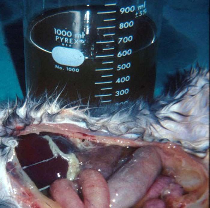
Following its appearance, FIP incidence steadily increased and it is currently one of the leading
infectious causes of death among young cats from shelters and catteries. The reason for the
sudden emergence of FIP is not known, but there are at least three possible explanations.
First, coronaviruses may have speciated into cats within the last half century. It is noteworthy
that FIP appeared within a decade of the initial descriptions of transmissible gastroenteritis
(TGE) of pigs in North America (Haelterman 1962). The causative virus of FIP is closely
related to TGEV of pigs and canine coronavirus (CCV) (Pedersen et al 1978), although they
are still genetically distinguishable (Motokawa et al 1996). However, recombinants between
these three viruses are known to occur ([Benetka et al 2006], [Herrewegh et al 1998],
[Pedersen et al 1984] and [Wesley 1999]). At least one strain of canine coronavirus can
induce mild enteritis in cats and enhance a subsequent infection with FIPV, indicating a
special closeness to feline coronaviruses (McArdle et al 1992). Therefore, CCV may be a
more likely parent of FECV in this scenario. A second explanation may involve a dramatic
but more subtle genetic change in the strains of FECV that predominated in the cat
population from the 1960’s onward. As will be explained below, the virus that causes FIP is
a simple mutation of the ubiquitous and not very noteworthy FECV. The strains of FECV that
existed before the appearance of FIP may have been resistant to the FIP mutation. A third
possibility involves changes in how cats were viewed as pets and their husbandry in this
modern era. There was a dramatic shift in the status, keeping, and breeding of cats as pets
after WWII. The numbers of pet cats greatly increased, purebreeding and cattery rearing
became increasingly popular, and more cats, and in particular kittens, found themselves in
shelters. These large multiple cat indoor environments are known to favor feline enteric
coronavirus (FECV) infection and FIP (reviewed by Pedersen et al 2008). Interestingly,
feline leukemia virus (FeLV) infection also became rampant among indoor multiple cat
households during this period, and FeLV infection was a significant cofactor for FIP until it
was pushed back into nature with testing, elimination/isolation, and eventual vaccination in
the 1970s and 1980s (Weijer et al 1986).

Causative agent

Feline infectious peritonitis virus (FIPV) is a mutant form (biotype) of FECV ([Pedersen et al
1981b], [Poland et al 1996] and [Vennema et al 1998]). Therefore, one cannot understand
FIP and FIPV without understanding FECV infection. FECV is a ubiquitous coronavirus
infection of cats. It is found worldwide and up to 40-80% of cats may be shedding the virus in
their feces at any given point in time (Reviewed by Pedersen et al 2008). The virus is
passed by the fecal-oral route and can survive for up to two weeks in litter dust. It is easily
passed from one premise to another on people’s bodies and clothing. FECV infection is
largely asymptomatic, causing a transient vomiting and mild diarrhea in a proportion of cats.
This enteric infection is rarely fatal in its own right. Cats may shed the virus for many years,

                                                     2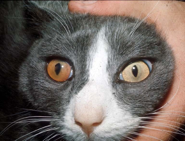
stop shedding after 6-9 months or so, or shed intermittently. Some cats that have recovered from the
infection can be reinfected, even with the same strain of FECV and their pattern of shedding
will resemble that of a primary infection.

The mutation responsible for the FIPV biotype is consistenly found in one of the 11 or more
genes that makeup the FECV genome, the 3c gene. The mutations are usually single
nucleotide polymorphisms (SNPs) or nucleotide deletions that inactivate all or parts of the
gene, thus preventing the production of a normal 3c protein. The specific deleterious
mutation in 3c is specific for each individual cat that develops FIP, even when affected
kittens are from the same litter (Vennema et al 1998; NC Pedersen, UC Davis, 2009,
manuscript in preparation). This explains why FIP cats do not seem to be infectious for other
cats. If cat-to-cat transmission existed, FIPV isolates from cats involved in the same
outbreaks would have identical 3c gene mutations. Loss of 3c gene function does not
prevent replication of the mutant (i.e., FIPV) in vivo or in vitro , but is thought to drastically
alter cell tropism by enhancing its internalization and replication in macrophages ([Dewerchin
et al 2005] and [Rottier et al 2005]). In contrast, the parent FECV preferentially infects a very
narrow cell type, the mature cells that are found at the tips of the intestinal vili (Pedersen et
al 1981b).

The FECV→FIPV mutation is more likely to occur during primary infection and in kittens,
because both conditions lead to a higher level of FECV replication (Pedersen et al 2008).
The higher the replication rate, the more likely mutations will occur. Very young cats also
have a decreased resistance to the mutation once it occurs (Pedersen et al. 2008). FIPV
mutants occurred in 20% of primary FECV infected cats in one study (Poland et al 1996).
However, FECV infection is often recurrent and there is some evidence that FIP may also
occur as a consequence of these recurrent bouts of virus replication. Addie et al (1995)
followed 56 cats deemed to have recurrent FECV infections based on decreasing and rising
antibody titers; 3 of these 56 animals subsequently developed FIP.

Epizootiology

FIP occurs wherever FECV is found ([Addie et al 1995], [Foley et al 1997a] and [Pedersen et
al 1981b, 1983a,b]) and is therefore worldwide and ubiquitous among virtually all cat
populations. FECV particularly endemic in multi-cat populations, such as catteries, shelters
and homes with numerous cats. It is made worse by a constant influx or new cats,
especially younger cats and kittens. FECV is also very prevalent among urban homeless
cats. One third of older cats and 90% of kittens and juveniles presented to shelters in
Sacramento, CA, USA were shedding FECV at the time of entry (Pedersen et al 2004). A

                                                       3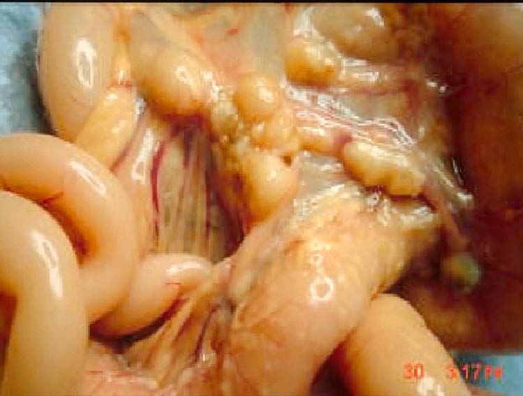
significant proportion of these cats were from the feral population, so it is fair to say that FECV is
enzootic among both indoor and outdoor cat populations. Similar findings have been
reported for most, but not all, other countries. Bell et al (2006a,b) found the seroprevalence
of FECV to be 34% among pedigree cats in the Sydney area and non-existent in feral cats
tested.

Extensive studies have been done on both natural ([Addie et al 2001], [Foley et al 1976b]
and [Pedersen et al 1981b]) and experimental FECV infections ([Pedersen et al 1981b] and
[Pedersen et al 2008]). Shedding has been shown to occur from the ileum, colon and rectum
(Herrewegh et al 1997). The same strain tends to persist in all cats within a given cattery or
region; however, an occasional cat in a group may be infected with more than one strain or a
different strain (Addie et al 2001). Virus shedding is somewhat associated with coronavirus
antibody titer levels; groups of cats with indirect fluorescent antibody titers of ≥1:100 or much
more likely to have coronavirus shedders than groups of cats with titers of≤1:25 (Pedersen
et al 2008).

Most deaths from FIP occur in cats 3-16 months of age and are uncommon after 3-5 years
([Pedersen 1976a] and [Pedersen 1983a]). FIP was the most common single cause of
disease in cats younger than two years of age in one study, while cancer was the most
common single disorder in the 2-8 year age range (Marioni-Henry et al 2004). The incidence
of FIP among a veterinary medical teaching hospital from 1986-1995 was 1:200 among new
feline visits, 1:300 among total cat accessions, and 1 of 100 accessions at diagnostic
laboratories (Rohrbach et al 2001). The incidence of FIP is significantly higher among kittens
and younger cats originating from pure breed catteries and shelters. An average of 1-5% of
young cattery or shelter cats in the US will die from FIP, with loses in catteries higher than
from shelters. Enzootics with >10% mortality were noted at least once in five years among
young cats and kittens originating from four typical catteries that were studied (Foley et al
1997a). Cave et al (2002) studied the causes of death among 274 sheltered and privately
owned pedigreed and non-pedigreed kittens from 1986-2000. Twenty-five per cent of all
kitten mortality was due to feline parvovirus (FPV). The main viral infections in 15-38 day
olds were feline herpesvirus and feline calicivirus. Feline infectious peritonitis caused the
death of 17/203 kittens in the postweaning (35-112 days of age) period, with an incidence of
8.4%.

FIP losses occur as enzootics or epizootics, with the former being more common. FIP losses
are sporadic, unpredictable and infrequent in the enzootic form; catteries with enzootic FIP
may not have any deaths for years, followed by several cases in rapid succession. The
disease may then disappear, only to reappear months or years later. Overall mortality from
enzootic disease over a 5 year or more periods is usually 1-5%. However, epizootics of 3-

                                                      4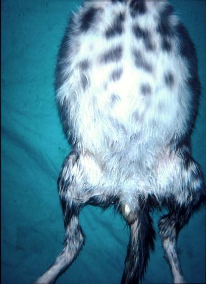
49% have been observed in groups of kittens raised in one cattery over a 4 year period and similar
explosive outbreaks have been seen in several other catteries ([Pedersen 1983a] and
[Potkay et al 1974]). Epizootics of FIP seldom last for more than 6-12 months before
returning to an enzootic state. Enzootic FIP is usually associated with persistence of the
same or similar strains of enteric coronaviruses within a population, while epizootics are
multifactorial and reflect increased population stresses, usually associated with overcrowding
and high kitten production; the unintentional use of genetically predisposed breeders;
possible bouts of horizontal transmission, or the introduction of a new strain of FECV.

Several risk factors for FIP have been identified in catteries (Foley et al 1997a). FIP in this
study was not significantly associated with particular catteries, mean cat number, mean age,
gender, cattery median coronavirus antibody titer, husbandry and quarantine practices,
caging and breeding practices, or prevalence of concurrent diseases. However, individual
cat age, individual cat coronavirus titer, overall frequency of fecal coronarvirus shedding, and
the proportion of cats in the cattery that were chronic FECV shedders, were associated with
increased risk of FIP. Deaths from FIP were more frequent in fall and winter, and on the
basis of analysis of cattery records, the number of deaths varied yearly. Epizootics (> 10%
mortality rate) were reported at least once in 5 years among the 4 catteries studied. One of
the most significant factors appeared to be genetic susceptibility, which accounts for up to
50% of the incidence (Foley and Pedersen 1996). Multi-cat households that have not
personally experienced FIP loses, but which relocated or sold cats that later develop FIP,
were no more likely to experience FIP over a 6 year period than households that had
experienced no prior FIP loses or catteries that had lost cats to FIP (Addie et al 1995). Thus,
every cattery with enzootic FECV infection is at risk of losing cats from FIP if they breed
enough kittens over a long enough time. The risk of developing FIP appears to decrease
over periods of 3 years in groups of cats, indicating an increase in population resistance
(Addie et al 1995).

The incidence of FIP in shelters seems to be directly correlated with the numbers and
density of young cats and the length of time that they are held before adoption; greater and
longer being much worse (K. Hurley, UCDavis, unpublished findings, 2008). Reports from
the US and Europe indicate an increased risk for young cats, purebreds, and intact males
and a decreased risk in spayed females (Rohrbach et al 2001). A report of 42 confirmed FIP
cases from Australia between 1990 and 2002 has shown FIP to be over-represented in
certain pure breeds (Burmese, Australian Mist, British Shorthair and Cornish Rex) and
under-represented in other breeds (Domestic shorthair, Persian) and a more even
distribution across the age spectrum (Norris et al 2005). The breed incidence of another
veterinary teaching hospital was determined over a 16-year period and the breed, gender

                                                     5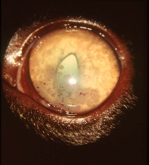
and reproductive status of affected cats were compared to the general cat population and to mixed
breed cats evaluated during the same period (Pesteanu-Somogyi et al 2006). As with
previous studies, sexually intact cats and purebreed cats were significantly more likely to be
diagnosed with FIP; males and young cats also had a higher prevalence of disease.
Abyssinians, Bengals, Birmans, Himalayans, Ragdolls and Rexes had a significantly higher
risk, whereas Burmese, Exotic Shorthairs, Manxes, Persians, Russian Blues and Siamese
cats were at decreased risk. Such studies indicate that the incidence of FIP among breeds
can vary greatly between countries and regions and FIP loses are probably more related to
bloodlines within a breed than to breeds themselves.

Clinical Features

Feline infectious peritonitis is mainly a disease of domestic cats. It has also been recognized
in the African lion, Mountain lion, Leopard, Cheetah, Jaguar, Lynx, Serval, Caracal,
European wild cat, Sand cat and Pallas cat ([Colby and Low 1970], [Colly 1973], [Fowler
1978], [Juan-Sallés et al 1998], [Pedersen 1983a], [Pfeifer et al 1983], [Poelma et al 1974],
[Theobold 1978], [Tuch et al 1974] and [Watt et al 1993]). An almost identical infectious
peritonitis has been described in interferon gamma deficient mice ([France et al 1999] and
[Kyuwa et al 1998a,b]) and in ferrets (Martinez et al 2008) infected with their respective
species of coronavirus.

Feline infectious peritonitis refers to the more common effusive (wet, non-parenchymatous)
form of the disease; a transmissible inflammatory condition of the visceral serosa and
omentum with exudation into the abdomen (Wolfe and Griesemer 1966). A second form of
the disease is characterized by granulomatous involvement of parenchymatous organs such
as the kidneys, mesenteric lymph nodes, bowel wall, liver, central nervous system and the
eyes ([Montali and Strandberg 1972] and [Pedersen 1976a]). Granulomatous FIP is called
"dry”, parenchymatous, or noneffusive because there is no inflammatory exudation into body
cavities.

The most common form of FIP is wet or effusive, although the proportion of cats with dry FIP
appears to be rising over the last few decades. Cats rarely manifest both forms of the
disease at the same time, and if they do, it is usually a transition stage from wet to dry or dry
to wet. Under experimental conditions, cats that develop dry FIP often have a brief episode
of effusive disease at the onset, while some cats with dry FIP may become more effusive in
the terminal stages of their illness. However, these transitions are much less noticeable in
nature.

                                                     6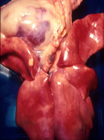
The incubation period (time from infection to disease) of effusive FIP is 2-14 days under experimental
conditions ([Evermann et al 1981], [Pedersen and Black 1983], [Pedersen and Floyd 1985]
and [Pedersen et al 1981a]). The incubation period for experimentally induced dry FIP is
several weeks longer. However, the actual incubation period in natural infections is
unknown, but there is evidence the infection may smolder in a subclinical state for weeks,
months and even years before overt signs are noticed. In retrospect, the clinical onset of FIP
may be preceded by a long history of vague ill health and stunted growth. Co-infections with
other feline pathogens may lower resistance to FIPV or may complicate the disease picture
(see proceeding discussion).

The earliest signs of overt FIP, besides a failure to thrive in young cats, include a
progressively worsening malaise, fluctuating fever, inappetance, and weight loss. Other
signs of disease are superimposed on these basic disease signs, depending on the form and
organ distribution of the inflammation (Table 1). Abdominal distension is the most common
physical finding in wet FIP and ranks higher than cardiovascular disease, neoplasia, hepatic
disease, and renal disease as causes of ascites in cats (Wright et al 1999) (Fig. 1). The
abdomen, besides being greatly enlarged, is often doughy feeling and painless on palpation,
and a fluid wave is easily induced on percussion. Upon opening, the abdomen is found to
contain up to a liter of a yellow-tinged, slight to moderately cloudy, mucinous fluid (Fig. 2).
Dyspnea can be a feature of cats with pleural involvement and thoracic effusions (Table 1).
Clinical signs due to ocular and central nervous system involvement are seen in less than
9% of cat with the wet form of FIP (Table 1).

There are several uncommon features of effusive FIP that warrant mention. Intact males
frequently develop scrotal enlargement due to extension of the peritonitis to the tunics
surrounding the testes and edema (Fig 1). A syndrome of hepatic lipidosis and extreme skin
fragility has been described in one cat with wet FIP (Trotman et al 2007). In-utero FIPV
infections have been observed in kittens born to queens that developed effusive FIP during
pregnancy; pneumonia, pleuritis and hepatitis were the principal lesions in affected kittens
(McKiernan et al 1981). Many cats with FIP have a generalized inflammation of the joint
linings (synovitis), due to immune complexing or the migration of infected
macrophage/monocytes into the synovial membrane. A cat with FIP may present, therefore,
with signs of fever and lameness. This can be mistaken for another type of infection or an
immune mediated polyarthritis. However, the more classical signs of FIP usually develop
soon after, making the cause of the lameness apparent.

As the name “dry FIP” implies, thoracic and abdominal effusions are either absent or too
scant to be detected other than at necropsy. Involvement of the eyes and/or CNS
predominates in 60% of the cats with dry FIP (Table 2). Signs referable to abdominal

                                                      7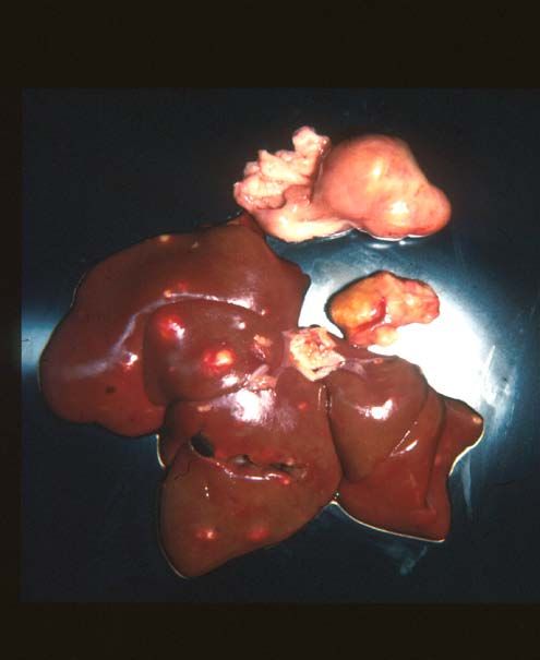
Table 1. Variability in clinical signs of effusive (wet) FIP
Clinical signs referable to
Involvement of the: % of affected cats
Peritoneal cavity 58.0
Peritoneal and pleural cavity 22.0
Pleural cavity 11.0
Peritoneal cavity and eyes 2.8
Peritoneal cavity and CNS 1.9
Peritoneal and pleural cavity, CNS 0.9
Peritoneal and pleural cavity, eyes 0.9
Pleural cavity, CNS and eyes 0.9
Peritoneal cavity, CNS, eyes 0.9
involvement are seen in 40% of animals, either with or without ocular and CNS disease
(Table 2). The abdominal lesions of dry FIP are much larger, fewer in number and less
widespread than the lesions of wet FIP. Lesions of dry FIP tend to extend downward from
the serosal or pleural surfaces into underlying parenchyma; hence the alternative name
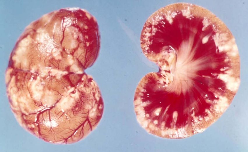
“parenchymatous FIP.” Abdominal lesions are frequently found in the kidneys (Fig. 3) and
mesenteric lymph nodes (Fig. 4, 5), and somewhat less frequently in the liver and hepatic
lymph nodes. Involvement of the wall of the caecum and colon with associated caeco-colic
lymphadenopathy is a specific form of dry FIP (Fig. 6) associated with signs typical of an
ulcerative colitis (i.e., soft, blood and mucus laden stools) ([Harvey et al 1996] and
[VanKruiningen 1983]). Abdominal lesions are often evident on palpation and sometimes
associated with local pain. About 10% of cats with dry FIP will have thoracic involvement, but
chest lesions are usually localized and only one part of a more systemic infection. Small
granulomas may involve the pleura and underlying lung parenchyma (Fig. 7). Involvement of
the pericardium has been described, and can lead to fluid distention of the pericardial sac,
cardiac tamponade and heart failure (deMadron 1986).
Central nervous system involvement is frequent in cats with dry FIP (Table 2). Over one half
of cats with inflammatory disease of the CNS have FIP, as well as one sixth of the total
number of cats showing CNS signs from any cause (Bradshaw et al 2004). FIP is also the
most common disease of the spinal cord in cats less than 2 years of age and is one of the
three leading causes, along with lymphosarcoman and vertebral neoplasia, of spinal disease
among cats of all ages (Marioni-Henry et al 2004). Most cats with CNS FIP are less than 2
years of age and often originate from large multiple cat households (Foley et al 1998).
8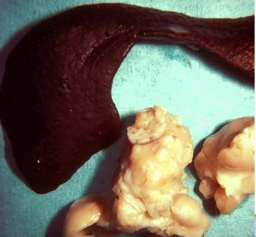
Central nervous system involvement in cats with dry FIP is varied in its clinical expression, dependent
on what other organs are involved (Table 2), its exact localization in the nervous system, and
severity. Signs referable to spinal cord involvement, such as posterior paresis,
incoordination, hyperesthesia, seizures and palsy of the brachial, trigeminal, facial and
sciatic nerves, have all been reported ([Holliday 1971], [Kornegay 1978], [Legendre and

Fig. 1. Grossly distended abdomen of a kitten with effusive feline infectious peritonitis. Note
the scrotal enlargement due to inflammation of the tunics.

                                                     9
Fig. 2. Over 600 ml of a yellow, mucinous effusion was removed from the abdomen at
necropsy. Note fibrin tags on liver and spleen and ground glass appearance of the serosa.
.
Fig. 3. Cross section of a kidney from a cat with dry FIP. Numerous granulomatous lesions
are seen on the capuslue of the kidney and extending downward into the parenchyma.
10Fig. 4. Enlarged mesenteric lymph node in a cat with the dry form of FIP. Note the residual fibrinous
plaque on the spleen. Such residual lesions support the concept that many cases of dry FIP
began as a brief bout of wet FIP.
Fig. 5. Mesenteric and hepatic lymph nodes and liver from a cat with nonefusive feline
infectious peritonitis. The lymph nodes are enlarged and involved with granulomatous
adenitis. The liver capsule contains raised, whitish foci 0.5-1 cm in diameter, extending into
the underlying parenchyma.
11Fig. 6. – Gross appearance of the caecum, colon and ceco-colic lymph nodes of a cat with
the intestinal form of dry FIP.
Fig. 7. Lungs and heart of above cat shown in Figure 5. A solitary pleural granuloma is
noted along the edge of a cranial lung lobe.
12Table 2. Variability in clinical signs of non-effusive (dry) FIP.
Clinical Signs Referable to
Involvement of: % of affected cats:
Peritoneal cavity 32.0
CNS 23.0
Eyes 15.0
CNS and eyes 8.5
Peritoneal cavity and eyes 7.4
Peritoneal and pleural cavities 4.3
Peritoneal and pleural cavities, CNS 3.2
Peritoneal and pleural cavities, eyes 2.1
Peritoneal cavity, CNS, eyes 2.1
Pleural cavity 1.1
Pleural cavity, CNS, eyes 1.1
Whitenack 1975], [Marioni-Henry et al 2005], [Pedersen 1976a]. [Slauson and Finn 1972],
[Quesnel et al 1997] and [Timmann et al 2007]). Hydrocephalus, secondary to disease of
the choroid and ependyma, has also been documented ([Fankhauser and Fatzer 1977],
[Hayashi et al 1980], [Krum et al 1975] and [Foley et al 1998]) and can lead to dementia,
personality changes (aggression, rage, hiding/withdrawal, etc.) or convulsive disorders.
Cerebellar-vestibular signs, such as nystagmus, head tilt or circling, have also been caused
by FIP.
Eye involvement, like CNS disease, is much more likely to occur in cats with dry than wet
FIP (Tables 1, 2). Uveitis and chorioretinitis are the predominant ocular manifestations of dry
FIP ([Campbell and Reed 1975], [Campbell and Schiessl 1978], [Doherty 1971], [Gelatt
1973], [Gillespie and Scott 1973] and [Slausen and Finn 1972]) (Figs 8, 9). FIP is also the
most frequent cause of uveitis/chorioretinitis in cats, with less common causes being FeLV-
associated lymphosarcoma; trauma; and lens-induced uveitis ([Goodhead 1996] and [Peffer
and Wilcock 1991]). Ocular disease in dry FIP occurs solely or in association with lesions in
the CNS or peritoneal cavity (Table 2). A change in the coloration of the iris is a frequent
early sign of ocular FIP (Fig. 8). Keratic precipitates on the back side of the cornea are
characteristic of FIP and are due to accumulations of fibrin, macrophages, and other
inflammatory cells (Fig. 9). Focal lesions akin to the granulomas of parenchymatous organs
may be apparent in the iris and distort the shape of the pupil (Figs. 8, 9).
13There are several uncommon, but interesting, manifestations of dry FIP. Granulomatous involvement
of the peripheral tissues resulted in priapism in a castrated cat (Rota et al 2007). Chronic
fibrinous and necrotizing orchitis may cause the scrotum and testicles to appear enlarged
([Foster et al 1996] and [Sigurdardóttir et al 2001]) and scrotal enlargement may be one of
the primary presenting complaints. Miscellaneous sites for lesions in noneffusive FIP include
the nasal passages, tongue and distal small intestine. Syringomyelia has been caused by
involvement of the fourth ventricle in one cat (Kitagawa et al 2007). Cutaneous lesions of a
granulomatous type have been reported in a FIV infected cat with FIP; the lesions were
thought to be due to a coronavirus induced vascultis (Cannon et al 2005). Toxic epidermal
necrolysis has also been observed ina cat with dry FIP ( NC Pedersen, UCDavis,
unpublished observation 2008). Non-puritic, slightly raised intradermal papules over the neck
and chest walls were observed in the terminal stages of a cat that had non-effusive,
progressing to effusive, FIP (Declercq et al 2008). FIPV was identified in skin lesions by
immunohistochemistry.
Fig. 8. Uveitis of the right eye in a cat with the dry form of FIP. The color of the iris has
changed, the anterior chamber is somewhat hazy, and there is a pigmented lesion in the
center of the cornea (a keratic precipitate). Note the irregularity in the shape of the right pupil
compared to the normal left pupil.
14Fig. 9. Keratic precipitates on the inner cornea of a cat with dry feline infectious peritonitis.
Note the reversed D-shape of pupil due to infiltration of the iris.
The lesions of dry FIP have also been mistaken for cancer, in particular lymphoma ([Kipar et
al 1999] and [Kornegay 1978]). One FeLV negative cat with FIP subsequently developed a
myeloproliferative disease (Madewell et al., 1978). Monoclonal gammopathies have been
observed in four cats with FIP (MacEwen and Hurvitz 1977) and a fifth cat with FIP was
observed to convert from a polyclonal- to monoclonal-gammopathy (Hurvitz 1982, as quoted
in Pedersen 1987).
FIP has many interesting interactions with other infectious agents. These agents may affect
immunity to FIPV, such as FeLV infecton. Alternatively, FIPV may induce an
immunosuppression that encourages opportunistic type pathogens. Concurrent FeLV
infection was seen in one-third to one-half of all FIP cases that were tested in the 1970s and
1980s ([Cotter et al 1973] and [Pedersen 1976a]). FeLV infection seems to have a specific
interfering effect on the ongoing immunity to FIPV. Cats that failed to develop FIP after
experimental infection developed FIP within 6-16 weeks after becoming FeLV viremic,
indicating that many FIP recovered cats maintain residual infections (Pedersen 1987a).
Mimicking the situation in the field, FIP also appears after natural exposure to both viruses
([Pedersen, 1987a] and [Pedersen et al 1977]). With the virtual elimination of FeLV from pet
cat populations in Westernized countries, this relationship has become much less common.
15Affected kittens with subclinical or clinical FIP are more susceptible to upper respiratory infections
caused by mycoplasma, Chlamydophila or herpesvirus, indicating that their resistance is not
normal. It is also known that cats with advanced feline immunodeficiency virus (FIV)
infection are more susceptible to FIP when exposed to FECV (Poland et al 1996). Systemic
toxoplasmosis, a relatively rare clinical infection of cats, has been described in two cats with
FIP. One report was from 1966 (Ward and Pedersen 1966) and a second report from 1995
(Toomey et al 1995). The occurrence of systemic toxoplasmosis in these two cats was
probably associated with reactivation of encysted organisms left from a much earlier self-
limiting infection. FIP is frequently associated with a lymphopenia, which is indicative of
immunosuppression. We have also noticed an increase in bacterial infections in cats with
FIP, some manifested as terminal sepsis or more chronic local infections of internal Organs
(NC Pedersen and P Pesavento, UCDavis, unpublished observations 2008). One young cat
had a chronic bacterial infection of the reproductive tract that was manifested early as a
purulent vaginal discharge and terminally as a supportive infection of the ovary and fallopian
tube, while a second cat had a secondary bacterial pneumonia. The stresses of pregnancy
and parturition, surgical procedures such as spaying, neutering and declawing have also
increased the incidence of FIP in young cats, suggesting that stresses occurring at a time
when the young cats are fighting an FIPV infection may tip the balance against the host
(Pedersen 1976a).
Hematologic, clinicopathologic and imaging Features
The diagnosis of FIP should be relatively simple, given its affinity for younger cats, its strong
tendency to involve catteries and shelters, the typical physical and historical findings, and
numerous characteristic laboratory abnormalities. Nonetheless, it somehow remains one of
the most difficult of diagnoses for many veterinarians. The truth is that veterinarians have
little trouble in placing FIP high, or at the top, of their diagnostic list, but have great difficulty,
and even reluctance, in confirming their diagnosis. However, it is not only the veterinarian
that has difficulties, but the owners as well are reluctant to give up without a definitive
diagnosis. It must be remembered that a diagnosis can be based on cumulative odds rather
than a single, simple, definitive test result. A young cat from a cattery or shelter with chronic
uveitis and/or neurologic signs, high serum proteins, hyperglobulinemia and
hypoalbuminemia, fluctuating antibiotic unresponsive fever, leukocytosis with a lymphopenia,
and an anemia of chronic disease can have no other disease than dry FIP based on odds
alone. Likewise, the same cat with similar history and laboratory findings, but with a yellow-
tinged, mucinous, inflammatory ascites instead of uveitis or neurologic signs is highly
unlikely to have any other disease than wet FIP. Results from a test that is incorrectly
touted as being definitive or highly indicative of FIP will only confuse the issue and induce
doubt when the diagnosis should not have been in doubt. Ultimately, FIP must be diagnosed
16by applying a workable knowledge of the disease with sensible weighing of signalment, history, clinical signs, clinicopathologic findings, serology and ante- or postmortem examination of affected tissues by histopathology and immunohistochemistry. Hemograms of cats with FIP often demonstrate several abnormalities, which tend to be similar regardless of the form of disease. A low-grade to moderately non-responsive anemia with hypoalbuminemia is typical of chronic disease. Other common hematologic changes include a leukocytosis with an absolute lymphopenia and neutrophilia, and hyperproteinaemia ([Paltrinieri et al 1998], [Paltrinieri et al 2001], [Pedersen 1976a] and [Sparkes et al 1991]). Serum total protein elevations are associated with increased levels of globulin and decreased levels of albumin. Cats with virus positive lymph nodes at necropsy were purportedly more likely to have globulin elevations (Paltrinieri et al 2001). Hypergammaglobulinemia is not directly related to feline coronavirus antibody titers (Paltrinieri et al 1998), suggesting that non-specific antibody globulins and/or complement factors are important contributors to the globulin elevation. An albumin globulin ratio
determined. A definitive diagnosis can be made by direct immunohistochemical examination of cells in
the fluid for viral antigens (Paltrinieri et al 1998,1999). Immunofluorescence is several times
more sensitive than immunoperoxidase. Real time RT-PCR analysis of cells from FIP
effusions will demonstrate high levels of viral RNA.

Hyperbilirubinemia, and less commonly hyperbiliverdinemia, frequently without jaundice, are
common in cats with FIP, especially the effusive form. In fact, FIP is the most common cause
of an icteric serum or plasma in a cat
between cats infected with FECV and FIPV, and although very high titers ( ≥1:1600) are highly
suggestive of FIP and negative titers tend to rule out FIP ([Hirschberger et al 1995] and
[Pedersen 1976c]), the overlap in titers between healthy coronavirus exposed and diseased
cats is so great that it has little definitive diagnostic value in individual animals ([Pedersen
1983a,b], [Paltrinieri et al 1998] and [Sparkes et al 1991]). Titers appear to rise
progressively in many cats as they go from a subclinical to clinical stage of disease
(Pedersen et al 1987a). However, progressive monitoring of titers is rarely done and titers
are usually measured when disease signs appear and titers levels have already plateaued.
Moreover, titers may also fall dramatically at the end, especially in cats with fulminating
effusive FIP.

The sensitivity and specificity of so-called FIP serology has been repeatedly questioned.
Hematology, antibody titers and serum protein electrophoresis from 48 cats (34 effusive and
14 noneffusive forms) affected with feline infectious peritonitis (FIP) were studied and
compared with those of 20 healthy cats (Sparkes et al 1991). In the effusive form, antibody
titers and protein electrophoresis in the effusions were analyzed. Seropositive animals
(antibody titer>1:100) were present among both the FIP infected (73%) and healthy cats
(70%). Sparkes et al (1991) also compared serologic data from 65 cats in which FIP was
considered as a differential diagnosis, but ended up having another disease. They found that
the presence of multiple abnormalities compatible with FIP increased the specificity (i.e.,
decreased false positives), but decreased the sensitivity of the diagnosis (i.e., increased
false negatives). However, there is no doubt that cats with very low or negative (≤1:25) feline
coronavirus antibody titers are less likely to have FIP, while cats with very high titers
(≥1:1600) are more likely. Given these limitations, this author recommends that FIP
serologic tests only be used as an aid in ruling in or out the possibility of FIP. The diagnosis
of FIP should never be made on antibody titers alone, a recommendation made by others
([Bell et al 2006b] and [Hartmann et al 2003]).

There have been many attempts to improve the specificity and sensitivity of antibody based
tests for FIP. One commercial test measures antibody titers to the 7b protein of feline
coronavirus based on the presumption that prototypic FECV (WSU-79-1683) lacks a 7b
protein, while the prototypic FIPV (WSU-79-1146) has an intact 7b protein. In theory, cats
with FIP should have antibodies to the 7b protein, while cats exposed to the common FECV
would not have such antibodies. Unfortunately, the lack of 7b protein in WSU-79-1683 is an
artifact of that specific isolate and field strains of FECV have an intact 7b gene (Herrewegh
et al 1995). Therefore, the 7b antibody test is no more specific or sensitive than the indirect
IFA test and this has been substantiated by published studies (Bell et al 2006b; Kennedy et
al 2008).

                                                      19
Because of the vagaries of FIP serology, FIP antibody testing should also not be used as a means to
control or eliminate FIP from catteries. Vast amounts of money are spent each year by
cattery owners on FIP testing. In almost all cases, the results are non-interpretable, even by
the veterinarians ordering the testing. Virtually all catteries having 6 to 8 cats and an active
breeding program will have FECV in their cattery and 50-80% or more of their animals
will have titers of 1:100 or greater (usually 1:25-1:1600). Cattery testing usually confirms
what is already known, that FECV is enzootic in the cattery. Antibody titers do not answer
the critical questions: 1) do any of the cat actually have FIP?, 2) are any of the cats
subclinically infected with FIPV, 3) will a particular cat develop FIP in the future?, and 4)
which cats are shedding FECV? Over- and mis-interpretation of various coronavirus
antibody tests result in considerable mortality from senseless euthanasia of healthy cats and
undue emotional and financial stress.
Serology has also been applied to the diagnosis of the neurologic form of FIP. Foley et al
(1998) observed what they believed to be specific feline coronavirus antibody production
within the CNS. They found that the most useful antemortem indicators of neurologic FIP
were positive IgG anti-coronavirus antibody titer in the cerebral spinal fluid (CSF), high
serum total protein concentration, and MRI findings suggesting periventricular contrast
enhancement, ventricular dilatation and hydrocephalus. The diagnostic value of positive anti-
coronavirus antibody titers in CSF was later questioned by Boettcher et al (2007). They
collected CSF from four clinical groups: 1) cats with FIP involving the CNS (n = 10); 2) cats
with FIP not involving the CNS (n=13); 3) cats with CNS disorders caused by diseases other
than FIP (n=29); and 4) cats with diseases other than FIP and not involving the CNS (n=15).
Cerebrospinal fluid was evaluated for concentrations of erythrocytes, leukocytes, and total
protein. Anti-coronavirus IgG was measured in CSF and serum by indirect
immunofluorescence assay. CSF IgG (1:32 to 1:4,096) was detected in 12 cats, including 6
cats with neurologic manifestation of FIP, 4 cats with FIP not involving the CNS, and 2 cats
with brain tumors. CSF IgG was detected only in cats with correspondingly high serum IgG
titers (1:4,096 -1:16,384) and significantly correlated with serum IgG titers. In another
attempt to measure local CNS antibody production in cats with FIP, Steinberg et al (2008)
used an albumin quotient and IgG index to determine whether proteins in the cerebrospinal
fluid were of blood or local origin. Neither the albumin quotient nor IgG index identified a
pattern consistent with intrathecal IgG synthesis in cats with the CNS form of FIP. The
conclusion of these various studies is that coronavirus antibodies will enter the CSF when
they are at very high levels in the serum; high serum titers are likely to be associated the dry
form of FIP (Pedersen, 1976a), most neurologic cases of FIP are of the dry type (Table 2);
therefore, positive feline coronavirus antibody titers in the CSF are likely to be associated
with FIP.
20Tests for the presence of FIPV RNA have been developed, but suffer from many of the same
weaknesses as serology. Some of the more popular PCR based tests are based on the lack
of the 7b gene in the FECV prototype WSU-79-1683 and its presence in the FIPV prototype
WSU-79-1146. As mentioned above, the lack of the 7b gene in this one isolate is an artifact,
probably of tissue culture adaptation. Therefore, PCR tests based on genetic differences
between WSU-79-1146 and WSU-79-1683 are invalid. Moreover, PCR tests are notoriously
susceptibility to laboratory contamination with the DNA products of amplification. Many
commercial laboratories do these procedures in an uncontrolled manner. In an attempt to
increase specificity and sensitivity, a PCR was developed that would only detect forms of the
viral RNA that were present during it replication stage (Simons et al 2004). The rationale was
that replicating forms of the viral RNA would only be found in the blood of cats with FIP. The
test was designed to amplify subgenomic mRNA of the highly conserved M gene. The test
was applied to 424 healthy and 651 cats suspected of having FIP. Almost one-half of the
diseased cats were positive for the replicating form of feline coronavirus mRNA in their
peripheral blood cells, whereas only 5% of healthy cats tested positive. Seventy five of 81
cats (93%) with post-mortem confirmed FIP tested positive, whereas 17 non-FIP cases all
tested negative. Such optimistic results were not found when the same test was applied to
another group of cats. In this study, 26 blood samples were collected from 25 healthy cats
and one cat with clinical signs suggestive of feline infectious peritonitis (FIP), namely, fever,
weight loss, enlarged abdomen, and ascites (Can-Sahna et al 2007). Blood samples were
then tested for replicating feline coronavirus messenger RNA by the procedure of Simons et
al (2004). Fourteen (54%) of the cats were positive for FCoV including the cat with clinical
disease, but a high rate of positivity was also observed among healthy cats , suggesting that
feline coronaviruses may be present in the blood samples from healthy cats as well as cats
with clinical FIP. This suggestion has been confirmed for healthy cats by others ([Kiss et al
2000] and [Meli et al 2004]). These conflicting findings call into question the value of PCR
for testing of blood. However, RT-PCR has accurately differentiated FIP effusions from
effusions of other causes (Hartmann et al 2003). The problem of laboratory contamination
with PCR products can be virtually eliminated by using a technique called real time PCR
(TaqMan).
Some clinicians and cattery owners will test for feline coronavirus in feces by PCR in an
attempt to identify FECV carriers within groups of cats and eliminate shedders from the
environment as a means to prevent FIP losses. FECV is shed at high levels in the feces, but
the carrier rate is 40-80% or higher in many multi-cat populations and shedding is
intermittent and infections recurrent (reviewed by Pedersen et al. 2008). PCR inhibitors have
been described in feces, so it is important to purify viral RNA using certain methods (Dye et
al. 2008). Such testing is expensive, and only a few laboratories can do it reliably on a
commercial basis. However, in the final analysis, it is virtually impossible to maintain a group
21of cats free of the virus, even if the infection can be eliminated, without strict quarantine facilities and
measures, as well restricting the movement of cats and people between cat populations (see
treatment and prevention).
Rivalta’s test has touted for the diagnosis of wet FIP (Hartmann et al 2003). A test tube is
filled with distilled water and one drop of 98% acetic acid is added, followed by a drop of the
peritoneal or pleural effusion. If the drop dissipates in the solution, the test is negative, and if
the drop retains its shape, the test is positive. A negative Rivalta’s test is reportedly 97%
accurate in ruling out FIP, while a positive test is 86% accurate in ruling in FIP. This author
sees no improvement in diagnostic value of this test over routine gross, microscopic, and
biochemical analysis of the fluid, but it is a simple and cheap supportive test.
Several indirect tests, usually based on the levels of certain inflammatory proteins or
byproducts (acute phase reactants), have been used to detect inflammatory conditions in
cats, and in particular FIP (reviewed by Paltrinieri 2008). Alpha-1-acid glycoprotein (AGP) is
an acute phase protein that increases in concentration in infectious and inflammatory
conditions. The serum and peritoneal fluid concentrations of AGP has been found useful in
the diagnosis of feline infectious peritonitis (FIP) ([Bence et al 2005] and [Saverio et al
2007]). AGP and amyloid A both increase a few hours after the inflammatory stimulus and
remain elevated for as long as the inflammation persists and have been evaluated in cats.
Serum AGP levels have also been used to study FIP in groups of cats (Paltrinieri et al 2007).
Serum AGP concentrations were observed to fluctuate over time in clinically healthy cats
from catteries with the highest prevalence of feline infectious peritonitis (FIP) and
significantly increased just before an outbreak of FIP. Although increased levels of AGP and
other inflammatory proteins are particularly common in cats with FIP (Saverio et al 2007),
they are not specific. Levels of AGP are usually high in cats with FIP and other inflammatory
diseases; moderate serum AGP levels (1.5-2 mg/ml) can discriminate cats with FIP from
other diseases that have a low pretest probability, while high serum AGP levels (>1.5- 3
mg/ml) are not usually seen in cats with diseases other than FIP ([Duthie et al 1997] and
[Saverio et al 2007]). However, the specificity of increased AGP levels has been questioned
by others (Duthie et al 1997). The potential value of raised levels of the acute phase
reactants, alpha 1-acid glycoprotein (AGP) and haptoglobin in the diagnosis of FIP was
examined in cats with confirmed FIP and in cats with other conditions. Levels of AGP greater
than 1.5 g/litre in serum, plasma or effusion samples were found to be of value in
distinguishing field cases of FIP from cats with similar clinical signs and differentiated these
two groups of cats more effectively than the albumin: globulin ratio. The concentration of
haptoglobin was higher in cats with FIP than in the group of healthy cats, but not sufficiently
to be of diagnostic value. Serum samples from feline immunodeficiency virus-infected cats
also had significantly elevated levels of AGP and haptoglobin, illustrating that raised levels of
22these inflammatory proteins are not pathognomonic for FIP. Modifications of AGP have also been
tested to increase its specificity (Ceciliani et al 2004). AGP in humans is heavily glycosylated
and undergoes several modifications of its glycan moiety during acute and chronic
inflammatory processes. Using human test modifications, feline AGP had very little L-fucose
residues on its surface and its branching degree was very low in normal and in several
pathological conditions. In contrast, feline AGP underwent several modifications during acute
FIP, including decreased expression of both alpha (2-6)-linked and alpha (2-3)-linked sialic
acid (76 and 44%, respectively) when compared to non-pathological feline AGP. The
possible role of some acute phase (inflammatory) proteins and immunoglobulins in both the
pathogenesis and diagnosis of feline infectious peritonitis (FIP) was also reported by
Giordano et al (2004). Serum protein electrophoresis and the concentration of haptoglobin
(Hp), serum amyloid A (SAA), alpha(1)-acid glycoprotein (AGP), IgG and IgM were
evaluated in healthy coronavirus exposed cats and cats with FIP. The highest concentration
of acute phase proteins was detected in affected cats, confirming the role of these proteins in
supporting a clinical diagnosis of FIP. Interestingly, healthy coronavirus exposed cats also
had increased acute phase proteins at the same time that members of the group developed
FIP appeared in the group. However, this increase persisted only in cats that developed FIP.
It is apparent from these various studies that levels of AGP, as well as other inflammatory
proteins, change dramatically in FIP, but that none of these changes are definitive in their
own right.
Magnetic resonance imaging (MRI) has proven useful in confirming the presence of
inflammatory neurologic disease indicated by CSF analysis (Negrin et al 2007).
Fourteen cats with inflammatory diseases affecting the CNS were reviewed, including eight
cats with FIP and and two cats with toxoplasmosis. Abnormalities affecting the CNS were
observed in MR images in 10/14 (71%) cats. Intracranial lesions appeared as slightly
hypointense foci in T1-weighted images in two (14%) cats, as hyperintense foci in T2-
weighted images in seven (50%) cats, and as hyperintense foci after intravenous
administration of a gadolinium-based contrast medium in 10 (71%) cats. In six cats with
lesions in T1- and/or T2-weighted images, additional lesions were visible in T1-weighted
images obtained after gadolinium-based contrast medium administration. In three cats,
lesions were visible only after contrast medium administration. MRI in this study did not
detect all cases of CNS inflammation in a population of cats with inflammatory cerebrospinal
fluid (CSF). However, it did add important information on the location of lesions, which can
be important in differentiating FIP from other inflammatory conditions. This substantiated the
conclusions of an earlier MRI study on cats with neurologic FIP (Foley et al 1998). Sixteen
domestic cats with confirmed neurologic FIP and 8 control cats with non-neurologic FIP were
studied pre- and ante-mortem. MRI imaging demonstrated periventricular contrast
enhancement, ventricular dilatation, and hydrocephalus in cats with neurologic FIP.
23The present gold standard for FIP diagnosis is immunohistochemistry on effusions or lesions
containing infected macrophages. Monoclonal or polyclonal antibodies that are highly feline
coronavirus specific, and that will react well with formalin fixed tissues, have been used
(Tammer et al 1995). Sections of lesions or cell pellets from ascetic or pleural fluids can be
directly examined for virus using fluorescein or horse radish peroxidase polyclonal or
monoclonal antibodies. Detection of coronavirus antigen in FIP effusions is very specific, but
less sensitive than detection of viral antigens in characteristic FIP parenchymal lesions.
Hirschberger et al (1995) identified antigen in 34/49 confirmed FIP effusions, whereas 50
effusions due to other causes were negative. The specificity of immunohistochemistry is a
factor of the poly- or mono-clonal antibodies used and the characteristic localization of FIPV
antigen within macrophages ([Paltrinieri et al 1998] and [Tammer et al 1997]). The sensitivity
of the test depends on having infected macrophages in the tissues or exudate cells on the
slides. For this reason, random biopsy of liver or kidney of cats with FIP often fail to yield
FIPV antigen by immunohistochemistry (Giordano et al 2005), and the same would be true
for real time PCR. Sensitivity is 5-10 times greater with fluorescein than horse radish
peroxidase staining, but the latter has the advantage of using formalin fixed and paraffin
embedded tissues, while the former requires frozen sections of tissues. Both can be used
with equal ease on slides of cells harvested from effusions and acetone fixed.
Virus isolation in tissue culture has not been yet possible for FECVs and is difficult for most
field strains of FIPV. Serotype II FIPVs seem to grow better in tissue culture, and will often
grow on both Crandell feline kidney (Crfk) and Felis catus whole fetus-4 (Fcwf-4 cells). The
latter cell line is of macrophage lineage Jacobse-Geels and Horzinek (1983) and is the
preferred cell line for the isolation of serotype I FIPVs ([Pedersen et al 1981a] and [Pedersen
and Floyd 1985]). The cost and low yield of cell culture isolation prohibits its routine
application to clinical diagnosis.
Following introduction of tests for detection of FeLV infection, one third or more of cats with
FIP were found to have concomitant FeLV infections (Cotter et al 1973, 1975). With
elimination of FeLV from many catteries and pet cat households, and the steady decline in
the incidence of FeLV in the entire cat population, the proportion of cats with FIP and
concurrent FeLV infections has greatly decreased. At the present, virtually all cases of FIP in
purebred cattery-bred cats are FeLV negative, and FeLV infection is detected in 10% or less
of domestic pet cats with FIP.
24Treatment and Prevention
It must be stated at the onset that no treatment has proven effective in curing cats of FIP, in
spite of the claims. Cats that develop FIP inevitably die of their disease in days, weeks or
months. The reason for these numerous false claims is uncertain, but spontaneous
remissions may account for at least some reports and misdiagnosis for the remainder. Cats
with ocular signs and no other systemic manifestations of FIP have occasionally gone into
remission with just symptomatic treatment. Cats with chronic fever, enlarged mesenteric
lymph nodes that were histologically compatible with FIP, and high coronavirus titers, have
gone into remission without treatment. Some cats without overt signs of FIP have
demonstrated fibrous lesions on the spleen and liver when necropsied for other reasons,
indicating a previous bout of FIP. Small quiescent lesions in the spleen and mesenteric
lymph nodes have also been found in otherwise healthy cats upon routine
ovariohysterectomies. Therefore, spontaneous remissions occur and at least some of these
natural responses may have fortuitously coincided with various treatments. Cures were first
reported with tylosin and prednisolone (Colgrove and Parker 1971), sparking a decade of
tylosin use for treatment of FIP. However, tylosin has no effect on FIP. Interestingly, a
significant proportion of cats with FIP are still treated with an antibiotic of one type or the
other. Some cats have gone into remission after use of prednisolone and phenylalanine
mustard or cyclophosphamide (Pedersen 1976a). Another cat was successfully “treated”
with prednisolone and phenylalanine mustard (Madewell et al 1978). No
immunosuppressive drug regimen has withstood the test of time.
A number of other equally dubious non-specific treatments have been used for FIP, almost
all with insufficient patient numbers, inadequate documentation of infection, or lack of
essential placebo controls and double blinding (Hartmann and Ritz 2008). FIPV is very
sensitive to human alpha and beta interferons in vitro (Weiss and Toivio-Kinnucan 1988).
Feline interferon omega also will inhibit FIPV in vitro and is commercially available in many
countries (Mochizuki et al 1994). Feline interferon omega reportedly induced complete or
partial remissions in two thirds of cats with FIP (Ishida et al 2004). However, in a larger and
double blinded study, this treatment was found to be totally ineffective (Ritz et al 2007).
Various immunosuppressants such as glucocorticoids and cyclophosphamide have been
used, but these drugs may prolong life but do not alter the fatal outcome (Hartmann and Ritz
2008). Immunostimulants, megadoses of vitamins, and numerous nutriceuticals have also
been advocated but found to be without merit. Pentoxyfiline, a tumor necrosis factor alpha
inhibitor (Zabel et al 1993), has been used on cats with FIP based on its benefit in treating
some types of human and feline vasculitis (Nichols et al 2001). TNF-alpha is upregulated in
FIP (Kiss et al 2004) and FIP is basically a vaculitis. However, this treatment has not proven
25beneficial on its own and has fallen from use. We have treated one FIP cat with feline IFN-γ and a
TNF-α inhibitor (pentoxfylline) with no beneficial affect.
Effective vaccines have been as elusive as effective treatments. Pedersen (1989)
hypothesized that the ideal FIPV vaccine should contain a live virus that would persist in the
body in a subclinical state, inducing a state of premonition immunity. However, a successful
vaccine against FIP has not been developed, even though most have been based on this
hyphothesis. Cats that survive infection with a progressively increasing dose of virus, starting
with sub-lethal levels, appear to develop a type of immunity (Pedersen and Black 1983).
However, this approach is not clinically applicable, because as many cats die as become
immune, and immunity appears to be tenuous. Some cats that appear to have resisted
disease have developed FIP months or years later, indicating the persistence of subclinical
infections. This finding was supported by a subsequent study by Baldwin and Scott (1997).
They first immunized cats intratracheally with a sublethal dose of virulent FIPV, followed by a
high dose of temperature attenuated virus. Cats demonstrated immunity to an aerosol
challenge with highly virulent FIPV but residual lesions were found upon necropsy
examination suggesting that immunity was either partial or of the premoniton type. Early
attempts to use an attenuated live FIPV strain, FIPV-Blackhigh passage, to induce immunity
failed to provide protection and even caused immune enhancement. A similar finding was
reported later for a virulence attenuated strain of FIPV-UCD1 (Kiss et al 2004). The
phenomenon of FIPV vaccine induced enchancement has been recently reviewed and is a
common them in many vaccine approaches (Huisman et al 2008).
A temperature sensitive mutant of FIPV-79-1146, administered intranasally, was later
developed and corporate studies showed a high degree of efficacy against challenge with
highly virulent FIPV-79-1146 ([Christianson et al 1989] and [Gerber et al 1990]). Immunity
was ascribed to a local IgA response and systemic cellular response measured by FIPV
induced lymphocyte proliferation. This vaccine is given as two intranasal doses, three or
more weeks apart, starting at 16 weeks or older. Presumably, efficacy could not be shown
for cats vaccinated and/or challenge-exposed at a younger age. This commercial vaccine
was studied in the field in a large single building shelter housing a thousand or more cats
([Postorino-Reeves et al 1992] and [Reeves et al 1992]). Five hundred FIV/FeLV negative,
feline coronavirus antibody negative cats were divided into two groups prior to being put into
this environment. One half got the intranasal live vaccine and one half was sham vaccinated.
The cats were than followed for 16 months. Overall deaths in both groups were the same.
However, two cats in the vaccinated group developed FIP during the 16 months and 8 non-
vaccinated cats succumbed to the disease. Protection in this study was not convincing and
the validity of using the vaccine only on coronavirus negative cats and measuring protection
over a limited period of time were questioned (Wolf 1997). Hoskins et al (1994) vaccinated
26You can also read