Advances in Clinical and Experimental Medicine
←
→
Page content transcription
If your browser does not render page correctly, please read the page content below
Advances
in Clinical and Experimental
Medicine

MONTHLY ISSN 1899-5276 (PRINT) ISSN 2451-2680 (ONLINE)   www.advances.umed.wroc.pl

2020, Vol. 29, No. 2 (February)

Impact Factor (IF) – 1.227
Ministry of Science and Higher Education – 40 pts.
Index Copernicus (ICV) – 155.19 pts.
Advances
in Clinical and Experimental
                   Medicine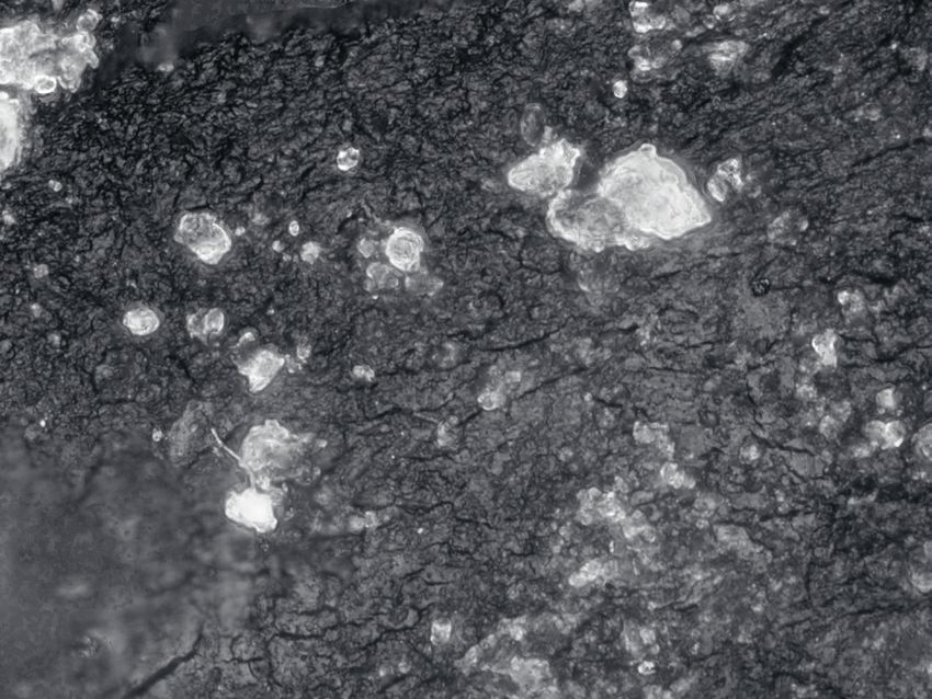
Advances
in Clinical and Experimental Medicine
ISSN 1899-5276 (PRINT)                                  ISSN 2451-2680 (ONLINE)                             www.advances.umed.wroc.pl

MONTHLY 2020                                            Advances in Clinical and Experimental Medicine is a peer-reviewed open access journal published
Vol. 29, No. 2                                          by Wroclaw Medical University. Its abbreviated title is Adv Clin Exp Med. Journal publishes original
                                                        papers and reviews encompassing all aspects of medicine, including molecular biology, biochemi-
(February)
                                                        stry, genetics, biotechnology, and other areas. It is published monthly, one volume per year.

                                                        Editor-in-Chief                                      Secretary
Editorial Office
                                                        Maciej Bagłaj                                        Katarzyna Neubauer
ul. Marcinkowskiego 2–6
50-368 Wrocław, Poland                                  Vice-Editor-in-Chief
Tel.: +48 71 784 11 36                                  Dorota Frydecka
E-mail: redakcja@umed.wroc.pl
                                                        Editorial Board                                      Piotr Ponikowski
                                                        Piotr Dzięgiel                                       Marek Sąsiadek
Publisher                                               Marian Klinger                                       Leszek Szenborn
Wroclaw Medical University                              Halina Milnerowicz                                   Jacek Szepietowski
Wybrzeże L. Pasteura 1                                  Jerzy Mozrzymas
50-367 Wrocław, Poland
                                                        Thematic Editors                                    Statistical Editors
© Copyright by Wroclaw Medical University,             Marzenna Bartoszewicz (microbiology)                Dorota Diakowska
   Wrocław 2020                                         Marzena Dominiak (dentistry)                        Leszek Noga
                                                        Paweł Domosławski (surgery)                         Lesław Rusiecki
Online edition is the original version of the journal   Maria Ejma (neurology)                              Technical Editorship
                                                        Jacek Gajek (cardiology)                            Joanna Gudarowska
                                                        Mariusz Kusztal                                     Paulina Kunicka
                                                           (nephrology and transplantology)                 Marek Misiak
                                                        Rafał Matkowski (oncology)
                                                        Ewa Milnerowicz-Nabzdyk (gynecology)                English Language Copy Editors
                                                        Katarzyna Neubauer (gastroenterology)               Eric Hilton
                                                        Marcin Ruciński (basic sciences)                    Sherill Howard Pociecha
                                                        Robert Śmigiel (pediatrics)                         Jason Schock
                                                        Paweł Tabakow (experimental medicine)               Marcin Tereszewski
                                                        Anna Wiela-Hojeńska
                                                           (pharmaceutical sciences)
                                                        Dariusz Wołowiec (internal medicine)

                                                        International Advisory Board                         Pavel Kopel (Czech Republic)
                                                        Reinhard Berner (Germany)                            Tomasz B. Owczarek (USA)
                                                        Vladimir Bobek (Czech Republic)                      Ivan Rychlík (Czech Republic)
                                                        Marcin Czyz (UK)                                     Anton Sculean (Switzerland)
                                                        Buddhadeb Dawn (USA)                                 Andriy B. Zimenkovsky (Ukraine)
                                                        Kishore Kumar Jella (USA)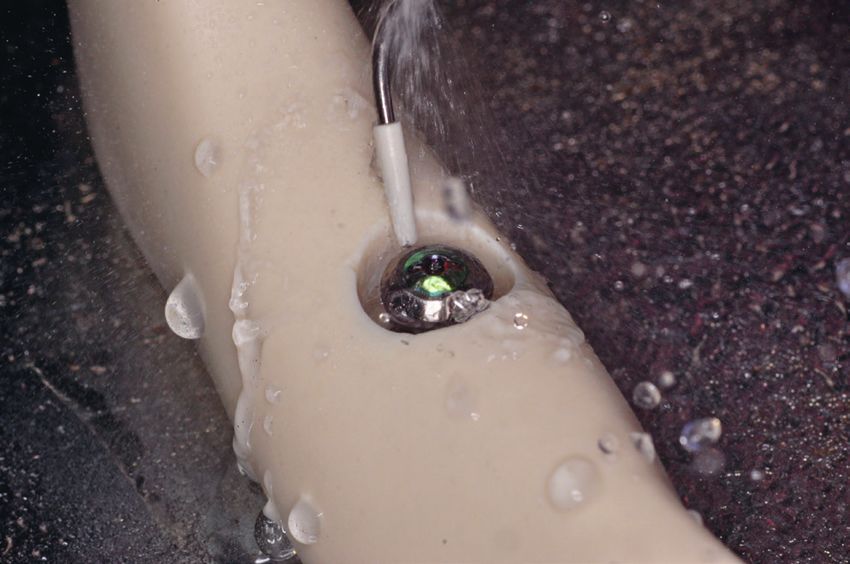
Editorial Policy
                            Advances in Clinical and Experimental Medicine (Adv Clin Exp Med) is an independent multidisciplinary forum for exchange of scientific and clinical information,
                            publishing original research and news encompassing all aspects of medicine, including molecular biology, biochemistry, genetics, biotechnology and other areas.
                            During the review process, the Editorial Board conforms to the “Uniform Requirements for Manuscripts Submitted to Biomedical Journals: Writing and Editing for
                            Biomedical Publication” approved by the International Committee of Medical Journal Editors (www.ICMJE.org/). The journal publishes (in English only) original
                            papers and reviews. Short works considered original, novel and significant are given priority. Experimental studies must include a statement that the experimental
                            protocol and informed consent procedure were in compliance with the Helsinki Convention and were approved by an ethics committee.

                            For all subscription-related queries please contact our Editorial Office:
                            redakcja@umed.wroc.pl

                            For more information visit the journal’s website:
                            www.advances.umed.wroc.pl

                            Pursuant to the ordinance No. 134/XV R/2017 of the Rector of Wroclaw Medical University (as of December 28, 2017) from January 1, 2018 authors are required
                            to pay a fee amounting to 700 euros for each manuscript accepted for publication in the journal Advances in Clinical and Experimental Medicine.

                            „Podniesienie poziomu naukowego i poziomu umiędzynarodowienia wydawanych czasopism naukowych oraz upowszechniania informacji o wynikach badań
                            naukowych lub prac rozwojowych – zadanie finansowane w ramach umowy 784/p-DUN/2017 ze środków Ministra Nauki i Szkolnictwa Wyższego
                            przeznaczonych na działalność upowszechniającą naukę”.
                                          Ministry of Science
                                          and Higher Education
                                          Republic of Poland

                                                                          Ministry
                  Indexed in: MEDLINE, Science Citation Index of
                                                              Expanded,
                                                                  Science Journal Citation Reports/Science Edition, Scopus,
 inistry of Science
                  EMBASE/Excerpta  Medica,  Ulrich’sTM        and Higher
                                                       International Periodicals Directory, Index Copernicus
nd Higher Education
 ublic of Poland
                                                                          Education
                                                                          Republic of Poland

                            Typographic design: Monika Kolęda, Piotr GilMinistry of Science
                   Ministry of Science
                        DTP: Wydawnictwo
                   and Higher  Education UMW                             and Higher Education
                                                                         Republic of Poland
                            Cover: Monika Kolęda
                   Republic of Poland

                            Printing and binding: EXDRUK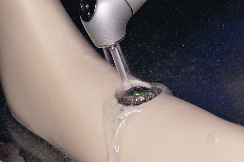
Advances
in Clinical and Experimental Medicine                                                                                          ISSN 1899-5276 (PRINT)
                                                                                                                              ISSN 2451-2680 (ONLINE)
MONTHLY 2020, Vol. 29, No. 2 (February)                                                                                     www.advances.umed.wroc.pl

Contents
Original papers
177     Paweł Kubasiewicz-Ross, Małgorzata Fleischer, Artur Pitułaj, Jakub Hadzik, Izabela Nawrot-Hadzik, Olga Bortkiewicz,
        Marzena Dominiak, Kamil Jurczyszyn
        Evaluation of the three methods of bacterial decontamination on implants with three different surfaces
183     Łukasz Smoliński, Tomasz Litwin, Karolina Kruk, Marta Skowrońska, Iwona Kurkowska-Jastrzębska, Anna Członkowska
        Cerebrovascular reactivity and disease activity in relapsing-remitting multiple sclerosis
189     Karolina Stokfisz, Anna Ledakowicz-Polak, Maciej Zagórski, Sławomir Jander, Katarzyna Przybylak, Marzenna Zielińska
        The clinical utility of remote ischemic preconditioning in protecting against cardiac surgery-associated
        acute kidney injury: A pilot randomized clinical trial
197     Bożenna Dembowska-Bagińska, Anna Wakulińska, Iwona Daniluk, Joanna Teisseyre, Irena Jankowska, Piotr Czubkowski,
        Ryszard Grenda, Wioletta Jarmużek, Wiesława Grajkowska, Jagoda Małdyk, Piotr Kaliciński
        Non-Hodgkin lymphoma after liver and kidney transplantation in children. Experience from one center
203     Joanna Małgorzata Przepiórka-Kosińska, Joanna Bartosińska, Dorota Raczkiewicz, Iwona Bojar, Jakub Kosiński, Dorota Krasowska,
        Grażyna Chodorowska
        Serum concentration of osteopontin and interleukin 17 in psoriatic patients
209     Wojciech Wilkoński, Lidia Jamróz-Wilkońska, Szczepan Zapotoczny, Janusz Opiła, Jerzy Krupiński, Jolanta Pytko-Polończyk
        The effects of alternate irrigation of root canals with chelating agents and sodium hypochlorite on the effectiveness
        of smear layer removal
215     Andrzej B. Hendrich, Paulina Strugała, Anna Dudra, Alicja Z. Kucharska, Anna Sokół-Łętowska, Dorota Wojnicz, Agnieszka Cisowska,
        Zbigniew Sroka, Janina Gabrielska
        Microbiological, antioxidant and lipoxygenase-1 inhibitory activities of fruit extracts of chosen Rosaceae family species
225     Rafał Januszek, Artur Pawlik, Bartłomiej Staszczak, Magdalena Jędrychowska, Jerzy Bartuś, Jacek Legutko, Dariusz Dudek,
        Andrzej Surdacki, Stanisław Bartuś
        Age and gender differences in clinical outcomes of patients with heavy-calcified coronary artery lesions treated
        percutaneously with rotational atherectomy
235     Dominika Ligia Wcisło-Dziadecka, Beniamin Grabarek, Celina Kruszniewska-Rajs, Joanna Magdalena Gola,
        Klaudia Simka, Urszula Mazurek
        Analysis of the clinical response and changes in the expression of TNF-α and its TNFR1 and TNFR2 receptors
        in patients with psoriasis vulgaris treated with ustekinumab
243     Tomasz Ociepa, Wioletta Posio, Marcin Sawicki, Tomasz Urasiński
        CIMT does not identify early vascular changes in childhood acute lymphoblastic leukemia survivors
251     Paulina Czechowicz, Małgorzata Małodobra-Mazur, Arleta Lebioda, Anna Jonkisz, Tadeusz Dobosz, Robert Śmigiel
        Polymorphisms of the MTHFR gene in mothers of children with trisomy 21 (Down syndrome) in a Polish population

Reviews
257     Krzysztof Wytrychowski, Anna Hans-Wytrychowska, Paweł Piesiak, Marta Majewska-Pulsakowska, Krystyna Rożek-Piechura
        Pulmonary rehabilitation in interstitial lung diseases: A review of the literature
265     Andrzej Stawarski, Paweł Maleika
        Neuroendocrine tumors of the gastrointestinal tract and pancreas: Is it also a challenge for pediatricians?

© Copyright by Wroclaw Medical University, Wrocław 2020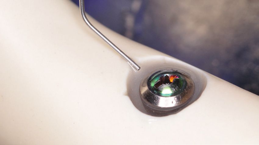
Original papers
Evaluation of the three methods of bacterial decontamination
on implants with three different surfaces
Paweł Kubasiewicz-Ross1,A,C,D, Małgorzata Fleischer2,B,C, Artur Pitułaj1,A–C, Jakub Hadzik1,A,D,F,
Izabela Nawrot-Hadzik3,E, Olga Bortkiewicz2,B,C, Marzena Dominiak1,F, Kamil Jurczyszyn1,A–C,E
1
Department of Oral Surgery, Wroclaw Medical University, Poland
2
Department of Microbiology, Wroclaw Medical University, Poland
3
Department of Biology and Pharmaceutical Botany, Wroclaw Medical University, Poland
A – research concept and design; B – collection and/or assembly of data; C – data analysis and interpretation;
D – writing the article; E – critical revision of the article; F – final approval of the article
Advances in Clinical and Experimental Medicine, ISSN 1899–5276 (print), ISSN 2451–2680 (online) Adv Clin Exp Med. 2020;29(2):177–182
Address for correspondence
Paweł Kubasiewicz-Ross
Abstract
E-mail: pawelkubasiewicz@wp.pl Background. The main goal of the treatment of the peri-implantitis is to decontaminate the surface
of the implant, thereby enabling further treatment involving, e.g., guided bone regeneration. Since new
Funding sources
None declared
implants of the rougher surface were introduced to the common dental practice, decontamination is even
more difficult.
Conflict of interest Objectives. The aim of the study was to evaluate 3 different methods of decontaminating implants with
None declared
3 different surfaces.
Material and methods. A total of 30 dental implants with 3 different surface types (machined, sandblasted,
Received on March 13, 2019 and acid-etched (SLA) and hydroxyapatite (HA)-coated) were used in the study. Each group of implants was
Reviewed on March 28, 2019
Accepted on September 25, 2019
coated with Escherichia coli biofilm and cultivated. Afterwards, the implants were transferred to the jaw model
and treated with a different method: sonic scaler mechanical debridement with a Woodpecker PT5 sonic
Published online on February 25, 2020 scaler (1st group), and mechanical debridement with sonic scaler and with the combination with chemical
agent Perisolv® (2nd group), and with Er:YAG laser treatment (3rd group). Each implant was treated with
the specific method and sent for further microbiological evaluation.
Results. The highest level of decontamination was achieved for machined-surface implants and the lowest
for HA-coated implants. The method with the highest biofilm reduction was the scaler and Perisolv® group.
The highest level of decontamination of HA-coated implants were achieved for Er:YAG laser irradiation method.
Conclusions. In the following paper, the superiority of combined chemical-mechanical method of decon-
taminating the surface of the implant on SLA and machined-surface implants was proved. On the contrary,
Er:YAG laser irradiation was reported as the best option for decontamination of the HA-coated implants.
In our opinion, it is a significant finding, revealing that the method of peri-implantitis management should be
Cite as considered in accordance to the type of the surface of the implant (customized to the surface of the implant).
Kubasiewicz-Ross P, Fleischer M, Pitułaj A, et al. E valuation
of the three methods of bacterial decontamination Key words: implant surface, implant surface treatment, bacterial coating
on implants with three different surfaces. Adv Clin Exp Med.
2020;29(2):177–182. doi:10.17219/acem/112606
DOI
10.17219/acem/112606
Copyright
© 2020 by Wroclaw Medical University
This is an article distributed under the terms of the
Creative Commons Attribution 3.0 Unported (CC BY 3.0)
(https://creativecommons.org/licenses/by/3.0/)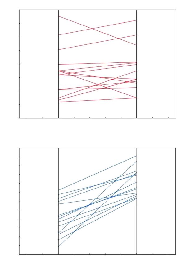
178                                                                                 P. Kubasiewicz-Ross et al. Decontamination methods of implants

Introduction                                                          metallic implants are coated with the bioactive compounds
                                                                      that accelerate bone formation or a rough surface is formed
   With the increasing number of patients treated with den-           directly on the metallic implants.11 Both techniques increase
tal implants, a corresponding number of post-treatment                the roughness of the surface of the implant, making os-
complications can be expected. The most common compli-                teointegration more favorable. However, as a result, it fa-
cation in dental implant therapy is peri-implantitis.1 It is de-      cilitates biofilm formation on dental implant surfaces.1,2,3,6
fined as an inflammatory reaction that affects the hard               To the best of our knowledge, there are very few studies
and soft tissue, which results in the loss of supporting bone         that evaluate various decontamination methods on different
and gingival pocket formation surrounding the function-               surfaces of the implants.
ing osseointegrated implant.2 This pathological condition
is caused by a polymicrobial aggressive biofilm that colo-
nizes the implant and abutment surface at the peri-implant            Material and methods
crevice level. It is reported that its prevalence can rise up
to 56%.1–3 Anaerobic Gram-negative organisms are most                   The study was conducted on a total number of 30 dental
commonly found in peri-implantitis-affected sites and in-             implants. Implants were divided into 3 equal groups with
clude in among others: Aggregatibacter actinomycetemcomi-             10 implants in each group. All the implants had the same
tans, Porphyromonas gingivalis, Peptostreptococcus micros,            length and diameter of L12Ø4 mm. The 1st group was ma-
Campylobacter rectus, Fusobacterium spp., and Prevotel-               chined-surface (M) implants (SGS Dental Implant System
la intermedia, although there are also studies reporting              Holding – Zn, St. Gallen, Switzerland). In the 2nd group,
the role of enteric rods (mostly Escherichia coli and Entero-         Denium Superline II (Dentium, Seoul, South Korea) sand-
bacter cloace) in this pathology, especially at its early stage.4,5   blasted and acid-etched dental implants (SLA) were used.
   Because of its complexity, peri-implantitis is still chal-         The 2rd group (HA) included the hydroxyapatite (HA)-coated
lenging to treat. Treatment involves decontamination and              dental implants (SGS Dental Implant System Holding – Zn).
guided bone and tissue regeneration techniques. The decon-
tamination process is especially difficult because the meth-          Bacterial cultivation
od applied can destroy the fragile surface of the implant.
For this purpose a number of mechanical interventions (e.g.,            Peri-implantitis is caused by Gram-negative and anaero-
abrasive air powder, Teflon and plastic curettes, ultrasonic          bic bacteria and E. coli were used as a model for Gram-
devices) and chemical agents (e.g., chlorhexidine, hydrogen           negative bacteria. The reasons we did so is that there are
peroxide) solely or in combination have been described                many studies involving bacterial adhesion and decontami-
as methods for implant surface decontamination. Although              nation carried out on dental implants with E. coli as bac-
all mentioned procedures result in compromise, a success-             teria of choice, as well as because it is a readily available
ful gold standard method has not been yet established.                and easily cultivated aerobic microorganism.
An acceptable cleaning technique must be able to debride
and detoxify the surface without traumatizing it. Decon-              Material
tamination with a laser, photodynamic therapy (PDT) and
the application of chlorhexidine (CHX) does not seem                    The McConkey’s medium (BioMaxima SA, Lublin,
to alter the surfaces of the dental implants. However, PDT            Poland); Sugar broth (BioMaxima SA); Saponin (Sigma-
can make an adhesive layer on the surface of the treated              Aldrich, St. Louis, USA); reference strain: E. coli ATCC
implants, which can facilitate new plaque formation.6,7               25922.
Recent studies have reported that lasers can also be used
in peri-implantitis management. Previously, high-power                Conduct of the experiment
CO2, diode and erbium lasers were used frequently, due
to their hemostatic properties, selective calculus ablation           Preparation of the inoculums
and bactericidal effects. However, high-power lasers can
cause an undesired increase of temperature and have been                The E. coli ATCC 25922 strain from McConkey’s medium
recently replaced by Er:YAG laser. Another disadvantage               was seeded into sugar broth and incubated at 37°C for 24 h.
of lasers is the high cost of equipment.8–10                          From the obtained culture in sugar broth, an inoculum with
   Dental implant surface decontamination has become even             a density of 0.5 on the McFarland Scale (MFa) was prepared.
more complicated since the introduction of dental implants
with improved osteoconductive properties. Machined-sur-               Implants coating
face implants, which have been used for decades, have been
replaced by implants characterized by a rougher surface.                The inoculum prepared in this way, in the amount
There are 2 main paths that can be followed in order to im-           of 500 µL, was inoculated with 50 mL of sugar broth. Then,
prove the osteoconductivity of the titanium implants. These           the implant was aseptically inserted and the whole was
approaches can be classified into the following 2 techniques:         incubated at 37°C for 24 h.
Adv Clin Exp Med. 2020;29(2):177–182 179
Preparation of implants for further tests
After this time, the implants were removed from the cul-
ture and rinsed 3 times, in each case, in 10 mL of sterile
saline to remove the plankton forms of the culture, leav-
ing only the biofilm formed by E. coli on the surface. Such
prepared implants were transferred to the Department
of Oral Surgery for further tests.
Model of the jaw
Before the decontamination process, each implant was
placed in peri-implantitis jaw model. The model was made
from acrylonitrile butadiene styrene (ABS) which is a com- Fig. 1. Sonic scaler mechanical debridement
mon thermoplastic polymer. According to the cumulative
interceptive supportive therapy (CIST) protocol,12 me-
chanical debridement and surgical operation classification
is needed when the bone loss depth is greater than 5 mm.
Following this standard, 6-millimiter bone loss depth was
defined in our model. The artificial bone defect was cre-
ated by removing of the material with the calibrated tre-
phine drill around the implant side.
Decontamination protocols
Every group of implants was decontaminated with 3 dif-
ferent methods. Before the decontamination process, each Fig. 2. Application of the Perisolv®
implant was placed in peri-implantitis model. Different
protocols of implant surface decontamination were used
in the study:
– Sonic scaler mechanical debridement with a Woodpecker
PT5 sonic scaler (Woodpecker, Guilin, China) (s). Each im-
plant was treated with a sonic device for 2 min alone (Fig. 1).
– Mechanical debridement with sonic scaler and with
the combination with chemical agent Perisolv® (Regedent
AG, Zurich, Switzerland). Each implant was pre-treated
with Perisolv® application for 30 s, then sonic scaler was
applied for 2 min (s+p) (Fig. 2).
– Er:YAG laser treatment. Implants were decontami-
nated with Er-YAG (LiteTouch™, Yokneam, Israel) laser
irradiation with a 1.3 × 17 mm tip, working up and down
continuously for 2 min, and the laser beam parameters
were set for 40 mJ, 0.80 W, 20 Hz (Er:YAG) (Fig. 3). Fig. 3. Er:YAG laser irradiation
Each implant was treated with the specific method and
sent for further microbiological evaluation. The procedure
for each implant was repeated 3 times and the results were Reax Control; Heidolph Instruments GmbH & CO. KG,
averaged. Schwabach, Germany). The obtained suspension of strains
(saponin solution and bacteria suspended in it, detached
Quantitative evaluation of microorganisms from the surface of the implant) was immediately cultured
present in the biofilm on the implants on McConkey’s medium. In the inoculation of bacteria,
undiluted suspension was used, and suspension with dilu-
surface tions from 1:10 to 1:1,000 inoculating volume: 10 L, 20 L,
Biofilm from the surface of the implants was removed 50 L, and 100 L. In order to obtain maximum separation
with the use of an aqueous saponin solution. The im- of the biofilm, the procedure of its removal was repeated
plants (each separately) were placed in 1 mL of 0.5% sapo- 3 times. Inoculated plates with McConkey’s medium were
nin solution and shaken for 1 min (2,500 rpm; Heidolph incubated at 37°C for 22–24 h.
180                                                                                         P. Kubasiewicz-Ross et al. Decontamination methods of implants

Reading the results                                                        120

                                                                           100
  After incubation, the colonies grown on the plates were
counted and the results obtained were given as the number                   80
of colony-forming units (CFU) per 1 mL. The percentage                                                                                                 M (S)

                                                                   R [%]
                                                                            60
of biofilm reduction R [%] after the tested factor of biofilm                                                                                          HA (S)
removal acted on was calculated according to the formula:                   40                                                                         SLA (S)

                 R = [(SC – S)/SC] • 100%,                                  20

  where SC (CFU/mL) – the total number of E. coli cells                         0
                                                                                    0       2          4          6      8        10         12
detached from the implant coating biofilm without the test                                                 No. of sample
factor acting (number of CFU/mL on the control implant);        Fig. 4. The results of scaler application on the decontamination
  S (CFU/mL) – the total number of E. coli cells detached       of the implant
from the implant coating biofilm, which remain after
the test factor acted.                                                  120
  In addition, to compare and reduce the measurement
                                                                        100
error, the degree of biofilm reduction was calculated after
the rejection of extreme values:                                           80

                R’ = [(S’C – S’)/S’C] • 100%,                   R [%]      60
                                                                                                                                                  M (S + P)
                                                                                                                                                  HA (S + P)
  where S’C (CFU/mL) – the total number of E. coli cells                   40                                                                     SLA (S + P)
detached from the implant coating biofilm without the test
                                                                           20
factor acting (number of CFU/mL on the control implant),
with no maximum or minimum value;                                           0
                                                                                0       2          4          6      8           10         12
  S’ (CFU/mL) – the total number of E. coli cells detached
                                                                                                       No. of sample
from the implant coating biofilm, which remain after
                                                                Fig. 5. The results of combined application of scaler and Perisolv
the test factor acted, with no maximum or minimum value.        application on the decontamination of the implant

Statistical analysis                                                    120

   Two-way analysis of variance (ANOVA) and Tukey’s post                100
hoc test were performed. All data is given as means ± stan-
                                                                           80
dard deviation (SD). A p-value
Adv Clin Exp Med. 2020;29(2):177–182 181
Table 1. Differences in the percentage of biofilm reduction between applied methods in relation to the implant surface
Biofilm M; M; Laser HA; HA; HA; Laser SLA; SLA; SLA; Laser
M; Scal
reduction Scal.+Perisolv Er:YAG Scaler Scal.+Perisolv Er:YAG Scal. Scal.+Perisolv Er:YAG
M;
98.7% 1.000182 P. Kubasiewicz-Ross et al. Decontamination methods of implants
studies in the field of decontamination which are similar 6. Saffarpour A, Nozari A, Fekrazad R, Saffarpour A, Heibati MN, Iran-
parvar K. Microstructural evaluation of contaminated implant surface
to our work on different surface implants. One of the men-
treated by laser, photodynamic therapy, and chlorhexidine 2 percent.
tioned studies was carried on SLA, TPS and HA implants. Int J Oral Maxillofac Implants. 2018;33(5):1019–1026.
After Er:YAG laser irradiation at pulse energies of 60 mJ 7. Mengel R, Buns CE, Mengel C, Flores-de-Jacoby L. An in vitro study
and 120 mJ and at a frequency of 10 pps led to bacterial of the treatment of implant surfaces with different instruments.
Int J Oral Maxillofac Implants. 1998;13(1):91–96.
reductions of 99.51% (SA), 98.39% (HA) and 99.6% (TPS) 8. Kuo HN, Mei HI, Liu TK, Liu TY, Lo LJ, Lin CL. In vitro laser treatment
at a pulse energy of 60 mJ, and 99.92% (SA), 99.85% (HA) platform construction with dental implant thread surface on bacte-
and 99.94% (TPS) at 120 mJ.20 rial adhesion for peri-implantitis. Biomed Res Int. 2017;2017:4732302.
9. Suzuki JB. Salvaging implants with an Nd:YAG Laser: A novel approach
to a growing problem. Compend Contin Educ Dent. 2015;36(10):756–761.
10. Arısan V, Karabuda ZC, Arıcı SV, Topçuoğlu N, Külekçi G. A randomized
Conclusions clinical trial of an adjunct diode laser application for the nonsurgical
treatment of peri-implantitis. Photomed Laser Surg. 2015;33(11):547–554.
11. Kuroda K, Okido M. Hydroxyapatite coating of titanium implants
The superiority of combined chemical-mechanical meth- using hydroprocessing and evaluation of their osteoconductivity.
od of decontaminating the surface of an implant on SLA Bioinorg Chem Appl. 2012;2012:730693.
and machined-surface implants was proved. On the con- 12. Shumaker ND, Metcalf BT, Toscano NT, Holtzclaw DJ. Periodontal
and periimplant maintenance: A critical factor in long-term treat-
trary, Er:YAG laser irradiation was reported as the best ment success. Comp Contin Educ Dent. 2009;30(7):388–390,392,394
option for decontaminating the HA-coated implants. passim; quiz 407,418.
13. Mellado-Valero A, Buitrago-Vera P, Solá-Ruiz MF, Ferrer-García JC.
Decontamination of dental implant surface in peri-implantitis treat-
ORCID iDs ment: A literature review. Med Oral Patol Oral Cir Bucal. 2013;1;18(6):
Paweł Kubasiewicz-Ross https://orcid.org/0000-0001-7305-7161 869–876.
Małgorzata Fleischer https://orcid.org/0000-0002-6610-3016 14. Subramani K, Wismeijer D. Decontamination of titanium implant
Artur Pitułaj https://orcid.org/0000-0002-9025-2628 surface and re-osseointegration to treat peri-implantitis: A litera-
Jakub Hadzik https://orcid.org/0000-0002-2353-3198 ture review. Int J Oral Maxillofac Implants. 2012;27(5):1043–1054.
Izabela Nawrot-Hadzik https://orcid.org/0000-0002-5797-7336 15. Meyle J. Mechanical, chemical and laser treatments of the implant
Olga Bortkiewicz https://orcid.org/0000-0001-6122-5359 surface in the presence of marginal bone loss around implants.
Marzena Dominiak https://orcid.org/0000-0001-8943-0549 Eur J Oral Implantol. 2012;5(Suppl):S71–81.
Kamil Jurczyszyn https://orcid.org/0000-0002-0667-7261 16. Blasi A, Iorio-Siciliano V, Pacenza C, Pomingi F, Matarasso S, Raspe
rini G. Biofilm removal from implants supported restoration using
different instruments: A 6-month comparative multicenter clinical
References study. Clin Oral Impl Res. 2016;27(2):e68–73.
1. Lindhe J, Meyle J. Peri-implant diseases: Consensus report of the sixth 17. Dennison DK, Huerzeler MB, Quinones C, Caffesse RG. Contaminated
European workshop on periodontology. J Clin Periodontol. 2008;35 implant surfaces: An in vitro comparison of implant surface coating
(8 Suppl):282–285. and treatment modalities for decontamination. J Periodontol. 1994;
2. Mahato N, Wu X, Wang L. Management of peri-implantitis: A system- 65(10):942–948.
atic review, 2010–2015. SpringerPlus. 2016;5:105. 18. Marotti J, Tortamano P, Cai S, Ribeiro MS, Franco JE, de Campos TT.
3. Khammissa RA, Feller L, Meyerov R, Lemmer J. Peri-implant mucosi- Decontamination of dental implant surfaces by means of photody-
tis and peri-implantitis: Bacterial infection. SADJ. 2012;67(2):70–74. namic therapy. Lasers Med Sci. 2013;28(1):303–309.
4. Medina CMA, Villa-Correa YA. Gram-negative enteric rods associat- 19. Eick S, Meier I, Spoerle F, et al. In vitro-activity of Er:YAG laser in com-
ed to early implant failure and peri-implantitis: Case report and sys- parison with other treatment modalities on biofilm ablation from
tematic literature review. Int J Odontostomat. 2015;9(2):329–336. implant and tooth surfaces. PLoS One. 2017;26;12(1):e0171086.
5. Leonhardt A, Dahlén G, Renvert S. Five-year clinical, microbiologi- 20. Kreisler M, Kohnen W, Marinello C, et al. Bactericidal effect of the
cal, and radiological outcome following treatment of peri-implan- Er:YAG laser on dental implant surfaces: An in vitro study. J Perio
titis in man. J Periodontol. 2003;74(10):1415–1422. dontol. 2002;73(11):1292–1298.Original papers
Cerebrovascular reactivity and disease activity
in relapsing-remitting multiple sclerosis
Łukasz SmolińskiA–F, Tomasz LitwinA–F, Karolina KrukB,F, Marta SkowrońskaA,B,F,
Iwona Kurkowska-JastrzębskaC,E,F, Anna CzłonkowskaA,C,E,F
Second Department of Neurology, Institute of Psychiatry and Neurology, Warszawa, Poland
A – research concept and design; B – collection and/or assembly of data; C – data analysis and interpretation;
D – writing the article; E – critical revision of the article; F – final approval of the article
Advances in Clinical and Experimental Medicine, ISSN 1899–5276 (print), ISSN 2451–2680 (online) Adv Clin Exp Med. 2020;29(2):183–188
Address for correspondence
Łukasz Smoliński
Abstract
E-mail: lsmolinski@ipin.edu.pl Background. In multiple sclerosis (MS), insufficient blood supply might worsen energy deficiency of the brain
tissue. Thus, cerebrovascular reactivity (CVR), which is the capacity of cerebral circulation to match blood
Funding sources
None declared
supply to metabolic demand, might be important in MS pathology.
Objectives. The objective of this study was to investigate the relationship of CVR to disease activity and
Conflict of interest neuroimaging markers of disease progression in patients with MS.
None declared
Material and methods. In 43 patients with relapsing remitting MS (RRMS) in clinical remission, 30 patients
with a relapse of MS and 30 healthy controls, we measured CVR with transcranial Doppler as a relative change
Received on February 25, 2019 in flow velocity after breath-holding (breath-holding index) and voluntary hyperventilation (hyperventilation
Reviewed on June 20, 2019
Accepted on November 25, 2019
index). All patients in remission underwent brain magnetic resonance imaging at baseline and 33 underwent
repeated imaging after 12 months, with various brain volume measurements taken.
Published online on February 19, 2020
Results. Cerebrovascular reactivity indices did not differ between patients in remission, patients with a relapse
and controls. In patients in remission, CVR did not differ between those with or without contrast-enhancing
lesions. In patients with a relapse, glucocorticoids significantly reduced both CVR indices. Cerebrovascular
reactivity was not related to brain volume, white matter lesion volume, percent brain volume change, and
the change in total white matter lesion volume.
Conclusions. In RRMS, CVR appeared normal and unrelated to disease activity. There was no substantial
association of CVR to brain atrophy and accumulation of white matter lesions.
Key words: multiple sclerosis, brain atrophy, cerebral blood flow, cerebrovascular reactivity, transcranial
Doppler ultrasonography
Cite as
Smoliński Ł, Litwin T, Kruk K, Skowrońska M, Kurkowska-
Jastrzębska I, Członkowska A. Cerebrovascular reactivity and
disease activity in relapsing-remitting multiple sclerosis.
Adv Clin Exp Med. 2020;29(2):183–188.
doi:10.17219/acem/114762
DOI
10.17219/acem/114762
Copyright
© 2020 by Wroclaw Medical University
This is an article distributed under the terms of the
Creative Commons Attribution 3.0 Unported (CC BY 3.0)
(https://creativecommons.org/licenses/by/3.0/)184 Ł. Smoliński et al. Cerebrovascular reactivity in MS
Introduction intravenous methylprednisolone (1 g for 3–5 days) due
to a relapse (an increase in Extended Disability Status
Multiple sclerosis (MS) is an inflammatory disease Scale (EDSS) score of at least 1 point), and 30 healthy
of the central nervous system, but neurodegenerative controls matched for age, sex and cardiovascular risk fac-
mechanisms are also implicated in the pathogenesis tors (Table 1). The study was approved by the Bioethics
of MS.1 For example, in MS, histotoxic hypoxia may cause Committee of our Institute, and all participants singed
energy deficiency of the brain tissues, which promotes informed consent before enrolment.
demyelination and axonal loss.2 Energy deficiency in MS
might be worsened by impaired regulation of cerebral Assessment of cerebrovascular reactivity
blood flow. Notably, many studies have found diffusely
reduced cerebral blood flow in patients with MS compared Cerebrovascular reactivity was assessed with transcrani-
with healthy age-matched controls.3 Moreover, evidence al Doppler ultrasonography (TCD) at around noon (11 AM
form magnetic resonance imaging (MRI) and histologic to 2 PM). We recorded blood flow velocity in the middle
studies shows that most white matter (WM) lesions in pa- cerebral artery with a 2-megaherz probe fixed with a head-
tients with MS are found in areas with the lowest blood band (DWL, Singen, Germany). Blood flow was monitored
perfusion, the so-called watershed zones.4–6 Similarly, in either the left or right middle cerebral artery, whichever
in an animal model of MS, demyelinating lesions tend had a better signal quality. The mean flow velocity (MFV)
to develop in areas with the worst blood supply.7 Thus, was calculated over 10–15 heart cycles. During TCD mea-
cerebrovascular reactivity (CVR), which is the capacity surements, we continuously recorded the end-tidal carbon
of cerebral circulation to match blood supply to metabolic dioxide concentration (EtCO2) with a capnometer (NMed,
demand, might be important in MS pathology. Cerebro- Beijing, China). The baseline MFV was recorded after about
vascular reactivity changes might be related to autonomic 5 min of resting. Then, we recorded the MFV after 2 min
dysfunction, which is common in MS. 8,9 Usually, CVR of hyperventilation (EtCO2 had to decrease by at least 10%
is estimated as a relative increase in cerebral flow after compared to baseline). After at least 3 min, when the MFV
increasing the systemic carbon dioxide concentration (CO2 and EtCO2 returned to baseline, we recorded the MFV after
inhalation, breath-hold).10,11 30 s of breath-hold. Cerebrovascular reactivity was estimat-
Reduced CVR may cause insufficient energy supply, par- ed with the breath-hold index (BHI) and a CO2-normalized
ticularly to areas with an increased energy demand, such hyperventilation index (HVΔCO2) as follows:
as damaged neurons or WM lesions infiltrated by meta- MFV after breat − hold − baseline MFV
bolically active immune cells.12 Cerebrovascular reactivity BHI = ;
baseline MFV × 30
impairment could amplify axonal loss and demyelination.
Indeed, during healthy aging, WM areas with the lowest HVΔCO2 =
CVR are most susceptible to demyelination.13 Similarly, baseline MFV − MFV after hyperventilation × 100%
= .
CVR impairment is associated with an increased risk of ce- baseline MFV × EtCO 2 change from baseline to hyperventilation
rebral ischemia.14
To date, few studies have investigated CVR in MS,15–17 Higher values of both BHI and HVΔCO2 indicated greater
and one group showed that CVR might be reduced CVR. In patients with a relapse, TCD measurements were
in MS.18,19 It remains unclear, however, whether clinical taken on the first and last day of intravenous glucocorti-
and neuroimaging disease activity is related to CVR in MS. coid treatment, before the first and after the last injection,
We also do not know whether low CVR increases the risks respectively. In patients in remission, TCD measurements
of brain atrophy and accumulation of demyelinating le- were taken within 1 h before MRI. Cerebrovascular reac-
sions. Because previous findings on CVR in MS are incon- tivity was assessed only once in patients in remission and
sistent and come from small studies, we checked whether controls. Atherosclerosis of the carotid arteries was ruled
CVR was impaired in MS in a larger group of patients. out by color Doppler ultrasound.
Moreover, we related CVR to clinical and neuroimaging
disease activity and longitudinal changes in brain volume Magnetic resonance imaging
and WM lesion volume. and image analysis
After the TCD study, patients in remission underwent
Material and methods brain MRI, as part of a routine clinical follow-up. With a 1.5 T
scanner (Philips, Eindhoven, the Netherlands), we acquired
Participants 3D T1-weighted images, before and after intravenous gado-
linium (Gd) injection, (TR, 25 ms; TE, 4.6 ms; field of view,
The study included 43 patients with RRMS in clinical 240 × 240 mm; voxel resolution, 0.937 × 0.937 × 1 mm) and
remission, for at least 3 months, who received interferon 2D FLAIR images (TR, 11,000 ms; TE, 140 ms; field of view,
beta in our clinic, 30 patients with RRMS who received 0.898 × 0.898 × 3 mm). The presence of Gd-enhancingAdv Clin Exp Med. 2020;29(2):183–188 185
(Gd(+)) lesions was assessed by an independent radiologist. treatment were compared with the dependent samples
Normalized brain volume (NBV) for head size, and normal- t-test. The Mann–Whitney U test was used to compare
ized volumes of grey matter (GM) and WM were measured CVR indices between patients with or without Gd(+) le-
based on the T1-weighted images with the SIENAX soft- sions. The Pearson coefficient or the Spearman coefficient
ware.20 The lesion-TOADS software was used to measure (rho) was calculated to study correlations between pairs
the total volume of WM lesions (TLV) based on the FLAIR of variables. A value of p < 0.05 was considered significant.
and T1-weighted images21; TLV was normalized for head All analyses were completed in the statistical package R
size based on scaling coefficients derived from SIENAX. (www.r-project.org).
Before running lesion-TOADS, we extracted brains from
whole-head images with the SPECTRE tool and registered
the T1-weighted images to the FLAIR images (rigid body Results
registration).21 During the study, 33 out of 43 patients
in remission had repeated MRI after 12 months (1 patient Clinical and imaging characteristics
entered secondary progressive MS, 3 patients were lost
to follow-up, 6 patients had less than 12 months of follow- There were no significant differences in age, proportion
up). For the 33 patients in remission who had follow-up of women, and the frequency of cardiovascular risk fac-
brain MRI after 12 months, we calculated the percentage tors between patients with MS in remission, patients with
brain volume change (PBVC), with the SIENA software, a relapse of MS and controls (Table 1). Among patients
and the TLV change, with lesion-TOADS.20 with MS in remission, the studied brain volumes corre-
lated negatively with EDSS, and disease duration correlated
Statistical analysis positively with TLV (Table 2).
Baseline anthropometric and clinical characteristics Cerebrovascular reactivity
were compared between the groups of participants with
one-way analysis of variance (ANOVA), the Mann–Whit- Neither of the 2 CVR indices, i.e., BHI and HVΔCO2, dif-
ney U test, and Fisher’s exact test, as appropriate. In pa- fered significantly between patients with MS in remission,
tients with a relapse of MS, the differences in CVR in- patients with a relapse of MS before intravenous glucocor-
dices before and after intravenous methylprednisolone ticoid treatment and controls (p = 0.56 for BHI; p = 0.1 for
Table 1. Clinical characteristics of patients with relapsing-remitting multiple sclerosis in remission, patients during a relapse and controls
Variable Controls (n = 30) Remission (n = 43) Relapse (n = 30) p-value
Age [years] 37.2 ±8.5 38.2 ±8.9 36.1 ±8.0 0.60a
Women, n (%) 23 (77) 33 (77) 24 (80) 0.96b
Disease duration [years] – 6.3 ±4.9 10.1 ±7.3 0.02c
Median EDSS (range) – 1.5 (0–6.0) 4.0 (2.0–4.0)186 Ł. Smoliński et al. Cerebrovascular reactivity in MS
Table 2. Correlations between cerebrovascular indices and clinical and imaging variables in patients with multiple sclerosis in remission (n = 43). Significant
correlations are in bold
Variable NBV GMV WMV TLV PBVC ∆TLV
r = –0.47 r = –0.59 r = –0.19 r = 0.25 r = 0.21 r = 0.18
Age
p = 0.001 p < 0.001 p = 0.22 p = 0.11 p = 0.25 p = 0.31
r = –0.29 r = –0.39 r = –0.09 r = 0.34 r = 0.01 r = 0.03
Disease duration
p = 0.06 p = 0.01 p = 0.58 p = 0.03 p = 0.99 p = 0.86
rho = –0.34 rho = –0.29 rho = –0.31 rho = –0.05 rho = 0.05 rho = 0.28
EDSS
p = 0.03 p = 0.06 p = 0.04 p = 0.73 p = 0.78 p = 0.11
r = –0.46 r = –0.53 r = –0.25 r = –0.16 r = 0.44
TLV –
p = 0.002 p < 0.001 p = 0.11 p = 0.38 p = 0.01
r = 0.21 r = 0.10 r = 0.10 r = –0.03 r = –0.12 r = –0.1
BHI
p = 0.17 p = 0.50 p = 0.50 p = 0.85 p = 0.52 p = 0.59
r = 0.15 r = 0.13 r = –0.03 r = –0.08 r = –0.21 r = 0.12
HVΔCO2
p = 0.33 p = 0.41 p = 0.86 p = 0.61 p = 0.25 p = 0.50
NBV – normalized brain volume; GMV – grey matter volume; WMV – white matter volume; TLV – total lesion volume; PBVC – percent brain volume change;
∆TLV – change in total lesion volume; EDSS – Extended Disability Status Scale; BHI – breath-hold indices; HVΔCO2, CO2 – normalized hyperventilation indices.
All brain volumes were measured in milliliters.
A B
2.0 7
1.8 p = 0.04
p = 0.04 6
1.6
1.4 5
1.2
4
HVΔCO2 [%]
BHI
1.0
3
0.8
0.6 2
0.4
1
0.2
0.0 0
controls MS remission MS relapse MS relapse controls MS remission MS relapse MS relapse
before steroids after steroids before steroids after steroids
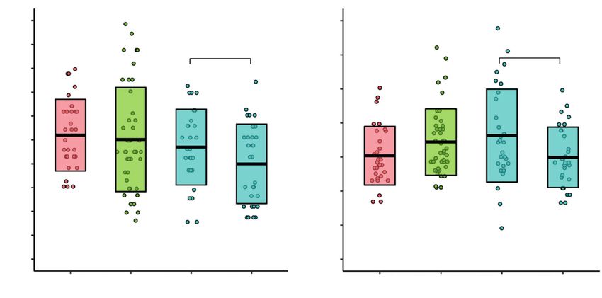
Fig. 1. (A) Breath-hold indices (BHI) and (B) CO2-normalized hyperventilation indices (HVΔCO2) in healthy controls (n = 30), patients with multiple sclerosis (MS)
in remission (n = 43), and patients with a relapse of MS before and after intravenous glucocorticoid treatment (n = 30). The middle bar represents the mean,
and the upper and lower bars represent standard deviations (SD)
HVΔCO2; Fig. 1). In patients with a relapse of MS, however, Discussion
both CVR indices decreased significantly after intravenous
glucocorticoid treatment (p = 0.04 for BHI and HVΔCO2; Our findings suggest that CVR is normal in RRMS and
Fig. 1). that it does not change during a relapse of MS or in patients
Among patients with remission, BHI and HVΔCO2 did with Gd(+) lesions. However, we found that treatment with
not differ between those with (n = 10) or without (n = 33) intravenous glucocorticoids reduced CVR in patients with
Gd(+) lesions (p = 0.20 for BHI, and p = 0.81 for HVΔCO2). a relapse of MS. In patients with MS in clinical remission,
Among patients in remission, neither of the 2 CVR indices CVR was not related to any of the brain volume measures,
correlated with the brain volumes studied, TLV, PBVC, including the longitudinal change in brain volume and
and TLV change (Table 2). Similarly, BHI and HVΔCO2 did WM lesion volume. Thus, it seems that there is no substan-
not correlate significantly with EDSS or disease duration tial relationship between CVR and diseases activity and
in patients in remission or relapse (data not shown). neuroimaging markers of disease progression in RRMS.Adv Clin Exp Med. 2020;29(2):183–188 187
Our findings are in line with those in most previous that we observed might be due to physiological variability
studies, which have reported normal CVR in patients with or becoming familiar with the procedure by participants.
MS. In the study by Uzuner et al. (n = 12), CVR measured Because CVR is a measure of cerebral metabolic reserve,
with TCD did not differ between patients with RRMS and we suspected that reduced CVR would be related to greater
controls. In contrast to our study, those investigators did brain atrophy and greater accumulation of WM lesions,
not find any significant effect of glucocorticoids on CVR.16 particularly because most WM lesions in MS occur in ar-
In another TCD-based study, Khorvash et al. reported that eas with reduced blood flow and reduced CVR.4–6,13 How-
CVR was higher in RRMS than in patients with migraines. ever, in our study, CVR was not related to brain volume
However, that study did not include healthy controls.15 reduction and the change in WM lesion volume.
Similar to our findings, Metzgen et al., who measured Our study had limitations. First, it included a relatively
CVR with blood oxygen level-dependent (BOLD) func- small group of patients. However, with over 70 patients with
tional MRI (fMRI) after CO2 inhalation, observed nor- RRMS, our study is the largest study on CVR in MS to date.
mal CVR in MS and did not find CVR to correlate with Additionally, the included number of patients allowed us
brain volume and WM lesion volume.17 Those investiga- to observe the well-established relationships in MS, such
tors showed that CVR was reduced in patients with MS as the correlation between EDSS, disease duration, and brain
and cognitive impairment, but this relationship is found volume. Because we did not include patients with progres-
in other diseases as well.22 To date, based on arterial spin sive MS, our findings may not hold true for these patients.
labeling (ASL) fMRI, only 1 group has reported reduced Second, we included patients with MS in remission who re-
CVR in MS. Moreover, that group found that CVR corre- ceived interferon beta only. Although the effect of interferon
lated negatively with WM lesion volume and GM atrophy.18 beta on CVR is unknown, a study among 5 patients with MS
ASL-based fMRI may be the best method to study CVR showed that interferon beta increased blood flow in the basal
impairment in MS; however, CVR measurements based ganglia.34 Moreover, interferon beta, similar to other disease-
on ASL and BOLD fMRI usually lead to similar conclu- modifying treatment, slows the rate of brain atrophy and
sions.23,24 Moreover, in Alzheimer’s disease, reduced CVR lesion accumulation in MS.35 Thus, the potential relationship
has been demonstrated with many techniques, including between CVR and brain atrophy along with lesion accumula-
BOLD, ASL and TCD.25 tion might be abolished by treatment with interferon beta.
Marshall et al. hypothesized that CVR might be reduced Third, some investigators regard breath-holding a less reli-
in MS due to habituation of cerebral vasculature to chroni- able hypercapnic stimulus than CO2 inhalation.36 Others,
cally increased nitric oxide concentrations.18 However, ni- however, have found these 2 stimuli equivalent for estimating
tric oxide seems essential for hypercapnia-induced cerebral CVR.37 Moreover, fMRI might be better than TCD for mea-
vasodilation,26 and consequently for CVR, and we were not suring CVR, but there is a good agreement between these
able to find any previous evidence that nitric oxide, when 2 approaches.38 In addition to breath-holding, we used vol-
chronically increased, like in MS,27 has an opposing effect.28,29 untary hyperventilation to measure CO2-normalized CVR.
In contrast, scavenging of nitric oxide by reactive oxygen spe- The relationships between CVR and other variables in our
cies (ROS) reduces vasodilation, which could occur in MS.30,31 study were consistent when assessed with the 2 CVR indices
Different effects of inflammation, such as increased (BHI, HVΔCO2). Apart from the use of 2 vasoactive stimuli
oxidative stress, might reduce CVR. For example, in pa- to measure CVR, the strengths of our study include enrol-
tients with diabetes, higher concentrations of inflamma- ment of patients in remission and a relapse of MS, measure-
tory markers were associated with reduced CVR.32 In our ment of CVR before and after glucocorticoid treatment, and
study, CVR was similar in patients with clinical remission longitudinal MRI analyses. We also measured CVR in all
and a relapse of MS, and Gd(+) lesions were not associated participants at the same time of the day, because CVR may
with reduced CVR. However, we did not measure inflam- decrease by over a third from morning to evening.29
matory markers in our study. We observed that treatment We conclude that CVR is normal and is not related
with glucocorticoids, which have anti-inflammatory ef- to disease activity in patients with RRMS. Moreover, CVR
fects, not only did not improve CVR, but significantly seems unrelated to the accumulation of WM lesions and
reduced it. Similarly, in patients with diabetes, reduced brain atrophy in these patients. It would be worthwhile
CVR was associated with increased concentrations of en- to verify our findings with fMRI-based CVR measure-
dogenous cortisol.32 We suspect that the glucocorticoid- ments, preferably in larger studies that would enroll pa-
induced reduction of CVR might be due to a direct effect tients with progressive MS.
of glucocorticoids on cerebral vessels. For instance, glu-
cocorticoids decrease endothelial synthesis of nitric oxide, ORCID iDs
and they increase the sensitivity of vascular smooth muscle Łukasz Smoliński https://orcid.org/0000-0003-1614-7069
cells to endogenous vasoconstrictions, such as norepineph- Tomasz Litwin https://orcid.org/0000-0003-2670-9651
rine.33 However, we measured CVR twice in patients with Karolina Kruk https://orcid.org/0000-0003-0149-5212
Marta Skowrońska https://orcid.org/0000-0002-0826-7821
a relapse only and not in those in remission or in con- Iwona Kurkowska-Jastrzębska https://orcid.org/0000-0001-6553-9080
trols. Therefore, the effect of glucocorticoids on CVR Anna Członkowska https://orcid.org/0000-0002-1956-1866188 Ł. Smoliński et al. Cerebrovascular reactivity in MS
References 20. Smith SM, Zhang Y, Jenkinson M, et al. Accurate, robust, and auto
mated longitudinal and cross-sectional brain change analysis. Neuro
1. Mahad DH, Trapp BD, Lassmann H. Pathological mechanisms in pro-
image. 2002;17(1):479–489. http://www.ncbi.nlm.nih.gov/pubmed/
gressive multiple sclerosis. Lancet Neurol. 2015;14(2):183–193. doi:10.
12482100. Accessed February 12, 2017.
1016/S1474-4422(14)70256-X
21. Shiee N, Bazin P-L, Ozturk A, Reich DS, Calabresi PA, Pham DL. A topo
2. Lassmann H. Multiple sclerosis pathology. Cold Spring Harb Perspect
logy-preserving approach to the segmentation of brain images with
Med. 2018;8(3):a028936. doi:10.1101/cshperspect.a028936
multiple sclerosis lesions. Neuroimage. 2010;49(2):1524–1535. doi:10.
3. D’haeseleer M, Hostenbach S, Peeters I, et al. Cerebral hypoperfu-
1016/j.neuroimage.2009.09.005
sion: A new pathophysiologic concept in multiple sclerosis? J Cereb
22. Catchlove SJ, Pipingas A, Hughes ME, Macpherson H. Magnetic res-
Blood Flow Metab. 2015;35(9):1406–1410. doi:10.1038/jcbfm.2015.131
onance imaging for assessment of cerebrovascular reactivity and its
4. Holland CM, Charil A, Csapo I, et al. The relationship between nor-
relationship to cognition: A systematic review. BMC Neurosci. 2018;
mal cerebral perfusion patterns and white matter lesion distribution
19(1):21. doi:10.1186/s12868-018-0421-4
in 1,249 patients with multiple sclerosis. J Neuroimaging. 2012;22(2):
23. Zhou Y, Rodgers ZB, Kuo AH. Cerebrovascular reactivity measured
129–136. doi:10.1111/j.1552-6569.2011.00585.x
with arterial spin labeling and blood oxygen level dependent tech-
5. Narayana PA, Zhou Y, Hasan KM, Datta S, Sun X, Wolinsky JS. Hypoper-
niques. Magn Reson Imaging. 2015;33(5):566–576. doi:10.1016/j.mri.
fusion and T1-hypointense lesions in white matter in multiple scle-
2015.02.018
rosis. Mult Scler. 2014;20(3):365–373. doi:10.1177/1352458513495936
24. Mandell DM, Han JS, Poublanc J, et al. Mapping cerebrovascular reac-
6. Haider L, Zrzavy T, Hametner S, et al. The topograpy of demyelin-
tivity using blood oxygen level-dependent MRI in patients with arterial
ation and neurodegeneration in the multiple sclerosis brain. Brain.
steno-occlusive disease: Comparison with arterial spin labeling MRI.
2016;139(Pt 3):807–815. doi:10.1093/brain/awv398
Stroke. 2008;39(7):2021–2028. doi:10.1161/STROKEAHA.107.506709
7. Desai RA, Davies AL, Tachrount M, et al. Cause and prevention of
25. Smoliński Ł, Członkowska A. Cerebral vasomotor reactivity in neu-
demyelination in a model multiple sclerosis lesion. Ann Neurol.
rodegenerative diseases. Neurol Neurochir Pol. 2016;50(6):455–462.
2016;79(4):591–604. doi:10.1002/ana.24607
doi:10.1016/j.pjnns.2016.07.011
8. Tantucci C, Bottini P, Fiorani C, et al. Cerebrovascular Reactivity and
26. Lavi S, Egbarya R, Lavi R, Jacob G. Role of nitric oxide in the regulation
Hypercapnic Respiratory Drive in Diabetic Autonomic Neuropathy. 2001.
of cerebral blood flow in humans chemoregulation versus mecha-
http://www.jap.org. Accessed June 2, 2019.
noregulation. Circulation. 2003;107(14):1901–1905.
9. Adamec I, Habek M. Autonomic dysfunction in multiple sclerosis. Clin
27. Smith KJ, Lassmann H. The role of nitric oxide in multiple sclerosis.
Neurol Neurosurg. 2013;115(Suppl 1):S73–S78. doi:10.1016/j.clineuro.
Lancet Neurol. 2002;1(4):232–241.
2013.09.026
28. Toda N, Ayajiki K, Okamura T. Cerebral blood flow regulation by nitric
10. Keage HAD, Churches OF, Kohler M, et al. Cerebrovascular function
oxide: Recent advances. Pharmacol Rev. 2009;61(1):62–97. doi:10.1124/
in aging and dementia: A systematic review of transcranial Dop-
pr.108.000547
pler studies. Dement Geriatr Cogn Dis Extra. 2012;2(1):258–270. doi:
29. Meadows GE, Kotajima F, Vazir A, et al. Overnight changes in the
10.1159/000339234
cerebral vascular response to isocapnic hypoxia and hypercapnia
11. Blair GW, Doubal FN, Thrippleton MJ, Marshall I, Wardlaw JM. Mag-
in healthy humans: Protection against stroke. Stroke. 2005;36(11):
netic resonance imaging for assessment of cerebrovascular reactiv-
2367–2372. doi:10.1161/01.STR.0000185923.49484.0f
ity in cerebral small vessel disease: A systematic review. J Cereb Blood
30. Sweazea KL, Lekic M, Walker BR. Comparison of mechanisms involved
Flow Metab. 2016;36(5):833–841. doi:10.1177/0271678X16631756
in impaired vascular reactivity between high sucrose and high fat diets
12. Campbell GR, Worrall JT, Mahad DJ. The central role of mitochondria
in rats. Nutr Metab (Lond). 2010;7(1):48. doi:10.1186/1743-7075-7-48
in axonal degeneration in multiple sclerosis. Mult Scler J. 2014;20(14):
31. Ohl K, Tenbrock K, Kipp M. Oxidative stress in multiple sclerosis:
1806–1813. doi:10.1177/1352458514544537
Central and peripheral mode of action. Exp Neurol. 2016;277:58–67.
13. Mandell DM, Han JS, Poublanc J, et al. Selective reduction of blood flow
doi:10.1016/j.expneurol.2015.11.010
to white matter during hypercapnia corresponds with leukoaraiosis.
32. Chung CC, Pimentel D, Jor’dan AJ, Hao Y, Milberg W, Novak V. Inflam-
Stroke. 2008;39(7):1993–1998. doi:10.1161/STROKEAHA.107.501692
mation-associated declines in cerebral vasoreactivity and cogni-
14. Silvestrini M, Vernieri F, Pasqualetti P, et al. Impaired cerebral vaso-
tion in type 2 diabetes. Neurology. 2015;85(5):450–458. doi:10.1212/
reactivity and risk of stroke in patients with asymptomatic carotid
WNL.0000000000001820
artery stenosis. JAMA. 2000;283(16):2122–2127. http://www.ncbi.nlm.
33. Yang S, Zhang L. Glucocorticoids and vascular reactivity. Curr Vasc
nih.gov/pubmed/10791504. Accessed August 6, 2018.
Pharmacol. 2004;2(1):1–12. http://www.ncbi.nlm.nih.gov/pubmed/
15. Khorvash F, Masaeli A, Shaygannejad V, Saadatnia M. Vasomotor reac-
15320828. Accessed June 23, 2018.
tivity comparison in multiple sclerosis patients with white matter
34. Mackowiak PA, Siegel E, Wasserman SS, Cameron E, Nessaiver MS,
lesions and nonmultiple sclerosis subjects with white matter lesions
Bever CC. Effects of IFN-β on human cerebral blood flow distribution.
in brain magnetic resonance imaging. Adv Biomed Res. 2016;5:23.
J Interf Cytokine Res. 1998;18(6):393–397. doi:10.1089/jir.1998.18.393
doi:10.4103/2277-9175.175916
35. Zivadinov R, Locatelli L, Cookfair D, et al. I nterferon beta-1a slows
16. Uzuner N, Ozkan S, Cinar N. Cerebrovascular reactivity in multiple
progression of brain atrophy in relapsing-remitting multiple scle-
sclerosis patients. Mult Scler J. 2007;13(6):737–741. doi:10.1177/
rosis predominantly by reducing gray matter atrophy. Mult Scler J.
1352458506074645
2007;13(4):490–501. doi:10.1177/1352458506070446
17. Metzger A, Le Bars E, Deverdun J, et al. Is impaired cerebral vaso-
36. Fierstra J, Sobczyk O, Battisti-Charbonney A, et al. Measuring cere-
reactivity an early marker of cognitive decline in multiple sclerosis
brovascular reactivity: What stimulus to use? J Physiol. 2013;591(23):
patients? Eur Radiol. 2018;28(3):1204–1214. doi:10.1007/s00330-017-
5809–5821. doi:10.1113/jphysiol.2013.259150
5068-5
37. Kastrup A, Krüger G, Neumann-Haefelin T, Moseley ME. Assessment of
18. Marshall O, Lu H, Brisset J-C, et al. Impaired cerebrovascular reactivity
cerebrovascular reactivity with functional magnetic resonance imag-
in multiple sclerosis. JAMA Neurol. 2014;71(10):1275–1281. doi:10.1001/
ing: Comparison of CO2 and breath holding. Magn Reson Imaging. 2001;
jamaneurol.2014.1668
19(1):13–20.
19. Marshall O, Chawla S, Lu H, Pape L, Ge Y. Cerebral blood flow modu-
38. Herrera CRC, Beltramini GC, Avelar WM, Lima FO, Li LM. Cerebral
lation insufficiency in brain networks in multiple sclerosis: A hyper-
vasomotor reactivity assessment using transcranial Doppler and
capnia MRI study. J Cereb Blood Flow Metab. 2016;36(12):2087–2095.
MRI with apnea test. Brazilian J Med Biol Res. 2016;49(11):e5437.
doi:10.1177/0271678X16654922You can also read