AN AUTOMATED SYSTEM FOR CLASSIFICATION OF DIABETIC RETINOPATHY USING FASTER-RCNN - IRJET
←
→
Page content transcription
If your browser does not render page correctly, please read the page content below
International Research Journal of Engineering and Technology (IRJET)                                         e-ISSN: 2395-0056
         Volume: 08 Issue: 06 | June 2021                   www.irjet.net                                             p-ISSN: 2395-0072

          AN AUTOMATED SYSTEM FOR CLASSIFICATION OF DIABETIC
                   RETINOPATHY USING FASTER-RCNN
            K. Sakthiyavathi1, S. Amutha2, A. Vigneshwari3, T. Devadharshini4, R. Kiruthiga5
    1Assistant    Professor, Department of Information Technology, Sri Manakula Vinayagar Engineering College,
                                                                Puducherry
     2Assistant Professor, Department of Information Technology, Sri Manakula Vinayagar Engineering College,

                                                                Puducherry
          3Department of Information Technology, Sri Manakula Vinayagar Engineering College, Puducherry
          4Department of Information Technology, Sri Manakula Vinayagar Engineering College, Puducherry
          5Department of Information Technology, Sri Manakula Vinayagar Engineering College, Puducherry

---------------------------------------------------------------------***----------------------------------------------------------------------
ABSTRACT: Diabetic Retinopathy is an eye disease caused as                and require trained ophthalmologists. The lack of skilled
a result of semipermanent polygenic disease. Because the                    clinicians also leaves a large proportion of patients untreated
disorder progresses it results in distortion and blurred vision.            and therefore receiving medical help too late, in part due to
The identification of DR stages color structure image needs                 poor adherence and access to the retina screening process.
dexterous clinicians to spot the presence of vital features that            However, early detection and prevention of DR progression
makes this a tough and time overwhelming task. As the DR                    are essential to reduce the rising threat of DR. Artificial
accompanies numerous stages and differing challenges, it's                  intelligence offers a better solution to this problem. Deep
tough to DR and it's tedious. Right now, build up a                         learning is used for an end-to-end assessment of medical
computerized division based mostly on order model for DR. At                images to generate a predicted output. The diagnostic use of
First, the original images are resized and green channels are               Fast RCNN algorithms is spreading in various medical
extracted from the structure pictures. Then, the Adaptive                   healthcare areas like radiology and pathology. In
Histogram Equalization (AHE) an image process technique is                  ophthalmology, groundbreaking work has recently been
employed to enhance the distinction of the image and enhance                conducted on the automation of DR detecting and prediction
the sides of the image. Later, The Faster R-CNN is utilized for             of various risk factors by Fast RCNN analysis of CFPs.
classifying the structure into totally different grades of DR.
This Faster R-CNN approach was found to be an efficient
algorithm concerning speed and accuracy. The approximate
accuracy of 89.5% was acquired from the Faster R-CNN.

Key Words: Fast Regional convolutional neural network
(FRCNN), Diabetic retinopathy, deep learning, segmentation

    I.   INTRODUCTION

Diabetic retinopathy (DR), a sight-threatening disease,
happens because of diabetics that bring about the harm of
                                                                            Diabetic Eye infection is mostly separated into non-
cells in the retina when the glucose level of the patient was
                                                                            proliferative (NPDR) and proliferative DR (PDR). In NPDR no
untypical. Around 29% of sugar patients with age above
                                                                            fresh blood vessels start in the retina, yet veins leak liquids
42years possess Diabetic eye disease, and among them, 4.3%
                                                                            or    blood-framing      Exudates,      hemorrhages,       and
have an extreme level of DR that leads to vision loss.
                                                                            microaneurysm. The size, shape, area, and appropriation of
Therefore, patients with diabetes are exposed to the danger
                                                                            those highlights show the development of DR. Non-
of this eye disease. If DR is untreated, blood & fluid leak from
                                                                            Proliferative DR is then separated as would be expected,
blood vessels of the retina leads to permanent vision loss.
                                                                            gentle, moderate, and extreme. In PDR close-off harmed
The underlying province of DR separating the clinical is
                                                                            veins cause the development of new strange vessels in the
finished by utilizing the procedure of fundus imaging the
                                                                            retina.
affected retinal structure of the eyes might be detected by
focusing the eye by a retina specialist or a trained grader.
But these methods are manual, which are time-consuming,

         © 2021, IRJET        |    Impact Factor value: 7.529           |     ISO 9001:2008 Certified Journal           |   Page 78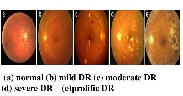
International Research Journal of Engineering and Technology (IRJET)                                e-ISSN: 2395-0056
        Volume: 08 Issue: 06 | June 2021              www.irjet.net                                         p-ISSN: 2395-0072

                                                                    highlights. At long last utilizing multiclass SVM and KNN
                                                                    classifier different unusual retinas are recognized. The
                                                                    preparation and testing are precisely ordered and its level of
                                                                    exactness is given in SVM classifier accomplish 85.60%
                                                                    precision and though KNN classifier accomplishes 55.17%
                                                                    exactness.

                                                                    In [11] Athira TR and Sivadas [2019] proposed an R-CNN
                    II. RELATED WORK
                                                                    (Regional Convolutional Neural Network) way to deal with
In [1] Nihel Zaabour, Alidouik [2020] proposed a strategy by        analyze DR from computerized fundus pictures. In our
utilizing fundus camera retinal pictures are gotten and were        exploration, we carried out another methodology where the
examined. The vein and the harmed territories are                   entire picture was portioned and just the areas of interest
distinguished and, in this way, hemorrhages are recognized          were taken for additional handling. Their strategy has
to group diabetic retinopathy. The request is done ward on          utilized 10 layers for R-CNN, prepared it on 130 fundus
the Random Forest system. For the ordinary instances of DR,         pictures, and tried it on 110 pictures. Every one of the
the announced exactness of order is 90% and for moderate            pictures was arranged into two gatherings with and without
and extreme it is 87.5%. Grouped DR as typical, non-                DR. This R-CNN (Regional Convolutional Neural Network)
proliferative (NPDR), and proliferative (PDR). The extraction       approach was discovered to be proficient as far as speed and
is finished by Histogram of Oriented Gradients (HOG) and by         precision. A precision of 93.8% was gotten from R-CNN.
factor examination the best component is chosen. Support
Vector Machine (SVM) and Random Forest learning are                 In [10] D. Sarwinda, T. Siswantining [2019] carried out
utilized to order various kinds of DR.                              identification utilizing a CNN calculation that has been
                                                                    generally perceived for applications, for example, picture
In [2] Harini R Sheela N [2016], creators examined a DR             preparing, design acknowledgment, and arrangement. It has
identification strategy utilizing FCM grouping and                  a few secret layers wherein the primary convolution layer is
morphological picture preparing. The picture resizing,              utilized to separate highlights and other important data from
CLAHE, contrast change, dim, and green divert extraction are        the picture. The yield is gotten from the SoftMax
remembered for pre-preparing. Picture acquired after the            convolutional layer. At first, the first fundus pictures are
pre-preparing was then given as a contribution to the               resized Since the pictures are gotten from various fundus
profound neural organization. Pre-handling assists with             cameras, they will have non-uniform enlightenments which
improving the nature of pictures by averaging. From non-            must be fused. The Green channel is removed from the hued
widened students and low-contrast pictures, microaneurysm           fundus picture. Extraction of the green channel gives better
and veins were distinguished. Numerical morphology pre-             differentiation among the most extreme and least forces in a
preparing was executed and a shade amended calculation              picture. The dataset for preparing the model is gotten from
was utilized for the vein identification. At that point, for        IDRID (Indian Diabetic Retinopathy Image Dataset) which
identification, the minima change and neighbourhood                 gives stamped and named fundus pictures of Diabetic sores.
thresholding was given to pre-handled fundus pictures. At           Every one of the pictures was characterized into two
last, recognized microaneurysms were thought about. The             gatherings i.e., typical and strange.
affectability and particularity of the proposed technique are
acquired about 81.66% and 99.99% individually.                      In [3] N. Chakrabarty et. al [2018] proposed a calculation for
                                                                    DR location that was finished utilizing ANN. The choice for
In [6] Swati Gupta and Karandikar [2015] proposed a                 screening DR changed into achieved the utilization of ANN
framework for computerized characterization of three kinds          through taking retinal photos utilizing the gathering focal
initial one is typical then the subsequent one is NPDR and          point. This technique gives 63% accuracy and a 57% review
the third is PDR. The retinal pictures are consequently             rate with less computational time. Attributes of flat and
identified by the extraction of veins, hard exudates, and           vertical Video Oculography (VOG) signals from non-
GLCM highlights. The pre-processing module is at first done         proliferative and proliferative patients were utilized for
by histogram evening out and contrast improvement                   recognition and arrangement. In [5] For the identification of
procedure. After those morphological activities are                 exudates, a novel wavelet-based technique was utilized. It
performed to recognize exudates and miniature aneurysms             gives an affectability of 86.67% and 83.05% particularity.
                                                                    For the distinguishing proof of retinal exudates bunching, a

       © 2021, IRJET       |   Impact Factor value: 7.529       |     ISO 9001:2008 Certified Journal         |   Page 79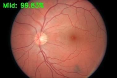
International Research Journal of Engineering and Technology (IRJET) e-ISSN: 2395-0056
Volume: 08 Issue: 06 | June 2021 www.irjet.net p-ISSN: 2395-0072
morphological methodology was utilized. To find the exudate B. Feature extraction and Classification using
territory contrast-restricted versatile histogram evening out Faster-RCNN
calculation was utilized for vein division.
Secondly the labelling of segmented images will undergo
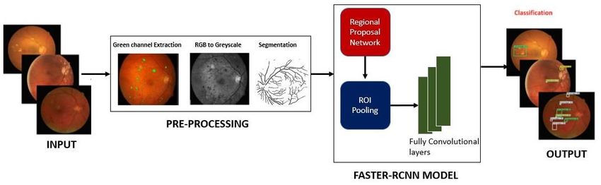
III. PROPOSED METHODOLOGY in the training phase using Faster-RCNN model. In the
proposed work, we have utilized the profound learning a
To overcome the restrictions of existing techniques, we have procedure named Faster-RCNN to remove the highlights
introduced the Deep learning technique dependent on Faster from the info pictures. To get the discriminative and
RCNN. The proposed technique recognizes the irregularities proficient set of highlights, it is important to choose such
of DR at the same time utilizing Faster RCNN. DR location a procedure that can naturally, get the highlights of
from fundus pictures is viewed as a two-way technique at information pictures without needing to utilize the
first, the fundus pictures from the dataset will get hand-coded key-highlights choice approaches. For
preprocessed by the technique for transformation from RGB highlight extraction, we have utilized profound learning
to grayscale because of the discovery of the neurons we a method named Faster-RCNN. The convolve-channels
increment the difference in the green fundus space of the of Faster RCNN empowers it to extricate the critical
pictures and the pictures will get preprocessed. The highlights of the information picture proficiently by
preprocessed picture will go through a division interaction. inspecting the design of the picture. Faster RCNN
At the point when the image is separated using the AHE comprises of Regional Proposal Network (RPN) and
technique, the checking of classes occurs. Then, Faster-RCNN Fast-RCNN. The completely convolutional module RPN
based characterization model will be worked by the can produce the item proposition of the information
appropriate preparing stage. When the model is made picture naturally, which is passed as information and
utilizing Faster RCNN, test input pictures can be given to refine by the Faster-RCNN module. The two modules
achieve appropriate yield. share the equivalent convolutional the layer which
permits the info picture to go through CNN as it were
A. Pre-Processing once to deliver and refine its item proposition. The
Faster-RCNN strategy can productively recognize and
Extract the unprocessed RGB images from the dataset and
order the various indications of DR by utilizing its
statistical feature of it (average, median, image resizing and
completely convolutional modules RPN and Fast-RCNN
skewness). RGB images are taken and they get initially
which work by supplanting the particular pursuit
preprocessed and get converted into grayscale and here the
calculation. RPN module works by utilizing less chosen
Adaptive Histogram Equalization method has been used to
windows and affirms the higher review rate, which
increase the contrast in green fundus area and with the help
assists with decreasing the cost of the proposed work.
of Morphological and Contour method to remove the edges
and noise in the green fundus. After completing the
preprocessed stage, the image will get segmented.
© 2021, IRJET | Impact Factor value: 7.529 | ISO 9001:2008 Certified Journal | Page 80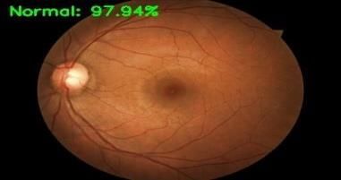
International Research Journal of Engineering and Technology (IRJET)                                     e-ISSN: 2395-0056
         Volume: 08 Issue: 06 | June 2021               www.irjet.net                                            p-ISSN: 2395-0072

1)Convolution layers: Faster RCNN has an aggregate of 13              explicitness is utilized to inspect that it correctly perceives
convolutional and relu layers alongside 4 pooling layers.             the individuals who are not gained of that sickness
These convolutional layers help the Faster-RCNN                       (genuine negative rate). Exactness is the level of which is
organization to compute the component guide of the info               ordered the occurrences unequivocally. Request exactness
picture which is subsequently imparted to the RPN module              is the extent of right conjectures to amount to assumptions
and related layers.                                                   made. It is regularly introduced as a rate by duplicating the
                                                                      outcome by 100. The equations used to figure precision
2) RPN: In this progression, the info object proposition is           are given in Eq
created. The RPN module comprises 3 x 3 completely
convolutional layers organization, which is utilized to make
the anchors and bouncing box relapse balances. This module
utilizes the SoftMax capacity to decide if the registered
secures are the piece of the frontal area or foundation               C)Results

3) Roi Pooling: This layer works by utilizing the registered          The outcome of any preprocessing strategy or procedure is
element     map     from    convolutional      layers    and          an improved picture with rich highlights. The Faster RCNN
recommendations from the RPN module to create the                     model takes less time likewise the exactness of preparing is
proposition highlight guides and offers them all related              acceptably contrasted with the ANN model. For both
layers of the organization.                                           measurable information and handled picture information,
                                                                      Faster RCNN gives great exactness for the hyperparameter
4) Classification: Finally, the characterization step is              change. Thought about an exceptionally appraised model for
performed to decide the class of the identified sores. It works       picture vision, a model prepared with 1000 pictures having
by utilizing the yield of the Roi pooling layer. The bouncing         an exactness of 75.5% with the VGGnet model. The model
box relapse is utilized to display the resultant area of the          prepared with CPU support had an effect on preparing time.
identified test box.                                                  For testing the model, 300 pictures have been thought of,
                                                                      with respect to result concerns both the typical retinopathy
    IV. EXPERIMENTAL RESULTS                                          pictures and different stages diabetic retinopathy pictures
                                                                      have anticipated consummately by the model. In figure point
A) Dataset                                                            by point boundaries of precision have been recorded.
To evaluate the effective detection and characterization of
DR Images, AHE, and FRCNN model is anticipated and
experimentation is done and contrasted and other existing
strategies utilizing fundus picture dataset. The dataset is
gotten from the Kaggle site which is open-source that
endeavors to assemble a DR identification model.

                Table 1. Dataset Description

                                                                      The proposed strategy results are assessed by utilizing the
                                                                      SE, SP, Acc, and mean IoU for all pictures of the test dataset.
                                                                      The proposed framework accomplished normal upsides of
                                                                      SE as 0.897, SP as 0.91, Acc as 0.89.5, F-measure as 0.90, and
                                                                      mean IOU as 0.88. Our proposed strategy shows great
B) Evaluation Metrics
                                                                      execution because of the precise limitation of sores by
   In clinical analysis, affectability is utilized to analyze         utilizing Faster RCNN.
the pictures that accurately perceive the occasions with
the illness (genuine positive rate), any place the

        © 2021, IRJET       |    Impact Factor value: 7.529       |     ISO 9001:2008 Certified Journal            |   Page 81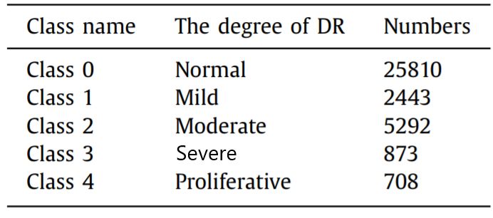
International Research Journal of Engineering and Technology (IRJET)                                       e-ISSN: 2395-0056
        Volume: 08 Issue: 06 | June 2021               www.irjet.net                                              p-ISSN: 2395-0072

Performance analysis                                                  Conference on Cognitive Computing and Information
                                                                      Processing (CCIP), pp. 1–4, IEEE, 2016
        Input Grades                  Accuracy                        [3] N. Chakrabarty, “A deep learning method for the
                                                                      detection of diabetic retinopathy,” in 2018 5th IEEE
        Proposed                      89.5                            Uttar Pradesh Section International Conference on
                                                                      Electrical, Electronics and Computer Engineering
        CNN                           82.36                           (UPCON), pp. 1–5, IEEE,2018

        VGGNet-19                     81.17                           [4] S. D. Kasurde and S. Randive, “An automatic
                                                                      detection of proliferative diabetic retinopathy,” in 2015
        ANN                           80.62                           International Conference on Energy Systems and
                                                                      Applications, pp. 86– 90,IEEE,2015.

Comparison with other methods                                         [5] N. Kasurde, D. K. Singh, and G. K. Singh, “Mobile
                                                                      phone based diabetic retinopathy detection system
                                                                      using ANN,” in 2017 4th IEEE Uttar Pradesh Section
                                                                      International Conference on Electrical, Computer and
                                                                      Electronics (UPCON), pp. 463– 467, IEEE.

                                                                      [6] Swati Gupta and Karandikar AM “Diagnosis of
                                                                      Diabetic Retinopathy using Machine Learning” in 2015
                                                                      Journal Research-and-Development.

                                                                      [7] M. Fraz, P. Remagnino, A. Hoppe, B. Uyyanonvara, A.
                                                                      R. Rudnicka, C. G. Owen, and S. A. Barman, "Blood vessel
                                                                      segmentation methodologies in retinal images -A
                                                                      Survey," Compute. Meth. Prog. Bio., vol. 108, pp. 407–
                                                                      433, Mar.2012.

                                                                      [8] J. Nayak, P. S. Bhat, U. R. Acharya, C. M. Lim, and M.
             V. CONCLUSION                                            Kagathi, "Automated identification of diabetic
                                                                      retinopathy stages using digital fundus images," J. Med.
                                                                      Syst., vol. 32, pp.107–115,2008.
In this project, we have introduced an effective picture
division-based grouping model to consequently fragments               [9] R. Pires, S. Avila, H. F. Jelinek, J. Wainer, E. Valle, and
and characterize the phases of DR. Here, Adaptive Histogram           A. Rocha, "Beyond lesionbased diabetic retinopathy: a
Equalization is utilized for dividing the pictures. At that           direct approach for retinal," IEEE J. Biomed, Health
point, a Faster RCNN based strategy is proposed for                   Inform., vol. 21, no. 1, pp. 193–200, Jan. 2017.
robotized DR location and limitation of DR injuries in retinal
                                                                      [10] D. Sarwinda, T. Siswantining, and A. Bustamam,
pictures. Our strategy can recognize various anomalies of DR          “Classification of diabetic retinopathy stages using
and results are appeared in the test results area. Besides, our       histogram of oriented gradients and shallow learning,”
proposed technique can likewise be applied to tackle the              in 2018 International Conference on Computer, Control,
diverse clinical picture limitation issues also. The                  Informatics and its Applications (IC3INA), pp. 83–87,
exploration work will be reached out by tending to other              IEEE 2018
retinal picture infections i.e., Cataract, Age-related Macular
                                                                      [11] Athira T R, Athira Sivadas, Aleena George, Amala
Edema degeneration, and so forth later on.                            Paul, Neethu Radha Gopan “Automatic detection of
                                                                      Diabetic Retinopathy using R-CNN” in May 5 2019
REFERENCES                                                            International Research Journal of Engineering and
                                                                      Technology.
[1] Nihel zaaboub1,2, ali douik “Early Diagnosis of
Diabetic Retinopathy using Random Forest Algorithm”                   [12] T. Shanthi, R.S. Sabeenian, “Modified Alexnet
5th International Conference on Advanced Technologies                 architecture for classification of diabetic retinopathy
or Signal and Image Processing, Sept 5 2020.                          images”, Dec. 2018.

[2] R. Harini and N. Sheela, “Feature extraction and                  [13] Kedir M. Adal_, Peter G. van Etten, Jose P. Martinez,
classification of retinal images for automated detection              Kenneth W. Rouwen, Koenraad A. Vermeer, Member,
of diabetic retinopathy,” in 2016 Second International                IEEE and Lucas J. van Vliet, Member, IEEE, “An

       © 2021, IRJET       |    Impact Factor value: 7.529        |     ISO 9001:2008 Certified Journal             |   Page 82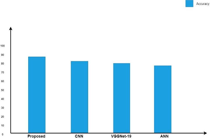
International Research Journal of Engineering and Technology (IRJET)                       e-ISSN: 2395-0056
        Volume: 08 Issue: 06 | June 2021             www.irjet.net                                 p-ISSN: 2395-0072

Automated System for the Detection and Classification
of Retinal Changes Due to Red Lesions in Longitudinal
Fundus Images,” IEEE Transcations on Biomedical
Engineering, 2017.

[14] Doshi D, Shenoy A, Sidhpura D, Gharpure P.
Diabetic retinopathy detection using deep convolutional
neural networks. 2016 international conference on
computing, analytics and security trends (CAST),
December 2016.

 [15] Bhatkar AP, Kharat GU. Detection of diabetic
retinopathy in retinal images using MLP classifier. 2015
IEEE international symposium on nano electronic and
information systems, December; 2015

       © 2021, IRJET      |    Impact Factor value: 7.529    |   ISO 9001:2008 Certified Journal    |   Page 83
You can also read