An Unexpected Consequence of Electronic Cigarette Use
←
→
Page content transcription
If your browser does not render page correctly, please read the page content below
An Unexpected Consequence of Electronic
                     Cigarette Use
                     Lindsay McCauley, Catherine Markin and Danielle Hosmer

                     Chest 2012;141;1110-1113
                     DOI 10.1378/chest.11-1334
                     The online version of this article, along with updated information and
                     services can be found online on the World Wide Web at:
                     http://chestjournal.chestpubs.org/content/141/4/1110.full.html

                      Chest is the official journal of the American College of Chest
                      Physicians. It has been published monthly since 1935.
                      Copyright2012by the American College of Chest Physicians, 3300
                      Dundee Road, Northbrook, IL 60062. All rights reserved. No part of
                      this article or PDF may be reproduced or distributed without the prior
                      written permission of the copyright holder.
                      (http://chestjournal.chestpubs.org/site/misc/reprints.xhtml)
                      ISSN:0012-3692

Downloaded from chestjournal.chestpubs.org at Naval Hospital Camp Pendleton on April 19, 2012
                        © 2012 American College of Chest Physicians
CHEST                                       Postgraduate Education Corner
                                                                                     PULMONARY AND CRITICAL CARE PEARLS

                    An Unexpected Consequence of Electronic
                    Cigarette Use
                    Lindsay McCauley, DO; Catherine Markin, MD, FCCP; and Danielle Hosmer, MD

                         CHEST 2012; 141(4):1110–1113               Laboratory Tests and Imaging Findings
                                                                       Laboratory findings showed a WBC count of 18.0
A   42-year-old woman was admitted to the hospital
    with a 7-month history of dyspnea, productive                   (3 103) with a normal differential and hemoglobin level
                                                                    of 11.2 g/dL. The chemistry panel and brain natri-
cough, and subjective fevers. She had been seen mul-
tiple times in the ED with similar complaints and had               uretic peptide levels were normal. Chest radiographic
received several courses of antibiotics.                            imaging showed new multifocal bilateral opacities.
   The patient had recently started using electronic                CT images (Fig 1) revealed extensive bilateral upper-
cigarettes (e-cigarettes), about 7 months prior, which              and lower-lobe patchy ground glass pulmonary opac-
coincided with the onset of her respiratory symptoms.               ities in a “crazy paving” pattern. Results of an HIV
Her past medical history also was significant for asthma,           test were negative. Results of a nasal Pertussis poly-
reported rheumatoid arthritis, fibromyalgia, schizoaf-              merase chain reaction swab were negative. Results
fective disorder, and hypertension. Her medications                 of urine Legionella antigen and serum Mycoplasma
included amlodipine, albuterol metered dose inhaler,                IgG and IgM tests were negative. Results of a
lovastatin, lisinopril, multiple vitamins, cyclobenzaprine,         hypersensitivity pneumonitis panel, extracted nuclear
citalopram, and multiple psychiatric medications.                   antigen panel, and tests for antinuclear antibody,
   The patient reported a recent exposure to fumi-
gation chemicals, as the result of a bedbug infes-
tation of her apartment building 2 weeks prior to
her hospitalization. She had no pets. There was no
other history of significant exposures, illicit drug
use, or recent travel. She denied any dysphagia or
aspiration.

Physical Examination
  On presentation, her vital signs were notable for mild
tachycardia and a pulse oximetric saturation of 94%
while breathing room air. Her physical examination
was normal except for bilateral rales.
Manuscript received May 27, 2011; revision accepted August 8,
2011.
Affiliations: From the Department of Internal Medicine
(Dr McCauley), the Department of Pulmonary Medicine
(Drs Markin and Hosmer), and the Department of Critical Care
Medicine (Dr Markin), Legacy Good Samaritan Medical Center,
Portland, OR.
Correspondence to: Danielle Hosmer, MD, Department of
Pulmonary Medicine, Legacy Good Samaritan Medical Center,
2222 NW Lovejoy St, Ste 411, Portland, OR 97210; e-mail:
dhosmer@lhs.org
© 2012 American College of Chest Physicians. Reproduction
of this article is prohibited without written permission from the
American College of Chest Physicians (http://www.chestpubs.org/     Figure 1. Representative CT images show the “crazy paving” pat-
site/misc/reprints.xhtml).                                          tern of patchy ground glass superimposed on interlobular septal
DOI: 10.1378/chest.11-1334                                          thickening. A, Bilateral upper lobes. B, Bilateral lower lobes.

1110                                                                                                   Postgraduate Education Corner

            Downloaded from chestjournal.chestpubs.org at Naval Hospital Camp Pendleton on April 19, 2012
                                    © 2012 American College of Chest Physicians
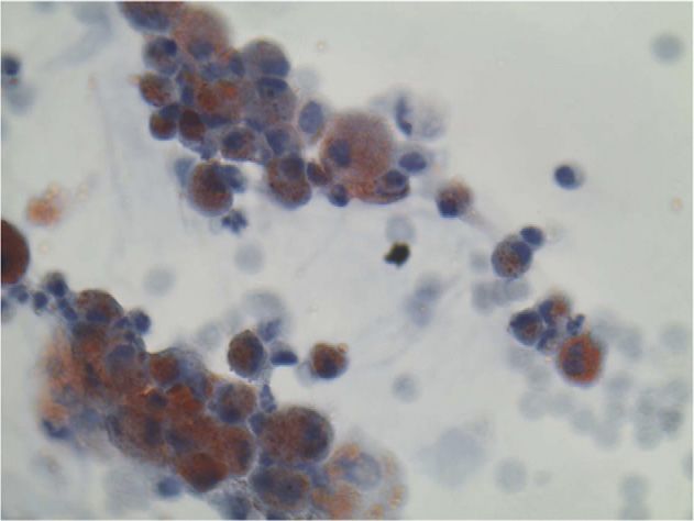
Figure 2. Photomicrograph of BAL sample shows lipid-laden
macrophages (Oil-Red-O stain, original magnification 3 100).

cyclic citrullinated peptide, and rheumatoid factor
were negative. A bird fancier’s panel showed trace
reactivity to pigeon and parrot droppings.
   Bronchoscopy and BAL were performed. The cell
count showed 48% neutrophils, 8% lymphocytes,
43% monocytes, and 1% eosinophils. Results of all
bacterial and viral cultures remained negative; fungal
cultures showed light growth of Candida. Results of a
viral DFA panel, Pneumocystis jeroveci DFA, and
Legionella antigen tests were negative. BAL cytologic
examination revealed abundant lipid-laden macro-
phages (Fig 2).

What is the diagnosis?

www.chestpubs.org                                                                   CHEST / 141 / 4 / APRIL, 2012   1111

           Downloaded from chestjournal.chestpubs.org at Naval Hospital Camp Pendleton on April 19, 2012
                                   © 2012 American College of Chest Physicians
Diagnosis: Exogenous lipoid pneumonia due laden macrophages in the sputum or BAL fluid. These
to e-cigarette use vacuolated macrophages stain orange with Sudan
stain or red with Oil-Red-O stain. No clear cytologic
Discussion profile has been found to be more suggestive of the
disease. Histologic examination shows an inflamma-
Lipoid pneumonia is a rare, primarily chronic tory landscape, similar to that seen with a foreign
inflammatory reaction secondary to the presence of body reaction. In severe disease, a proliferative fibro-
lipid substances in the lungs, with subsequent uptake sis and disorganization of the pulmonary architecture
by alveolar macrophages and accumulation in the can occur. A biopsy may be necessary to confirm the
interstitium. The endogenous form occurs when fat is diagnosis in certain cases.
deposited into the lung tissue in vivo, typically from Once the diagnosis has been identified, all efforts
proximal obstructive lesions, fat embolism, necrotic should be made to avoid recurrent oil exposures and
tissue, lipid storage disease, or hyperlipidemia. The stop aspiration. Expectorants and repeat therapeutic
exogenous form develops from inhaling or aspirat- BAL have not been shown to offer any benefit. Sys-
ing lipids, such as those seen in animal, vegetable, or temic corticosteroids have been recommended; how-
mineral oil. Classically, exogenous lipoid pneumonia ever, they lack proven efficacy. Therefore, their use
is associated with aspiration of mineral oil-based lax- should be limited to severe cases.
atives in the pediatric population or with occupa- For this patient, the suspected source of her exog-
tional exposures. The incidence is also higher in older enous lipoid pneumonia was recurrent exposure to
patients with underlying debility, achalasia, reflux, glycerin-based oils found in e-cigarette nicotine vapor.
and other neuromuscular disorders of the pharynx Since the 1980s, there has been an ever-increasing
and esophagus. development of electronic nicotine-delivery systems.
Most patients are asymptomatic; however, symp- The e-cigarette comprises a plastic tube and a battery-
toms may include cough, dyspnea, fever, weight loss, powered electronic heating device that vaporizes a
chest pain, pleurisy, hemoptysis, chills, and night sweats. liquid nicotine cartridge. E-cigarettes are advertised
Findings on physical examination may be normal as an alternative to smoked tobacco and as a smoking
or nonspecific, such as tachypnea and adventitious cessation aide.
breath sounds. Thus, a high clinical suspicion is However, health analysis and empirical research
required to make the diagnosis of exogenous lipoid on e-cigarettes is sparse. Recent evaluation of the
pneumonia. nicotine solution and vapor content of e-cigarettes
Depending on the severity of the disease, exoge- found primary components of propylene glycol, glyc-
nous lipoid pneumonia may present with hypoxia or erin, and nicotine. Other chemicals identified in trace
respiratory alkalosis. Results of pulmonary function amounts include N-nitrosamines, diethylene glycol,
tests typically show a restrictive ventilatory defect polycyclic aromatic hydrocarbons, anabasine, myo-
and/or diffusion impairment, but they may be normal. smine, and b-nicotyrine. Many of these compounds
The most frequent chest radiographic findings are are carcinogenic and harmful to humans.
extensive bilateral alveolar consolidations and ground Vegetable glycerin is often added to the nicotine
glass opacities in the dependent portions of the lungs. solutions used in e-cigarettes to make the visual smoke
However, unilateral involvement may be seen, affect- when the solution is vaporized. Glycerin is produced
ing the right and left lungs equally. Adenopathy is by heating palm or coconut oil; however, it can also
rare. Fibrosis may occur and lead to volume loss. be produced from animal fat and soap through a fatty-
Solid lesions may also develop, resembling broncho- acid splitting operation.
genic carcinoma. As discussed, most cases of exogenous lipoid pneu-
High-resolution CT imaging plays an important monia are associated with aspiration of mineral oil
role in the diagnosis of lipoid pneumonia. The most or lipid-based preparations. There is one published
frequent findings are bilateral posterior and lower- case of exogenous lipoid pneumonia due to inhaling
lobe-predominant alveolar consolidation, ground glass vaporized weed oil. Other cases have been reported
opacities, and the “crazy paving” pattern. Consolidated involving inhalation of crack cocaine mixed with
areas are typically hypodense (2 30 to 2 75 HU), petroleum jelly. To our knowledge, there are no prior
with similar attenuation to the surrounding adipose published cases of exogenous lipoid pneumonia due to
tissue. The use of CT scan angiography may help the use of glycerin-based e-cigarettes. Importantly, this
confirm these findings, with the consolidated lung case highlights harm caused by the nicotine-solution
having a considerably lower attenuation than the carrier and the delivery system of the e-cigarette.
enhancing vessels. Prior discussion regarding the safety of e-cigarettes
A key component to making a true diagnosis of has primarily focused on nicotine and other carci-
exogenous lipoid pneumonia is the presence of lipid- nogenic components. Certainly, the risk of lipoid
1112 Postgraduate Education Corner
Downloaded from chestjournal.chestpubs.org at Naval Hospital Camp Pendleton on April 19, 2012
© 2012 American College of Chest Physicianspneumonia adds another dimension to the super- Acknowledgments
charged social, political, and medical debate surround- Financial/nonfinancial disclosures: The authors have reported
ing the regulation and legality of e-cigarette use. to CHEST that no potential conflicts of interest exist with any
companies/organizations whose products or services may be dis-
cussed in this article.
Clinical Course Other contributions: This work was performed at Legacy Good
Samaritan Medical Center, Portland, OR.
The patient was instructed to avoid the use of
e-cigarettes, and, subsequently, her symptoms improved.
A follow-up chest radiograph was normal, and pul- Suggested Readings
monary function testing showed mild diffusion impair-
Gondouin A, Manzoni P, Ranfaing E, et al. Exogenous lipid
ment but no obstructive or restrictive defects. pneumonia: a retrospective multicentre study of 44 cases in
France. Eur Respir J. 1996;9(7):1463-1469.
Clinical Pearls Vethanayagam D, Pugsley S, Dunn EJ, Russell D, Kay JM,
Allen C. Exogenous lipid pneumonia related to smoking weed
1. Exogenous lipoid pneumonia is a chronic inflam- oil following cadaveric renal transplantation. Can Respir J.
matory reaction to the deposition of lipid substances 2000;7(4):338-342.
in the lung, typically as a result of aspiration or inha- Cottin V, Chalabreysse L, Cordier JF. Exogenous lipid pneumonia.
Respiration. 2008;76(4):442-443.
lation of oil-based products. Gurell MN, Kottmann RM, Xu H, Sime PJ. Exogenous lipoid
2. Chest CT imaging typically shows bilateral pneumonia: an unexpected complication of substance abuse.
alveolar consolidation and ground glass opacities, Ann Intern Med. 2008;149(5):364-365.
including the “crazy paving” pattern, in the depen- Basset-Léobon C, Lacoste-Collin L, Aziza J, Bes JC, Jozan S,
dent areas of the lungs. Courtade-Saïdi M. Cut-off values and significance of Oil Red
O-positive cells in bronchoalveolar lavage fluid. Cytopathology.
3. The presence of lipid-laden macrophages in 2010;21(4):245-250.
sputum or BAL fluid helps to confirm the diagnosis. Betancourt SL, Martinez-Jimenez S, Rossi SE, Truong MT,
4. The symptoms and pathologic changes often Carrillo J, Erasmus JJ. Lipoid pneumonia: spectrum of clin-
completely resolve with the cessation of exposure; ical and radiologic manifestations. AJR Am J Roentgenol. 2010;
however, severe cases can progress to fibrosis and 194(1):103-109.
Flouris AD, Oikonomou DN. Electronic cigarettes: miracle or
chronic respiratory failure. menace? BMJ. 2010;340:c311.
5. Many public health authorities, including the Trtchounian A, Williams M, Talbot P. Conventional and electronic
US Food and Drug Administration, caution that the cigarettes (e-cigarettes) have different smoking characteristics.
risks and benefits of e-cigarettes have not been ade- Nicotine Tob Res. 2010;12(9):905-912.
quately studied. This case demonstrates an important Yamin CK, Bitton A, Bates DW. E-cigarettes: a rapidly growing
Internet phenomenon. Ann Intern Med. 2010;153(9):607-609.
heretofore unrecognized (as far as we know) health Cahn Z, Siegel M. Electronic cigarettes as a harm reduction
risk of e-cigarette use: exogenous lipoid pneumonia strategy for tobacco control: a step forward or a repeat of past
due to glycerin-based e-cigarettes. mistakes? J Public Health Policy. 2011;32(1):16-31.
www.chestpubs.org CHEST / 141 / 4 / APRIL, 2012 1113
Downloaded from chestjournal.chestpubs.org at Naval Hospital Camp Pendleton on April 19, 2012
© 2012 American College of Chest PhysiciansAn Unexpected Consequence of Electronic Cigarette Use
Lindsay McCauley, Catherine Markin and Danielle Hosmer
Chest 2012;141; 1110-1113
DOI 10.1378/chest.11-1334
This information is current as of April 19, 2012
Updated Information & Services
Updated Information and services can be found at:
http://chestjournal.chestpubs.org/content/141/4/1110.full.html
References
This article cites 10 articles, 6 of which can be accessed free at:
http://chestjournal.chestpubs.org/content/141/4/1110.full.html#ref-list-1
Permissions & Licensing
Information about reproducing this article in parts (figures, tables) or in its entirety can be
found online at:
http://www.chestpubs.org/site/misc/reprints.xhtml
Reprints
Information about ordering reprints can be found online:
http://www.chestpubs.org/site/misc/reprints.xhtml
Citation Alerts
Receive free e-mail alerts when new articles cite this article. To sign up, select the
"Services" link to the right of the online article.
Images in PowerPoint format
Figures that appear in CHEST articles can be downloaded for teaching purposes in
PowerPoint slide format. See any online figure for directions.
Downloaded from chestjournal.chestpubs.org at Naval Hospital Camp Pendleton on April 19, 2012
© 2012 American College of Chest PhysiciansYou can also read