Article Middle ear cholesteatoma in 11 dogs - Dermato clinica
←
→
Page content transcription
If your browser does not render page correctly, please read the page content below
Article

Middle ear cholesteatoma in 11 dogs
Valentina Greci, Olga Travetti, Mauro Di Giancamillo, Rocco Lombardo, Chiara Giudice,
Barbara Banco, Carlo M. Mortellaro

          Abstract — Middle ear cholesteatoma is a rare condition in dogs with chronic otitis. Otorrhea, otodinia, and pain
          on temporomandibular joint palpation are the most common clinical signs. Neurological abnormalities are often
          detectable. Computed tomography reveals the presence of an expansive and invasive unvascularized lesion involv-
          ing the tympanic cavity and the bulla, with little or no contrast enhancement after administration of contrast
          mediu. Video-otoscopy may detect pearly growth or white/yellowish scales in the middle ear cavity. Surgery is the
          only therapy but is associated with a high risk of recurrence.

          Résumé — Cholestéatome de l’oreille moyenne chez 11 chiens. Le cholestéatome de l’oreille moyenne est une
          affection rare et potentiellement sous-estimée chez les chiens présentant une anamnèse d’otite chronique. L’otorrhée,
          l’otodynie et la douleur à la palpation de l’articulation temporo-mandibulaire sont les signes cliniques les plus
          courants. Des anomalies neurologiques sont souvent détectables. Une tomographie par ordinateur révèle la présence
          d’une lésion étendue et envahissante non vascularisée affectant la cavité tympanique et la bulle avec peu ou pas
          d’augmentation de contraste après l’administration d’un milieu de contraste. La vidéo-otosocopie peut détecter
          une lésion de croissance nacrée ou des écailles blanc-jaune dans la cavité de l’oreille moyenne. La chirurgie est la
          seule thérapie mais elle est associée à un risque élevé de récurrence.
                                                                                                    (Traduit par Isabelle Vallières)
          Can Vet J 2011;52:631–636

                            Introduction                                  Cholesteatoma is an epidermoid cyst lined by a pluristrati-

M
                                                                      fied keratinizing epithelium containing keratin debris and is
        iddle ear cholesteatoma is a serious complication of
                                                                      characterized by independent and progressive growth, causing
        chronic otitis media in human beings and dogs (1–4).
                                                                      destruction of adjacent tissue, especially bone (7). The term
In dogs, it is rarely reported and is possibly underdiagnosed
                                                                      cholesteatoma is a misnomer because it is not a tumor (suffix-
(2–6). Neither breed nor sex predisposition has been described.
                                                                      “oma”), it does not contain fat (-“stea”-) or cholesterol crystal
A history of chronic otitis, otodinia, pain on temporomandibu-
                                                                      (-“chol”-), but cholesteatoma is still used for describing this
lar joint (TMJ) palpation, and discomfort when opening the
                                                                      disease (7–10).
mouth are the most common clinical findings. Neurological
                                                                          The etiopathogenesis of cholesteatoma is controversial and
abnormalities, including head tilt, facial palsy, and ataxia, can
                                                                      many theories have been proposed (1,7–9,11). In dogs, it is sug-
be detected on examination or can represent the major reason
                                                                      gested that middle ear cholesteatoma develops according to the
for referral (2,3,5,6).
                                                                      migration theory or the invagination theory (2,3). According to
                                                                      the former, migration of the stratified squamous epithelium from
Dipartimento di Scienze Cliniche, Sezione di Chirurgia (Greci,
                                                                      the external auditory meatus into the infected middle ear cavity
Lombardo, Mortellaro), Sezione di radiologia e Chirurgia
                                                                      through a perforated ear drum may produce the necessary condi-
Sperimentale (Travetti, Di Giancamillo), Dipartimento
                                                                      tions for development of cholesteatoma. According to the latter,
di Patologia Animale, Igiene e Sanità Pubblica, Sezione di
                                                                      the pars flaccida and occasionally the pars tensa of the tympanic
Anatomia e Patologia Aviare (Giudice, Banco); Facoltà di
                                                                      membrane retract into the middle ear because of inflammation,
Medicina Veterinaria, Università degli Studi di Milano 7, 20133,
                                                                      negative pressure, or both, and afterwards the pocket is slowly
Milano, Italy.
                                                                      filled by keratin thus establishing cholesteatoma (7,11).
Address all correspondence to Dr. Valentina Greci; e-mail:                The only possible treatment is surgery and the goal of sur-
valentina.greci@unimi.it                                              gery is to remove all keratin debris and stratified squamous
Reprints will not be available from the authors.                      epithelium, and to control infection. Despite surgery, the risk
Use of this article is limited to a single copy for personal study.   of recurrence is high (3,7).
Anyone interested in obtaining reprints should contact the                This paper reports the main clinical, imaging, and pathologi-
CVMA office (hbroughton@cvma-acmv.org) for additional                 cal findings, and the surgical outcome for 11 dogs diagnosed
copies or permission to use this material elsewhere.                  with and treated for middle ear cholesteatoma.

CVJ / VOL 52 / JUNE 2011                                                                                                              631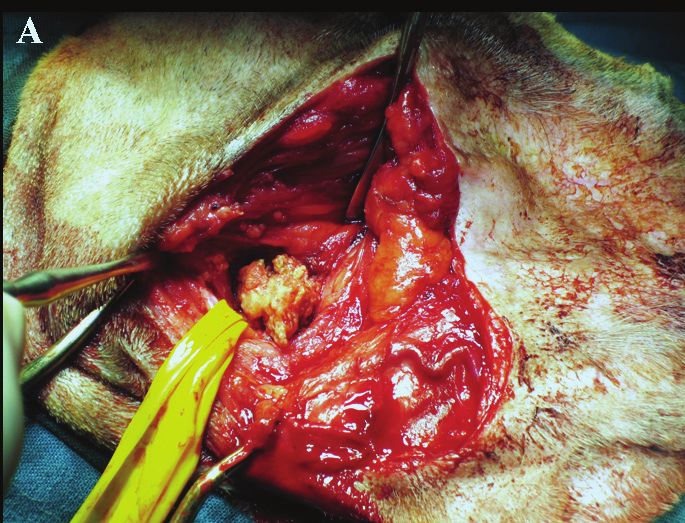
Table 1. Signalment, clinical signs, duration of clinical signs, site localization, surgical technique and findings, histopathology and outcome
of the 11 dogs diagnosed with middle ear cholesteatoma
Duration of Choles-
Age clinical signs Neurological teatoma Histo-
Case Breed Sex (y) (wk) Clinical signs abnormalities location Surgery pathology Follow-up
1 Pug M 8 6 Bilateral otorrhea, Left head tilt, Bilateral TECALBO, Keratin 48 mo, no
otodinia, shaking left facial keratin scales recurrence
head/ear paw palsy
A R T I C LE
2 Cross-breed M 5 16 Otorrhea, otodinia, Left TECALBO, 3 layers 28 mo,
discomfort opening keratin scales recurrence at
the mouth, shaking 5 mo (VBO)
head/ear paw,
dysphagia
3
Flat-coated M 10 4 Otorrhea, otodinia, Left head tilt, Left TECALBO, 3 layers 13 mo,
retriever shaking head/ear paw left facial keratin scales recurrent
palsy, ataxia, disease not
confirmed
4 Poodle Mn 5.5 24 Otodinia, discomfort Right TECALBO, Keratin 39 mo,
opening the mouth, keratin scales recurrent
dysphagia, shaking disease at
head/ear paw 13 mo (VBO)
5
Afghan M 8 12 Draining tract at Left TECALBO, Keratin 34 mo,
greyhound surgical site, keratin scales recurrences
otodinia, discomfort at 2 (LBO),
opening the mouth, 26 (VBO)
shaking head/ear paw and 36
(VBO) mo
6 Weimaraner Fs 9 4 Otorrhea, otodinia, Right TECALBO 3 layers 2 wk
shaking head/ear paw video-assisted, and CG
keratin scales
7
Cocker M 6 3 Otorrhea, otodinia, Left head tilt, Left TECALBO Keratin 28 mo,
spaniel discomfort opening left facial video-assisted, no recurrence
the mouth palsy, ataxia mass-like lesion
8 Schnauzer M 5.5 8 Otorrhea, otodinia, Left TECALBO, 3 layers 27 mo,
shaking head/ear paw mass-like no recurrence
lesion
9 Cross-breed Fs 9 16 Otorrhea, otodinia, Left head tilt, Left TECALBO, Keratin 22 mo,
discomfort opening left facial keratin scales and CG recurrence at
the mouth, shaking palsy, ataxia 10 (VBO)
head/ear paw and 14 mo
(response to
medical
management)
10
Golden F 4.5 16 Otorrhea Left head tilt, Left VBO, 3 layers 12 mo,
retriever left facial keratin scales and CG no recurrence
palsy, ataxia
11 Labrador M 9 28 Otodinia, discomfort Left TECALBO, 3 layers 13 mo,
retriever opening the mouth mass-like lesion and CG no recurrence
M — male intact; Mn — male neutered; F — female intact; Fs — female spayed; CG — cholesterol granuloma.
Materials and methods performed were total ear canal ablation and lateral bulla oste-
otomy (TECALBO) and ventral bulla osteotomy (VBO).
Eleven dogs were identified that had a histologic diagnosis of
During surgery, swabs for bacterial culture and susceptibility
cholesteatoma. The dogs had been presented to the School of
and samples for histological examination were collected from
Veterinary Medicine, Università degli Studi di Milano, Italy,
the middle ear cavity. Both fresh samples and samples in 10%
between January 2001 and July 2007 for investigation of chronic
formalin were submitted for histopathology. Follow-up consisted
otitis unresponsive to topical and systemic therapy. Medical
of clinical examination and telephone interviews.
records were reviewed for signalment, history, and physical
examination, radiological and video-otoscopy findings, micro-
biological results, histopathological features, treatment, and Results
outcome. Middle ear cholesteatoma was bilateral in 1 dog and Signalment and clinical findings
unilateral in 10 dogs. Two dogs were medium-sized cross-breeds, and 9 dogs belonged
All dogs underwent radiological investigation and video- to specific breeds (pug, flat-coated retriever, poodle, Afghan grey-
otoscopy under general anesthesia. The surgical procedures hound, cocker spaniel, schnauzer, weimaraner, golden retriever,
632 CVJ / VOL 52 / JUNE 2011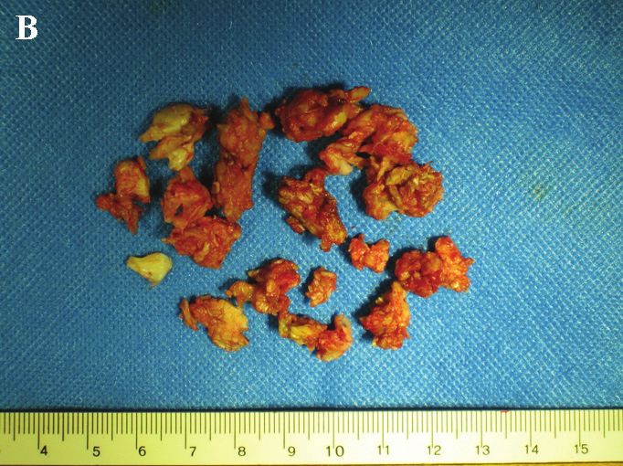
Labrador retriever). Seven dogs were intact males, 1 dog was a
castrated male, 2 dogs were spayed females, and 1 dog was an
intact female. Mean age was 7.2 y (range: 4.5 to 10 y).
    All dogs had a history of chronic recurrent otitis externa, unre-
sponsive to topical or systemic therapy over the last 3 to 30 wk
(mean duration 13.3 wk). In all dogs the main signs were paw-
ing at the ear and head shaking. During physical examination,

                                                                                                                                              A R T I C LE
all but one of the dogs showed algic reaction at palpation of the
bulla (otodinia) and TMJ. Otorrhea was present in 8 dogs and
6 dogs showed discomfort when opening the mouth. Two dogs
exhibited dysphagia. Neurological abnormalities characterized by
head tilt, facial paralysis, and ataxia were detected in 5 dogs. One
dog, 2 mo prior to presentation, underwent Zepp surgery (lateral
ear canal resection) and at the time of referral had a draining
tract beneath the surgical opening. Details are shown in Table 1.

Imaging
The main findings on computed tomography (CT) scan in
10 dogs (11 ears) were enlargement of the middle ear cavity,
complete loss of air contrast within the middle ear cavity, and
absence or minimal positive enhancement after IV injection
of contrast medium (Figure 1B). Sclerosis of the petrosal bone
and involvement of the epitympanic recess were also detected
in all dogs, and TMJ sclerosis (Figure 1B) was detected in all
but one dog. On CT scan, the bulla wall showed signs of lysis,
sclerosis, or both, in 2, 6, and 3 dogs, respectively. The ear
canal was completely occluded in 4 ears, partially occluded in
3 ears, and patent in 4 ears. Ventrodorsal open mouth and lat-
eral 30° degree views of 1 dog revealed enlargement of 1 bulla,
loss of air contrast within the middle ear cavity, sclerosis of the
tympanic wall and petrosal bone, unremarkable contralateral
bulla, and patent ear canals.
                                                                        Figure 1. A — Transverse slice obtained at the level of the
   Video-otoscopy revealed total occlusion of the horizontal            ossicular chain shows, at bone setting, complete loss of air
canal (end-stage otitis — ESO) in 4 ears, moderate stenosis of          contrast associated with soft tissue density within the middle ear
the horizontal canal in 4, and no changes in 4. The ear drum            cavity (black arrow); enlargement of the tympanic cavity and bulla
                                                                        with mild osteolysis and sclerosis of the latero-ventral aspect
was ruptured in 8 ears. Pearly growths that differed in shape and       of the bulla (white arrow), and sclerosis of the temporal bone
size were found protruding from the middle ear cavity in 3 ears,        (arrowhead); absence of air contrast in the horizontal tract of the
and white/yellow scales were found within the middle ear cavity         ear canal (compared with contralateral ear canal); disappearance
                                                                        of the ossicular chain and mild erosion of the promontory.
in 2 ears (Figure 2 A,B,C).                                             B — Transverse slice obtained at the level of the TMJ at bone
                                                                        setting; note the severe osteosclerosis of the right mandibular
Surgery                                                                 fossa (white arrow).
Ten dogs (11 ears) underwent TECALBO whilst 1 dog (1 ear)
underwent VBO because it was a show dog. In 9 ears, the                 Histopathology
middle ear cavity was filled with a spongy yellowish material           Histopathology identified keratin debris in 6 ears; in the other
resembling keratin debris, whilst in 3 ears a mass-like lesion was      6 ears there was a cystic lesion lined by a multilayered, intensely
removed (Figure 3 A,B,C). In all dogs, the bulla was enlarged           hyperplastic, keratinizing epithelium (up to 25 layers thick)
with irregular surface and had small irregular cavities hiding          and containing abundant amorphous lamellar keratin debris
additional debris. In 2 dogs (2 ears), video-assisted curettage of      (Figure 4). In 4 ears, a cholesterol granuloma was also found
the middle ear cavity was performed.                                    within fibrous stromal tissue surrounding and sustaining a
                                                                        cystic lesion.
Bacterial culture and susceptibility
On aerobic culture of the middle ear bacteria were recovered            Follow-up
from 8 of the 12 ears. More than 1 species of organism was              Mean follow-up lasted 26.5 mo (2 wk to 48 mo). Surgical
isolated from 1 ear. Bacteria isolated were Staphylococcus inter-       short-term complications such as facial palsy and dryness of the
medius (3 ears), Proteus mirabilis (2 ears), Pseudomonas aeruginosa     nostrils developed in 2 dogs and spontaneously resolved within
(1 ear), Escherichia coli (1 ear), and Klebsiella pneumoniae (1 ear).   4 mo after surgery. Schirmer tear tests (STT; Essex Pharma
Multiple antibiotic resistances were detected.                          GmbH, Munich, Germany) were normal (15 mm/min) in 1 dog

CVJ / VOL 52 / JUNE 2011                                                                                                               633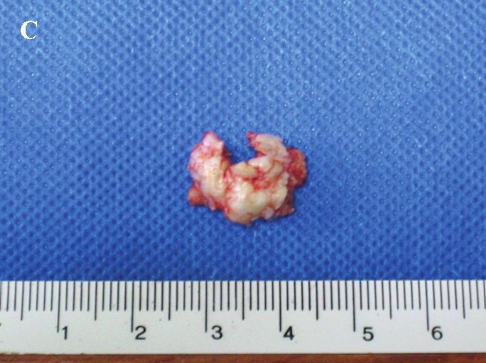
A R T I C LE

                                                                                Figure 3. Gross appearance of middle ear cholesteatoma
                                                                                during surgery. A — Spongy aspect of the keratinous material; the
                                                                                material spontaneously protruded after enlargement of the bulla
                                                                                opening; B — All material removed from the middle ear cavity
                                                                                in the same ear, note the keratinous appearance; C — Cyst-like
                                                                                aspect of cholesteatoma after surgical removal.

                                                                                and 10 mm/min in the other dog. Dryness of the nostril was
                                                                                attributed to temporary neurogenic dysfunction of the parasym-
                                                                                pathetic fibers of the facial nerve. Damage to those fibers most
                                                                                likely occurred during the curettage of the middle ear cavity.
                                                                                Dogs with neurological abnormalities showed persistent head tilt
                                                                                after surgery whilst facial palsy and ataxia resolved. One dog was
                                                                                lost to follow-up at 48 mo, but was reported to be free of any
                                                                                clinical signs on the last clinical and telephone follow-up. One
                                                                                dog died of an unrelated cause 2 wk after surgery; postmortem
                                                                                CT of the bullae showed a patent middle ear cavity and, on nec-
               Figure 2. Various otoscopic appearances of middle ear            ropsy, the bulla surface was clean and smooth. Six dogs (7 ears)
               cholesteatoma. A — White scales filling the middle ear cavity;   (58.33%) had resolution of middle ear disease. Recurrence of
               B — A yellow spongy pearly growth protruding from the middle
               ear cavity; C — A pinkish pearly growth filling the middle ear   cholesteatoma was suspected in 5 dogs (5 ears) (41.66%). Mean
               cavity and protruding in the horizontal canal.                   time for recurrence was 7.5 mo (2 to 13 mo).

               634                                                                                                         CVJ / VOL 52 / JUNE 2011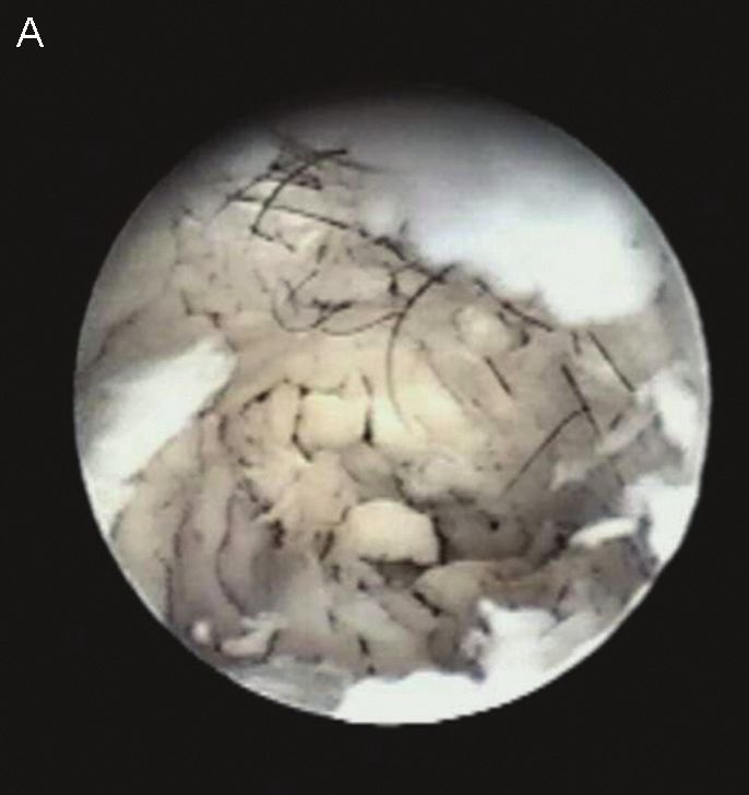
17 mo after surgery the dog was still free of clinical signs. Only
1 dog was available for follow-up of video-assisted surgery and
at 27 mo after surgery was still free of clinical signs.
Discussion
Middle ear cholesteatoma is a serious and rarely reported condi-
tion associated with chronic otitis in dogs. There is a history of
A R T I C LE
chronic recurrent otitis which becomes unresponsive to topi-
cal or systemic therapy; there is no breed predisposition, but
there is a predominance of males (2–6). As in humans, there is
no explanation for the male predominance (8). Although the
clinical signs are nonspecific, in this study, algic reaction at
TMJ palpation was detected in all but one dog. On CT scan,
cholesteatoma showed distinctive features that should be con-
sidered almost pathognomonic: presence of an expansive and
invasive unvascularized lesion involving the tympanic cavity,
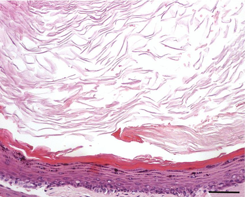
Figure 4. Section of cholesteatoma. Abundant lamellar lytic and/or sclerotic changes of the bulla wall, and absent or
eosinophilic debris (keratin) fills a cyst lumen lined by a minimal contrast enhancement (3–7). Moreover, TMJ sclerosis
multilayered squamous epithelium characterized by diffuse was detected in all but one dog.
intense hyperplasia and orthokeratotic hyperkeratosis.
Hematoxylin and eosin, 1003 (bar 1 cm = 60 mm). To our knowledge, the otoscopic appearance of middle ear
cholesteatoma has not previously been reported in dogs. During
video-otoscopy, the detection of a pearly growth lesion or
Clinical signs at the time of suspected recurrence were oto- white/yellow scales in or protruding from the middle ear cavity
dinia (5 dogs), head tilt (3 dogs), facial palsy (2 dogs), ataxia should alert the clinician to the possible presence of middle ear
(2 dogs), discomfort opening the mouth (3 dogs), para-aural cholesteatoma (7).
swelling (2 dogs), and draining tract at the surgical incision site The only possible treatment for middle ear cholesteatoma is
(2 dogs). Recurrence was confirmed in 4 dogs, 2 of which had surgery, but cholesteatoma tends to recur after surgery (2,3,7).
multiple recurrences, whilst 1 dog was euthanized and necropsy TECALBO is the most frequently performed surgical technique
was not permitted. Computed tomographic scans on the other (2,3,5,6), whilst VBO has been suggested as the treatment of
4 dogs showed enlargement of the middle ear cavity, remodeling choice by Venker-Van Haagen (4), but outcomes have not been
of the bulla due to previous surgery and active disease, and pres- reported. Ventral bulla osteotomy may be preferable when the
ence of soft tissue density in the middle ear cavity. A course of ear canal appears normal or mildly changed, whilst TECALBO
7 to 10 d of empirical antibiotic and anti-inflammatory therapy should be selected when the owner is not dedicated to ongoing
was initiated, but there was no clinical improvement, and the management of otitis externa or ESO is present (2,3,12,13).
patients were submitted for surgery. A combination of the 2 techniques has been performed only
Two dogs were successfully treated by VBO and at 32 and once (3) but could potentially decrease the incidence of recur-
43 mo after surgery had no signs of recurrence. One dog was rence because of the wider exposure of the middle ear cavity for
diagnosed with recurrent middle ear cholesteatoma at 2, 26, and curettage. In humans, video-assisted surgery has been reported
36 mo, and otitis media at 14 mo after the first surgery. This to reduce the incidence of recurrence (14–16).
dog was treated by LBO once and VBO 3 times, and at 7 mo The histological diagnosis of cholesteatoma is made by the
after the last surgery was still exhibiting discomfort opening the presence of the 3 components, keratin debris, the epithelium,
mouth, had head tilt, and a draining tract at the surgical incision and the subepithelial connective tissue; the presence of keratin
site, and was on and off empirical antibiotic. The other dogs arranged as masses or flakes is highly suggestive of an underly-
had recurrent cholesteatoma at 10 mo after surgery and were ing cholesteatoma and final diagnosis relies on CT and surgical
treated by VBO. Four months later, otodinia, ataxia, and head findings (7). In veterinary medicine, the presence of keratin
tilt recurred and cholesteatoma was confirmed on CT-guided alone or keratinic masses in the middle ear has been considered
cytology and histology. The owner declined surgical treatment adequate for establishing a diagnosis of aural cholesteatoma
and the dog was treated empirically with anti-inflammatory (3,17); CT features and surgical findings in this study further
dosage of steroids and broad-spectrum antibiotics. Clinical signs supported the diagnosis of middle ear cholesteatoma. Moreover,
resolved and at 17 mo after the second surgery the dog was still in this study, a cholesterol granuloma was found concurrently
free of clinical signs. with middle ear cholesteatoma in 4 dogs; one of these dogs had
Although free of clinical signs, the show dog underwent CT recurrent cholesteatoma but not recurrent cholesterol granu-
and video-otoscopic examination at 7 mo after surgery. The loma. Cholesterol granuloma is recognized as a complication
CT scan was characterized by post-surgical remodeling of the of otitis media and may be seen in association with middle ear
bulla and presence of radiodense tissue in the middle ear cav- cholesteatoma (5,7). Microscopically, cholesterol granuloma is
ity; video-otoscopy showed the absence of the ear drum, and composed of cholesterol crystals, hemosiderin, multinucleated
easily removable keratin scales within the middle ear cavity. At foreign body giant cells, and granulomatous tissue embedded
CVJ / VOL 52 / JUNE 2011 635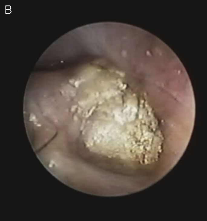
in fibrous tissue with inflammatory cells, but does not contain                 2. Little CJ, Lane JG, Gibbs C, Pearson GR. Inflammatory middle ear
                                                                                                  disease of the dog: The clinical and pathological features of cholestea-
               epithelium. Hemorrhage is usually evident and plays a role in
                                                                                                  toma, a complication of otitis media. Vet Rec 1991;128:319–322.
               the pathogenesis. Surgery is an effective treatment (7,18,19).                  3. Hardie EM, Linder KE, Pease AP. Aural cholesteatoma in twenty dogs.
                  Bacteria cultured from the middle ear cavity on first diagnosis                 Vet Surg 2008;37:763–770.
                                                                                               4. Venker-Van Haagen AJ. The ear. In: Schlütersche, ed. Ear, Nose, Throat
               and recurrence were those typically associated with otitis media
                                                                                                  and Tracheobronchial Diseases in Dogs and Cats. 1st ed. Hannover:
               (20). However, 5 samples were negative on culture (3,5); the                       Schlütersche Verlagsgesellschaft mbH & Co, 2005:1–50.
               authors could not find an explanation for the failure to recover                5. Davidson EB, Brodie HA, Breznock EM. Removal of a cholesteatoma
                                                                                                  in a dog, using a caudal auricular approach. J Am Vet Med Assoc 1997;
               bacteria, given the previous history of chronic otitis and the
                                                                                                  211:1549–1553.
A R T I C LE

               presence of middle ear disease in all dogs (20,21).                             6. Jacques D, Bouvy B. Un cas de cholestéatome auriculaire chez un
                  Recurrence of cholesteatoma is not a rare event and occurred                    chien traité par ablation totale du conduit auditif associée à une
                                                                                                  ostéotomie latérale de la bulle tympanique. Prad Méd Chir Anim Comp
               in 1/4 dogs at 12 mo after surgery in 1 study and in 10/19 dogs
                                                                                                  1999;34:67–72.
               at a mean time of 11.3 mo in another study (2,3). In this                       7. Ferlito A, Devaney KO, Rinaldo A, et al. Clinicopathological consulta-
               study, recurrence was suspected in 5 dogs and occurred at the                      tion. Ear cholesteatoma versus cholesterol granuloma. Ann Otol Rhinol
                                                                                                  Laryngol 1997;106:79–85.
               mean time of 7.5 mo. A minimum follow-up time of 12 mo
                                                                                               8. Olszweska E, Wagner M, Bernal-Sprekelsen M, et al. Etiopathogenesis
               should be recommended. It is likely that such a high incidence                     of cholesteatoma. Eur Arch Otorhinolaryngol 2004;261:6–24.
               of recurrence may be due to the persistence of keratin debris                   9. Friedmann I. Epidermoid cholesteatoma and cholesterol granuloma;
                                                                                                  experimental and human. Ann Otol Rhinol Laryngol 1959;68:57–79.
               or the perimatrix hidden in the epitympanic recess and in the
                                                                                              10. Friedmann I. The comparative pathology of otitis media, experimental
               newly formed small cavities in the bulla wall detectable during                    and human. II. The histopathology of experimental otitis of the guinea-
               surgery (2,22).                                                                    pig with particular reference to experimental cholesteatoma. J Laryngol
                                                                                                  Otol 1955;69:588–601.
                  Successful management of recurrent disease with prolonged
                                                                                              11. Persaud R, Hajioff D, Trinidade A, et al. Evidence-based review of
               intermittent antibiotic treatment has been reported (3). Although                  aetiopathogenic theories of congenital and acquired cholesteatoma.
               all dogs in this study underwent medical management with                           J Laryngol Otol 2007;121:1013–1019.
                                                                                              12. Mason LK, Harvey CE, Orsher RJ. Total ear canal ablation combined
               broad-spectrum antibiotics and anti-inflammatory drugs before
                                                                                                  with lateral bulla osteotomy for end-stage otitis in dogs. Results in thirty
               surgery, resolution of clinical signs after medical management                     dogs. Vet Surg 1988;17:263–268.
               occurred in only 1 dog at the second confirmed recurrence of                   13. Beckman SL, Henry WB Jr, Cechner P. Total ear canal ablation combin-
                                                                                                  ing bulla osteotomy and curettage in dogs with chronic otitis externa
               cholesteatoma. Interestingly, the only dog that underwent CT
                                                                                                  and media. J Am Vet Med Assoc 1990;196:84–90.
               and video-otoscopy when free of clinical signs, showed on both                 14. Tarabichi M. Endoscopic management of cholesteatoma: Long-term
               procedures findings suggestive of persistence of middle ear cho-                   results. Otolaryngol Head Neck Surg 2000;122:874–881.
                                                                                              15. Tarabichi M. Endoscopic management of limited attic cholesteatoma.
               lesteatoma. However, given the absence of clinical signs, the dog
                                                                                                  Laryngoscope 2004;114:1157–1162.
               was not subjected to surgery.                                                  16. Ajalloueyan M. Experience with surgical management of cholesteatomas.
                  In conclusion, middle ear cholesteatoma in dogs is a rare                       Arch Otolaryngol Head Neck Surg 2006;132:931–933.
                                                                                              17. Sturges BK, Dickinson PJ, Kortz GD, et al. Clinical signs, magnetic
               complication of chronic otitis and otitis media with a tendency
                                                                                                  resonance imaging features, and outcome after surgical and medical
               to recur despite surgery. Owners should be advised that a sec-                     treatment of otogenic intracranial infection in 11 cats and 4 dogs. J Vet
               ond surgery or on-going antibiotic therapy for management of                       Intern Med 2006;20:648–656.
                                                                                              18. Cox CL, Payne-Johnson CE. Aural cholesterol granuloma in a dog.
               otitis media may be necessary. The use of CT scan for middle
                                                                                                  J Small Anim Pract 1995;36:25–28.
               ear disease assessment can improve diagnosis of this disease,                  19. Fliegner RA, Jubb KV, Lording PM. Cholesterol granuloma associated
               especially in light of the tomographic distinctive features of                     with otitis media and destruction of the tympanic bulla in a dog. Vet
                                                                                                  Pathol 2007;44:547–549.
               middle ear cholesteatoma. Video-assisted surgery should be
                                                                                              20. Palmeiro BS, Morris DO, Wielmet SP, Shofer FS. Evaluation of out-
               further investigated in dogs. Investigation of the post-surgical                   come of otitis media after lavage of the tympanic bulla and long-term
               tomographic aspects of the middle ear cavity in dogs treated                       antimicrobial drug treatment in dogs: 44 cases (1998–2002). J Am Vet
                                                                                                  Med Assoc 2004;225:548–553.
               for cholesteatoma may aid in understanding the post-surgical
                                                                                              21. Murphy KM. A review of techniques for the investigation of otitis
               behavior of the disease and in detecting predictive changes for                    externa and otitis media. Clin Tech Small Anim Pract 2001;16:236–241.
               recurrence in asymptomatic patients.                           CVJ             22. Stangerup SE, Drozdziewicz D, Tos M, Hougaard-Jensen A. Recurrence
                                                                                                  of attic cholesteatoma: Different methods of estimating recurrence rates.
                                          References                                              Otolaryngol Head Neck Surg 2000;123:283–287.

                1. Goycoolea MV, Hueb MM, Muchow D, Paparella MM. The theory
                   of the trigger, the bridge and the transmigration in the pathogenesis of
                   acquired cholesteatoma. Acta Otolaryngol 1999;119:244–248.

               636                                                                                                                                 CVJ / VOL 52 / JUNE 2011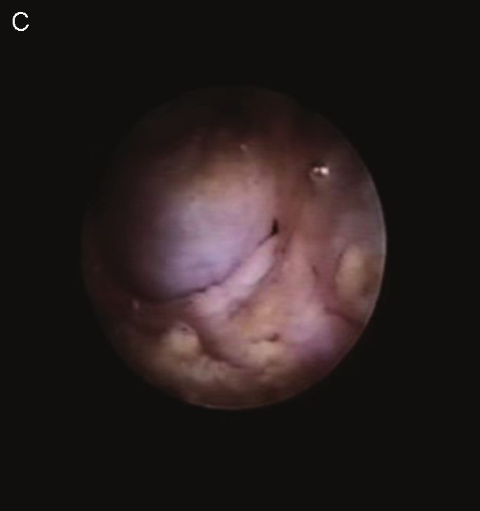
You can also read