Atypical Microbiological Feature of Infectious Endophthalmitis on Jeju Island: A 10-Year Study at a Single Tertiary Referral Center - Hindawi ...
←
→
Page content transcription
If your browser does not render page correctly, please read the page content below
Hindawi
Journal of Ophthalmology
Volume 2021, Article ID 6620926, 10 pages
https://doi.org/10.1155/2021/6620926

Research Article
Atypical Microbiological Feature of Infectious
Endophthalmitis on Jeju Island: A 10-Year Study at a Single
Tertiary Referral Center

          Joong Hyun Park ,1 Dong Yoon Kim ,2 Ahnul Ha ,1 Dae Joong Ma ,3 Hye Jin Lee ,1
          Jinho Jeong ,1 and Jin Young Kim 1
          1
            Department of Ophthalmology, Jeju National University Hospital, Jeju National University School of Medicine, Jeju,
            Republic of Korea
          2
            Department of Ophthalmology, Chungbuk National University Hospital, Chungbuk National University College of Medicine,
            Cheongju, Republic of Korea
          3
            Department of Ophthalmology, Hallym University Kangnam Sacred Heart Hospital, Hallym University College of Medicine,
            Seoul, Republic of Korea

          Correspondence should be addressed to Jin Young Kim; muse1016@naver.com

          Received 12 December 2020; Revised 12 February 2021; Accepted 15 February 2021; Published 2 March 2021

          Academic Editor: Alessandro Meduri

          Copyright © 2021 Joong Hyun Park et al. This is an open access article distributed under the Creative Commons Attribution
          License, which permits unrestricted use, distribution, and reproduction in any medium, provided the original work is
          properly cited.

          Background. To analyze the microbiological causes of infectious endophthalmitis on an isolated island over ten years. Methods. A
          retrospective review of the medical records of 49 eyes clinically diagnosed with infectious endophthalmitis between January 2009
          and December 2018 was done. The subjects were recruited from a single tertiary referral center on Jeju Island. The baseline
          characteristics of all subjects were investigated, and a culture examination was performed. The isolated pathogens were analyzed to
          determine how their microbiological features differed from those in regions with different geographical conditions. Results. Of the
          49 eyes, causative microorganisms were identified in 27 eyes (55.1%). Bacteria were found in 24 cases and fungi in 3 cases. Among
          the exogenous causes, Staphylococcus species (Staphylococcus aureus, S. epidermidis, and S. hominis) were the most common
          pathogens (7 cases). Achromobacter xylosoxidans was the second most common causative pathogen (4 cases) followed by
          Moraxella species (3 cases). The most frequent endogenous origin was due to Klebsiella pneumoniae (6 cases). The subjects were
          divided into two groups according to the treatment results and analyzed for predisposing factors related to visual outcomes. The
          presence of diabetes mellitus (P � 0.038) and initial visual acuity (P ≤ 0.001) were significant predisposing factors for visual
          outcomes. Conclusion. The causative microorganisms of endogenous endophthalmitis on Jeju Island were not different from those
          reported previously. However, isolated exogenous microorganisms were different from those reported in other studies from
          inland areas. A high incidence and atypical clinical features of Achromobacter xylosoxidans and Moraxella in exogenous
          endophthalmitis were observed, reflective of the distinct climatic features of Jeju Island: high humidity and temperature.
          Therefore, considering the causative microorganisms of exogenous endophthalmitis, it may be assumed that the causative
          microorganisms of exogenous endophthalmitis and its clinical manifestations differ according to the region.

1. Introduction                                                         causative organism directly invades the eye. While post-
                                                                        cataract endophthalmitis and posttraumatic endoph-
Infectious endophthalmitis is one of the most fatal com-                thalmitis constitute the majority of exogenous
plications of ophthalmic diseases, characterized by severe              endophthalmitis seen worldwide, endophthalmitis can also
intraocular infection originating from either exogenous or              occur in conjunction with intravitreal injections, keratitis,
endogenous factors. In exogenous endophthalmitis, the                   bleb after filtering surgery, and scleral buckle [1, 2].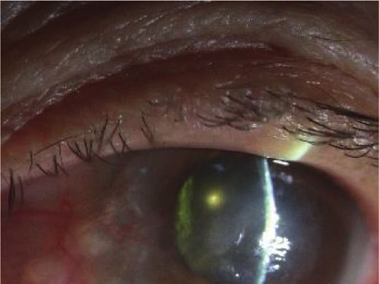
2                                                                                                  Journal of Ophthalmology

Postoperative cases accounted for 40–80%, and posttrau-         and visual outcomes of exogenous and endogenous
matic cases showed an incidence of 2–15% out of the total       endophthalmitis in different climates in Korea over a pe-
endophthalmitis cases in countries like England, Australia,     riod of ten years.
Korea, and Brazil, whereas the incidence of posttraumatic
endophthalmitis was higher in developing countries in-
cluding India, Egypt, and Thailand [1, 3–5]. Endogenous         2. Materials and Methods
endophthalmitis is less common than exogenous endoph-
thalmitis and is transmitted hematogenously from distant        This study was approved by the Institutional Review Board
foci of infection within the body [6].                          of Jeju National University Hospital (IRB No. 2019-01-011)
    The clinical features of infectious endophthalmitis vary    and was conducted in accordance with the Declaration of
according to the causative microorganism [7, 8]. Therefore,     Helsinki. The primary objective was to report the micro-
identifying the causative organism through culture is           biological features of infectious endophthalmitis on Jeju
critical. The frequency of causative microorganisms in          Island. The secondary objective was to determine the ra-
endophthalmitis varies depending on the risk factors for        tionale for the distribution of causative organisms of in-
endophthalmitis and its geographic locations. In post-          fectious endophthalmitis, which is different from that of
cataract endophthalmitis, Staphylococcus has been reported      other regions in Korea.
to be the most frequently isolated exogenous pathogen
globally including in Korea [9–12]. In other studies,
Streptococcus viridans and Pseudomonas aeruginosa were          2.1. Subjects. Patients with infectious endophthalmitis that
referred to as common exogenous pathogens of post-              occurred and was diagnosed on Jeju Island were included in
cataract endophthalmitis [13–15]. In contrast, the fungus       this study. Based on the origin of infectious endophthalmitis,
was found to be a common cause of postoperative                 it was classified as either exogenous or endogenous.
endophthalmitis in tropical areas including India [16–18].      Endophthalmitis originating from a source within the body
Bacillus species are known as major pathogens in post-          was defined as endogenous, and that due to direct inocu-
traumatic endophthalmitis, causing fulminant infections         lation of an organism from outside was defined as exoge-
with poor prognosis [19]. In a prospective study on             nous. Exclusion criteria were as follows: (a) patients referred
postintravitreal anti-VEGF endophthalmitis in England,          from other regions; (b) patients with diseases mimicking
coagulase-negative staphylococci and Staphylococcus epi-        infectious endophthalmitis (noninfectious uveitis, blebitis,
dermidis presented in higher frequencies [20]. Keratitis can    sterile endophthalmitis after intravitreal injection or sur-
spread to cause endophthalmitis; a case series in Florida       gery); (c) patients with diseases (corneal opacity, progressed
found that 53% of keratitis-related endophthalmitis was         cataract [≥ moderate stage), glaucoma, and comorbid retinal
caused by molds contrary to an incidence of 17% of in a         diseases such as diabetic retinopathy, retinal vein or artery
similar case series from New Jersey, assumed to be at-          occlusion, and age-related macular degeneration) that could
tributed to the high humidity of the Florida region [21, 22].   significantly affect visual acuity; and (d) patients with follow-
Endophthalmitis is sometimes known to occur after               up duration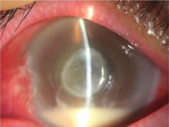
Journal of Ophthalmology                                                                                                                       3

2.4. Treatment Modality and Indication. Management in-                    Table 1: Baseline characteristics of patients in this study.
cluded intravitreal injection (vancomycin 1.0 mg/0.1 mL,              Characteristics
ceftazidime 2.25 mg/0.1 mL), topical fortified antibiotics,
                                                                      Age (years)                                              65.98 ± 17.03a
and intravenous or oral broad-spectrum antibiotics; intra-            Sex (male: female)                                           26 : 23
vitreal voriconazole (100 μm/0.1 mL) was administered when
                                                                      Underlying disease, n (%)
fungal infection was suspected. Pars plana vitrectomy was
                                                                        Diabetes                                                 12 (24.5)
performed for patients with (a) invisible fundus due to                 Hypertension                                             36 (73.5)
severe infection and inflammation, (b) no improvement
                                                                      Initial BCVA, LogMAR                                      0.39 ± 0.21a
after initial treatment, (c) visual acuity ≤ counting fingers, or
                                                                      Culture-positive, n/total (%)                             27/49 (55.1)
(d) severe clinical features at the initial visit. After initiating
                                                                        Bacteria, n (%)                                          24 (88.9)
treatment, the treatment regimen was modified according to               Fungus, n (%)                                             3 (11.1)
the results of the antibiotic sensitivity test and culture.
                                                                      BCVA � best collected visual acuity. LogMAR � logarithm of the minimum
                                                                      angle of resolution. a Values are presented as mean ± standard deviation.

2.5. Environmental Factors’ Investigation. To compare the
differences in environmental factors between Jeju Island and               Table 2: Causative factors in infectious endophthalmitis.
other inland areas, data from the Korea Meteorological                Factors                   No. of identified cases/total cases (%)
Administration during the same study period (10 years,
                                                                      Exogenous                              19/39 (48.7)
2009–2018) in Korea were analyzed. The following envi-                  Cataract surgery                     13/24 (54.2)
ronmental factors were investigated: average annual tem-                Trauma                                2/8 (25.0)
perature, average annual precipitation, average annual                  Other surgery                         2/4 (50.0)
relative humidity, and annual sunshine hours.                           Infectious keratitis                  2/3 (66.7)
                                                                      Endogenous                              7/9 (77.8)
                                                                        Liver abscess                         6/7 (85.7)
                                                                        Septic pneumonia                      1/1 (100)
2.6. Data Analysis. The initial and final visual acuities were
                                                                        Urinary tract infection                0/1 (0)
estimated through manifested refraction and converted to
                                                                      Unknown                                 1/1 (100)
the logMAR scale for statistical analysis. The visual acuity of
                                                                      Total                                  27/49 (55.1)
no light perception, light perception, hand motion, and
counting fingers were set as 3.0, 2.5, 2.3, and 2.0, respectively
[27]. Treatment failure was defined as final visual acuity              3.2. Isolated Microorganisms. Table 3 shows the microbial
≥logMAR 1.0 or cases with evisceration due to uncontrolled            distribution, treatment, and treatment success rate of ex-
inflammation and severe pain. Based on these criteria, we              ogenous and endogenous endophthalmitis cases. Among the
divided the subjects into two groups: treatment success               exogenous endophthalmitis cases, Staphylococcus spp.
group and treatment failure group. The predisposing factors           showed the highest frequency (7/20, 35.0%). As a solitary
that could influence visual outcomes were analyzed.                    cause of exogenous endophthalmitis, S. epidermidis and
    Statistical analyses were performed using SPSS version            Achromobacter xylosoxidans were the most common path-
23 (SPSS Inc., Chicago, IL, USA), and variables were                  ogens, with 4 cases each (4/20, 20.0%). They were followed
compared using independent t-tests and Pearson’s chi-                 by Moraxella species (3/20, 15.0%), S. aureus (2/20, 10.0%),
square tests. Statistical significance was set at P value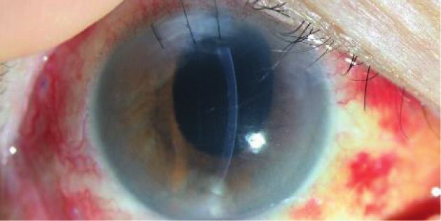
4                                                                                                      Journal of Ophthalmology

                   Table 3: Identified organisms and treatment results in exogenous and endogenous endophthalmitis.
                                                                                                       The number of
Causative organism                                                                No.
                                                                                           PPV/EVI      Injections   Success (%)
Exogenous                                                                          20
                                                    Staphylococcus epidermidis      4         3/1            8          3 (75.0)
                         Staphylococcus species        Staphylococcus aureus        2         2/0            3          1 (50.0)
                                                      Staphylococcus hominis        1         2/0            4          1 (100)
G (+) bacteria
                      Achromobacter xylosoxidans                                    4        12/0           49          4 (100)
                         Streptococcus species          Streptococcus oralis        1        2/0            6            0 (0)
                                                     Streptococcus intermedius      1        1/0            2           1 (100)
                           Moraxella species                                        3         3/1            6          1 (33.3)
G (−) bacteria
                        Pseudomonas aeruginosa                                      2        1/2            3            0 (0)
Filamentous                Fusarium species                                         2        2/1            8           1 (50.0)
Endogenous                                                                          7
G (−) bacteria           Klebsiella pneumoniae                                      6        6/0            10         2 (33.3)
Yeast                       Candida species                                         1        1/0             3           0 (0)
                                  Total                                            27                                  14 (51.9)
PPV � pars plana vitrectomy. EVI � evisceration.

annual precipitation (top place), higher average annual              4. Discussion
relative humidity (fourth place), and the lowest annual
sunshine hours (first place) over 10 years in Korea (Figure 3).       The primary finding of this study is a unique distribution of
                                                                     exogenous pathogens in endophthalmitis, which is different
                                                                     from most previous reports. A previous study reported
3.4. Clinical Outcomes. The treatment success and failure            significantly higher incidences of Pseudomonas and Asper-
rates were compared and summarized in Table 4. The mean              gillus species as causes of postoperative endophthalmitis in
age in the treatment success and treatment failure groups            India with tropical climatic conditions [17]. Furthermore,
were 62.90 ± 17.90 years and 72.13 ± 13.67 years, respec-            Ramos et al. [28] revealed an association between Pseudo-
tively. In both groups, there were more males than females,          monas aeruginosa and high humidity, temperature, and
and the male-to-female ratio in the treatment success and            precipitation. In addition, some studies found that the
treatment failure groups was 17 : 15 (53.1% males) and 9 : 8         frequency of Gram-negative bacteria such as Enterobacter
(52.9% males), respectively. The culture positivity rate in          cloacae and Klebsiella pneumoniae increased with an in-
the treatment success group was higher (40.6%) than that in          crease in temperature [29, 30]. Jeju Island has a warm and
the treatment failure group (35.3%); however, the differ-             humid climate throughout the four seasons, unlike other
ence was not significant (P � 0.715). Hypopyon was ob-                inland regions in Korea. Therefore, this study focused on
served more commonly in the treatment failure group                  determining how this unique feature of Jeju Island might
(66.7%) than in the treatment success group (56.3%);                 contribute to the differences in microbial features from other
however, no significant difference was found between the               regions in Korea.
two groups (P � 0.566). The two groups were compared                      Endogenous endophthalmitis is known to be largely
according to the number of treatments, such as intravitreal          affected by individual chronic underlying diseases and
injection and/or vitrectomy. The mean number of intra-               medical conditions such as diabetes mellitus, infectious
vitreal injections was 3.77 ± 4.95 in the treatment success          endocarditis, and liver cirrhosis [6]. In Western countries
group, and 2.80 ± 1.93 in the treatment failure group, and           including the United States and Europe, Gram-positive
this difference was statistically insignificant (P � 0.472).           bacteria and fungi are common, whereas Klebsiella pneu-
There were no significant differences in the number of                 moniae is the most common cause of endogenous
vitrectomies (P � 0.153).                                            endophthalmitis in East Asia, including Korea [31]. This
    There were two main predisposing factors related to the          variation, which has not yet been revealed, is thought to be
visual outcomes of infectious endophthalmitis in the                 associated with intravenous drug abuse in Western coun-
comparison between the two groups. First, the initial best-          tries, genetic differences between racial groups, and a high
corrected visual acuity (BCVA) was the most important                incidence of cholangiohepatitis in East Asia [31, 32]. This
factor in determining the final visual outcome. The initial           study showed similar results to previous reports in Korea
visual acuity presented as logMAR in the treatment success           showing a predominance of Klebsiella pneumoniae as the
and treatment failure groups was 1.71 ± 0.76 and 2.47 ± 0.23         causative microorganism in endogenous endophthalmitis
(P ≤ 0.001), respectively. Moreover, diabetes mellitus was           [25, 31, 32].
relatively common in the treatment failure group (47.1%),                 The Endophthalmitis Vitrectomy Study indicated that
which had a higher risk than the treatment failure group did         Staphylococcus epidermidis was the most commonly isolated
(P � 0.038).                                                         exogenous pathogen followed by Staphylococcus aureus,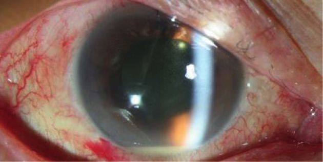
Journal of Ophthalmology 5
(a) (b) (c)
(d) (e) (f )
(g) (h) (i)
(j) (k)
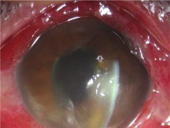
Figure 1: Representative cases of endophthalmitis caused by Achromobacter xylosoxidans. (a) The corneal edema, anterior chamber cell
reaction, hypopyon, and pupillary membrane were found at the first visit. (b) In spite of pars plana vitrectomy and intravitreal injection, the
endophthalmitis has aggravated. (c) Intraocular lens was removed and partial lens capsule was removed by the outcome and quiet state
persisted. (d) After ciliary sulcus fixation of the intraocular lens, cellular reactions have recurred. (e) The Gram-negative rod Achromobacter
xylosoxidans was revealed from blood agar plate culture. (f ) Microphotograph of Achromobacter xylosoxidans with gram staining (×1000).
(g) Surgical procedure showing removal of the intraocular lens. (h) The remnant lens capsule is being removed completely using
microforceps. (i) After additional vitrectomy, repeated intraocular lens removal, and en bloc delivery of the lens capsule, recurrence has
stopped thereafter. (j) Another patient showed the anterior chamber reaction and severe pupillary fibrotic membrane from the initial
presentation. (k) Completely healed state after repeated intravitreal injections and vitrectomies, including en bloc delivery of the lens
capsule.
Streptococcus species, and Enterococcus species [33]. Other introduced, and the sources of detection included blood,
studies have also shown Staphylococcus epidermidis to be the urine, sputum, swimming pools, and water tanks. Of the 14
most common pathogen [9, 34] while Torabi et al. [13] strains observed, 13 presented positive growth at 37°C except
revealed that Streptococcus viridans was the most common for one that was incubated at room temperature [36].
pathogen. Kim et al. [14] and Dave et al. [15] reported that Moreover, a high incidence of Moraxella species was also
Pseudomonas aeruginosa was the most commonly isolated found, which is a unique aspect of this study. Moraxella
pathogen. Unlike previous studies, this study’s results species are normal flora microorganisms known to flourish
demonstrated a high frequency of Achromobacter xylosox- under moist and high-temperature conditions, and Larsen
idans (4/20, 20.0%) and Moraxella species (3/20, 15.0%) as et al. [37] reported that the colonies tended to spread at 37°C
causes of exogenous endophthalmitis. in a moist chamber. This pathogen, with low virulence, exists
Achromobacter xylosoxidans is a very rare cause of in- in the upper respiratory tract, genitourinary tract, and
fectious endophthalmitis. This microorganism adapts well in conjunctiva, known to cause ocular diseases such as infec-
humid and warm climates [35]. According to Holmes et al., tious keratitis, conjunctivitis, and endophthalmitis [38, 39].
several strains of Achromobacter xylosoxidans were According to a Korean study on infectious keratitis,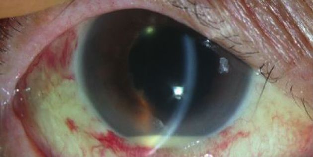
6                                                                                                                   Journal of Ophthalmology

                 (a)                                 (b)                               (c)                                  (d)

                 (e)                                 (f )                              (g)                                  (h)

Figure 2: Representative cases of endophthalmitis caused by Moraxella species. In both cases, infectious keratitis was the predisposing
factor. (a, b) Corneal edema, central infiltration, and epidefect were observed along with significant hypopyon at the initial visit. (c, d)
Regressed central infiltration and epidefect after intravitreal antibiotic injection. (e, f ) Conjunctival chemosis and injection, 4°/c corneal
infiltration with epidefect, pupillary membrane, and hypopyon were seen. (g, h) Pars plana vitrectomy and intravitreal antibiotic injection
were performed, and the endophthalmitis has cleared up.

                       Average annual temperature                      Average annual precipitation
                           0.0–12.9°C             15.0–15.9°C               800–900 mm               1400–1599 mm
                           13.0–13.9°C            16.0–16.9°C               1000–1199 mm             1600–1799 mm
                           14.0–14.9°C                                      1200–1399 mm
                                            (a)                                                (b)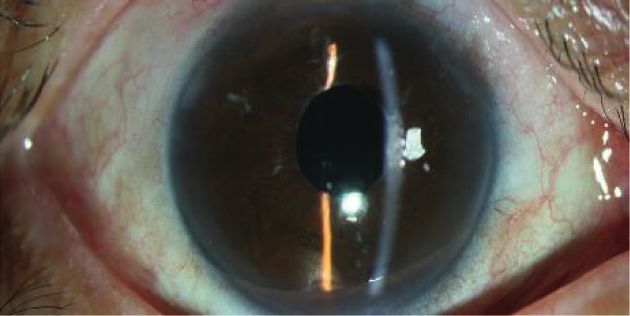
Journal of Ophthalmology                                                                                                                                7

                        Average annual relative humidity
                                                                            Average annual sunshine hours
                            55.0–55.9%            65.0–69.9%
                            60.0–64.9%            70.0–74.9%                    1800–1999 hr          2200–2399 hr
                                                                                2000–2199 hr          2400–2599 hr
                                               (c)                                                       (d)

Figure 3: The average temperature, the annual precipitation, the average humidity, and the annual sunshine hours for 10 years (2009–2018)
in South Korea. The graph showed that Jeju Island had the highest average temperature (1st place), the highest annual precipitation (1st
place), higher average relative humidity (4th place), and the lowest annual sunshine hours (1st place) for 10 years in the country.

                             Table 4: The comparison between treatment success and treatment failure groups.
                                                                                              No. of cases (%)
Variables
                                              Treatment success (32 eyes)                         Treatment failure (17 eyes)                     P-value
Age                                                 62.90 ± 17.90                                         72.13 ± 13.67                           0.086a
Sex                                                                                                                                               0.990b
  Male                                                  17 (53.1)                                               9 (52.9)
  Female                                                15 (46.9)                                               8 (47.1)
Diabetes mellitus                                                                                                                                 0.038c
  Yes                                                    5 (15.6)                                               8 (47.1)
  No                                                    27 (84.4)                                               9 (52.9)
Hypertension                                                                                                                                      0.801b
  Yes                                                   12 (37.5)                                               7 (41.2)
  No                                                    20 (62.5)                                              10 (58.8)
Hypopyon                                                                                                                                          0.566b
  Yes                                                   18 (56.3)                                              11 (66.7)
  No                                                    14 (43.7)                                              6 (35.3)
Culture results                                                                                                                                   0.715b
  positive                                              13 (40.6)                                                6 (35.3)
  negative                                              19 (59.4)                                               11 (64.7)
Initial BCVA (log MAR)                                 1.71 ± 0.76                                             2.47 ± 0.23                        ≤0.001a
Treatment number
  Vitrectomy                                           1.42 ± 1.30                                             0.85 ± 0.80                        0.153a
  Intravitreal injection                               3.77 ± 4.95                                             2.80 ± 1.93                        0.472a
PPV � pars plana vitrectomy. BCVA � best-corrected visual acuity. a Independent t-test;   b
                                                                                              Pearson’s chi-square test; c Fisher’s exact test.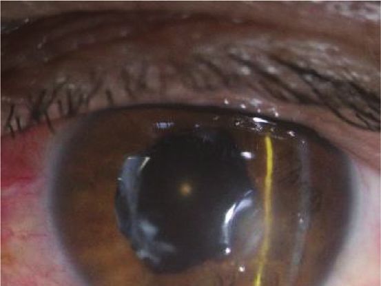
8                                                                                                     Journal of Ophthalmology

infection by Moraxella species was only observed in 0.3% of        the assumption that more bacterial loads may be present in
the total 689 cases [40], and only a few cases of infectious       culture-positive cases. Diabetes mellitus and initial visual
endophthalmitis associated with a bleb at the delayed onset        acuity were the only factors associated with the treatment
have been described [41, 42]. These characteristics of Ach-        success rate in this study.
romobacter xylosoxidans and Moraxella species match the                To the best extent of the author’s knowledge, this is the
climate of Jeju Island. It is an isolated island surrounded by     first study on endophthalmitis on Jeju Island. In addition,
the ocean, with high all-year-round temperature and hu-            the data collection over ten years from a single tertiary
midity. In addition, the sunlight exposure time was the            center could aid in strengthening the analysis of the
lowest in Korea, which is the optimum condition for the            features of endophthalmitis on Jeju Island. Moreover, it
growth of these bacteria (Figure 3).                               can be proposed that exogenous endophthalmitis might
     Achromobacter xylosoxidans displayed a notable treat-         be affected by environmental factors around the causative
ment process compared to the other pathogens detected in           pathogens. This study has some limitations. First, Jeju
our study. Few cases of severe recurrent endophthalmitis           Island is a popular tourist spot, and some of the subjects
caused by Achromobacter xylosoxidans have been reported            were from other regions that deteriorated the distinc-
[43]. Owing to the characteristics of Achromobacter xylo-          tiveness of the microbiome in endophthalmitis on Jeju
soxidans in creating a biofilm, and remaining in the capsular       Island. Furthermore, owing to its retrospective design,
bag and intraocular lens, a high tendency of recurrence was        some of the patient’s data were missing or distorted. This
observed despite simple pars plana vitrectomy and intra-           report showed the environmental effects of exogenous
vitreal antibiotic injections [44, 45]. Two cases in the current   pathogens; however, the control groups from another
study underwent pars plana vitrectomy six and four times           region with different geographic locations were not in-
each and were administered with intravitreal injections 23         cluded. Further data collection and comparative inves-
and 18 times, respectively. After several recurrences, radical     tigation of districts with different climatic conditions
capsulectomy was performed along with intraocular lens             might yield more significant results.
removal to terminate the episode. For the other two cases of
infectious endophthalmitis caused by Achromobacter xylo-
soxidans, pars plana vitrectomy, radical capsulectomy, and         5. Conclusions
intraocular lens removal were the initial treatment modal-
ities, and complete remission was achieved (Table 3). The          This study investigated and reported the microbiology, in-
average final BCVA was 0.43, which ranged from 0.2 to 0.7,          cidence, and clinical features of Jeju Island for the first time.
and all four cases were successfully treated.                      This decade-long study from a single tertiary center dem-
     There has been evidence that humidity and temperature         onstrated atypical distribution in microorganisms of exog-
were associated with the incidence of Pseudomonas aeru-            enous endophthalmitis, distinguishable from most previous
ginosa [28], and an increasing rate of Gram-negative species       studies. High incidences of rare pathogens as causes of
was found at high temperatures [29, 30]. Unlike endogenous         endophthalmitis were detected, and the climatic features of
endophthalmitis, which arises from a specific focus of in-          Jeju Island have met the thriving conditions of these
fection in the body, the source of exogenous endoph-               pathogens. Endophthalmitis is an ophthalmic emergency,
thalmitis mainly originates from periocular normal flora.           and that can deteriorate ocular structures in a short time in
Being exposed to the outside environment, the incidence            the absence of accurate diagnosis and appropriate treatment.
and distribution of normal flora could possibly be affected          If unfavorable progress is observed despite adequate treat-
and changed by the humid and warm climate of Jeju Island,          ment, atypical pathogens of endophthalmitis regarding
thus justifying the rare presentation of Achromobacter             climatic and geographic features of the region should be
xylosoxidans and Moraxella species in this study.                  suspected. These findings will be of particular interest to
     Considering the predisposing factors of infectious            ophthalmologists and will contribute to the future treatment
endophthalmitis in our study, cataract surgery was the most        of infectious endophthalmitis on Jeju Island.
common cause (24/49, 48.9%) followed by endogenous
cause (18.4%) and trauma (16.3%). Of the eight cases of
trauma-induced infectious endophthalmitis, six patients            Data Availability
were injured during farm work; therefore, it can be pre-
                                                                   The datasets included in this study are available from the
sumed that the higher proportion of traumatic causes was
                                                                   corresponding author upon reasonable request.
due to the relatively large number of agricultural workers on
Jeju Island. In contrast, the most common cause of en-
dogenous endophthalmitis was a liver abscess. This result is       Disclosure
similar to that of many previous studies in East Asia and
Korea [25, 31, 32].                                                This work was presented at “The 121st Annual Meeting of the
     Factors associated with final visual acuity have been          Korean Ophthalmological Society” in 2019 based on the link
reported to be initial visual acuity, diabetes mellitus, the       http://www.ophthalmology.org/abstract/2019_121/eng/ and
presence of microorganisms, and hypopyon in previous               “The Association for Research in Vision and Ophthalmol-
studies [33, 46, 47]. Ho et al. [9] identified culture-negativity   ogy” in 2018 based on the link https://www.arvo.org/annual-
as a positive prognostic factor for better visual acuity, under    meeting/meeting-info/past-annual-meetings/.
Journal of Ophthalmology                                                                                                                     9

Conflicts of Interest                                                  [11] J. Y. Jung, B. Y. Ko, and B. Y. Kim, “Factors associated with a
                                                                            poor visual result in acute endophthalmitis after cataract
The authors do not have any potential conflicts of interest                  surgery,” Journal of the Korean Ophthalmological Society,
relevant to this manuscript.                                                vol. 49, no. 8, pp. 1242–1247, 2008.
                                                                       [12] N. E. Lee and J. M. Park, “Clinical results of bacterial
                                                                            endophthalmitis: bacterial culture and visual acuity out-
Authors’ Contributions                                                      comes,” Journal of the Korean Ophthalmological Society,
                                                                            vol. 52, no. 10, pp. 1173–1181, 2011.
Park JH and Kim DY wrote the original draft and performed              [13] H. Torabi, S.-A. Tabatabai, and A. Khodabande, “Treatment
the conceptualization, formal analysis, and investigation                   outcomes of post cataract surgery endophthalmitis in a ter-
under the supervision of the corresponding author, Kim JY.                  tiary referral center in Iran,” Journal of Current Ophthal-
Ma DJ, Lee HJ, Ha A, and Jeong J performed data curation,                   mology, vol. 30, no. 2, pp. 152–155, 2018.
methodology, and validation. And after completing the                  [14] J. Y. Kim, S. J. Wang, C. J. Park, and S. B. Lee, “Risk factors
draft, reviewing and editing were done by Kim DY and Kim                    leading to enucleation or evisceration in endophthalmitis,”
JY. Joong Hyun Park and Dong Yoon Kim contributed                           Journal of the Korean Ophthalmological Society, vol. 48, no. 10,
equally to this work.                                                       pp. 1362–1368, 2007.
                                                                       [15] V. P. Dave, R. R. Pappuru, M. Tyagi, A. Pathengay, and T. Das,
                                                                            “Endoscopic vitrectomy in endophthalmitis: initial experience
Acknowledgments                                                             of 33 cases at a tertiary eye care center,” Clinical Ophthal-
                                                                            mology, vol. 13, p. 243, 2019.
This work was supported by a reasearch grant from Jeju
                                                                       [16] R. K. Forster, R. L. Abbott, and H. Gelender, “Management of
National University Hospital in 2017.                                       infectious endophthalmitis,” Ophthalmology, vol. 87, no. 4,
                                                                            pp. 313–319, 1980.
References                                                             [17] D. Y. Kunimoto, T. Das, S. Sharma et al., “Microbiologic
                                                                            spectrum and susceptibility of isolates:,” American Journal of
 [1] M. L. Durand, “Bacterial and fungal endophthalmitis,”                  Ophthalmology, vol. 128, no. 2, pp. 240–242, 1999.
     Clinical Microbiology Reviews, vol. 30, no. 3, pp. 597–613,       [18] A. R. Anand, K. L. Therese, and H. N. Madhavan, “Spectrum
     2017.                                                                  of aetiological agents of postoperative endophthalmitis and
 [2] A. Meduri, A. De Maria, A. A. Severo, and P. Aragona,                  antibiotic susceptibility of bacterial isolates,” Indian Journal of
     “Infectious conjunctivitis caused by pseudomonasaeruginosa             Ophthalmology, vol. 48, no. 2, pp. 123–128, 2000.
     in infected and extrused scleral buckles,” BMJ Case Reports,      [19] J. J. Miller, I. U. Scott, H. W. Flynn Jr et al., “Endophthalmitis
     vol. 13, Article ID e232296, 2020.                                     caused by Bacillus species,” American Journal of Ophthal-
 [3] S. Sharma, T. R. Padhi, S. Basu, S. Kar, A. Roy, and T. Das,           mology, vol. 145, no. 5, pp. 883–888, 2008.
     “Endophthalmitis patients seen in a tertiary eye care centre in   [20] D. A. M. Lyall, A. Tey, B. Foot et al., “Post-intravitreal anti-
     Odisha: a clinico-microbiological analysis,” Indian Journal of         VEGF endophthalmitis in the United Kingdom: incidence,
     Medical Research, vol. 139, pp. 91–98, 2014.                           features, risk factors, and outcomes,” Eye, vol. 26, no. 12,
 [4] A. Gharamah, A. Moharram, M. Ismail, and A. Al-Hussaini,               pp. 1517–1526, 2012.
     “Bacterial and fungal endophthalmitis in Upper Egypt: related     [21] C. R. Henry, H. W. Flynn, D. Miller, R. K. Forster, and
     species and risk factors,” Asian Pacific Journal of Tropical            E. C. Alfonso, “Infectious keratitis progressing to endoph-
     Biomedicine, vol. 2, no. 8, pp. 655–659, 2012.                         thalmitis,” Ophthalmology, vol. 119, no. 12, pp. 2443–2449,
 [5] C. Bhoomibunchoo, T. Ratanapakorn, S. Sinawat,
                                                                            2012.
     T. Sanguansak, K. Moontawee, and Y. Yospaiboon, “Infec-
                                                                       [22] M. Malihi, X. Li, S. Patel et al., “Infectious keratitis-associated
     tious endophthalmitis: review of 420 cases,” Clinical Oph-
                                                                            endophthalmitis,” Retina, vol. 37, no. 4, pp. 662–666, 2017.
     thalmology (Auckland, N.Z.), vol. 7, pp. 247–252, 2013.
                                                                       [23] J. F. English, R. Barry, and R. W. Essex, “Postoperative
 [6] A. A. Okada, R. P. Johnson, W. C. Liles, D. J. D’Amico, and
                                                                            endophthalmitis following chandelier-assisted scleral buckle
     A. Sullivan Baker, “Endogenous bacterial endophthalmitis,”
                                                                            for primary repair of rhegmatogenous retinal detachment,”
     Ophthalmology, vol. 101, no. 5, pp. 832–838, 1994.
 [7] A. E. Kuriyan, K. D. Weiss, H. W. Flynn Jr et al.,                     Acta Ophthalmologica, vol. 97, no. 1, pp. e130–e131, 2019.
     “Endophthalmitis caused by streptococcal species: clinical        [24] T. Sakono, H. Otsuka, H. Shiihara, N. Yoshihara, and
     settings, microbiology, management, and outcomes,” Amer-               T. Sakamoto, “Acute bacterial endophthalmitis after scleral
     ican Journal of Ophthalmology, vol. 157, no. 4, pp. 774–780,           buckling surgery with chandelier endoillumination,” Ameri-
     2014.                                                                  can Journal of Ophthalmology Case Reports, vol. 8, pp. 7–10,
 [8] X.-B. Yang, Y.-Y. Liu, Z.-X. Huang, Y. Mao, L. Zhao, and               2017.
     Z.-P. Xu, “Clinical analysis of 1593 patients with infectious     [25] J. Wong, T. K. Chan, H. M. Lee, and S. P. Chee, “Endogenous
     endophthalmitis,” Chinese Medical Journal, vol. 131, no. 14,           bacterial endophthalmitis an East Asian experience and a
     p. 1658, 2018.                                                         reappraisal of a severe ocular affliction,” Ophthalmology,
 [9] I.-V. Ho, G. Fernandez-Sanz, S. Levasseur et al., “Early pars          vol. 107, no. 8, pp. 1483–1491, 2000.
     plana vitrectomy for treatment of acute infective endoph-         [26] M. W. Johnson, B. H. Doft, S. F. Kelsey et al., “The
     thalmitis,” Asia-Pacific Journal of Ophthalmology, vol. 8,              endophthalmitis vitrectomy study: relationship between
     pp. 3–7, 2019.                                                         clinical presentation and microbioloaic spectrum,” Ophthal-
[10] E. H. Leung, A. E. Kuriyan, H. W. Flynn, D. Miller, and                mology, vol. 104, pp. 261–272, 1997.
     L. C. Huang, “Persistently vitreous culture-positive exogenous    [27] C. Lange, N. Feltgen, B. Junker, K. Schulze-Bonsel, and
     bacterial endophthalmitis,” American Journal of Ophthal-               M. Bach, “Resolving the clinical acuity categories “hand
     mology, vol. 165, pp. 16–22, 2016.                                     motion” and “counting fingers” using the freiburg visual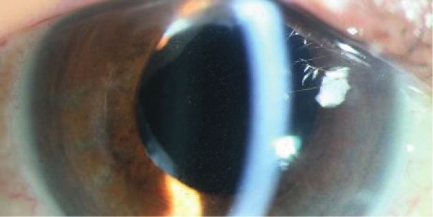
10                                                                                                                 Journal of Ophthalmology

       acuity test (FrACT),” Graefe’s Archive for Clinical and Ex-          [44] R. M. Donlan and J. W. Costerton, “Biofilms: survival
       perimental Ophthalmology, vol. 247, no. 1, pp. 137–142, 2009.             mechanisms of clinically relevant microorganisms,” Clinical
[28]   G. P. Ramos, J. L. Rocha, and F. F. Tuon, “Seasonal humidity              Microbiology Reviews, vol. 15, no. 2, pp. 167–193, 2002.
       may influence Pseudomonas aeruginosa hospital-acquired                [45] V. M. Villegas, A. Emanuelli, H. W. Flynn et al., “Endoph-
       infection rates,” International Journal of Infectious Diseases,           thalmitis caused by achromobacter xylosoxidans after cataract
       vol. 17, pp. 757–761, 2013.                                               surgery,” Retina, vol. 34, no. 3, p. 583, 2014.
[29]   M. R. Eber, M. Shardell, M. L. Schweizer, R. Laxminarayan,           [46] S.-J. Yoo, S.-W. Cho, and J.-W. Kim, “Clinical analysis of
       and E. N. Perencevich, “Seasonal and temperature-associated               posttraumatic endophthalmitis,” Journal of the Korean
       increases in gram-negative bacterial bloodstream infections               Ophthalmological Society, vol. 45, pp. 69–78, 2004.
       among hospitalized patients,” PLoS One, vol. 6, Article ID           [47] J.-H. Cheng, Y.-H. Chang, C.-L. Chen, Y.-H. Chen, D.-W. Lu,
       e25298, 2011.                                                             and J.-T. Chen, “Acute endophthalmitis after cataract surgery
[30]   E. N. Perencevich, J. C. McGregor, M. Shardell et al., “Summer            at a referral centre in Northern Taiwan: review of the causative
       peaks in the incidences of gram-negative bacterial infection              organisms, antibiotic susceptibility, and clinical features,” Eye,
       among hospitalized patients,” Infection Control & Hospital                vol. 24, no. 8, p. 1359, 2010.
       Epidemiology, vol. 29, no. 12, pp. 1124–1131, 2008.
[31]   S. Lee, T. Um, S. G. Joe et al., “Changes in the clinical features
       and prognostic factors of endogenous endophthalmitis,”
       Retina, vol. 32, no. 5, pp. 977–984, 2012.
[32]   H. W. Lim, J. W. Shin, H. Y. Cho et al., “Endogenous
       endophthalmitis in the Korean population,” Retina, vol. 34,
       no. 3, pp. 592–602, 2014.
[33]   E. Vitrectomy, “Results of the endophthalmitis vitrectomy
       study,” Archives of ophthalmology, vol. 113, pp. 1479–1496,
       1995.
[34]   X. Lu, H. Xia, C. Jin et al., “Prognostic factors associated with
       visual outcome of salvageable eyes with posttraumatic
       endophthalmitis,” Scientific Reports, vol. 9, pp. 1–8, 2019.
[35]   K. Marion-Sanchez, K. Pailla, C. Olive, X. Le Coutour, and
       C. Derancourt, “Achromobacter spp. healthcare associated
       infections in the French West Indies: a longitudinal study
       from 2006 to 2016,” BMC Infectious Diseases, vol. 19, p. 795,
       2019.
[36]   B. Holmes, J. J. Snell, and S. P. Lapage, “Strains of Achro-
       mobacter xylosoxidans from clinical material,” Journal of
       Clinical Pathology, vol. 30, no. 7, pp. 595–601, 1977.
[37]   J. L. Larsen, N. Bille, and N. C. Nielsen, “Occurrence and
       possible role of Moraxella species in pigs,” Acta Pathologica et
       Microbiologica Scandinavica. Section B: Microbiology and
       immunology, vol. 81, no. 2, pp. 181–186, 1973.
[38]   S. J. LaCroce, M. N. Wilson, J. E. Romanowski et al.,
       “Moraxella nonliquefaciens and M. osloensis are important
       Moraxella species that cause ocular infections,” Microor-
       ganisms, vol. 7, no. 6, p. 163, 2019.
[39]   S. Das, M. Constantinou, M. Daniell, and H. R. Taylor,
       “Moraxella keratitis: predisposing factors and clinical review
       of 95 cases,” British Journal of Ophthalmology, vol. 90, no. 10,
       pp. 1236–1238, 2006.
[40]   J. Y. Kim, K. C. Yoon, Y. G. Park, N. C. Cho, and I. C. You,
       “Age-related clinical analysis of infectious keratitis in two
       tertiary centers,” Journal of the Korean Ophthalmological
       Society, vol. 51, no. 7, pp. 927–934, 2010.
[41]   R. M. Lipman and T. A. Deutsch, “Late-onset Moraxella
       catarrhalis endophthalmitis after filtering surgery,” Canadian
       Journal of Ophthalmology. Journal Canadien D’ophtalmologie,
       vol. 27, no. 5, pp. 249-250, 1992.
[42]   R. L. Bergren, W. S. Tasman, R. T. Wallace, and L. J. Katz,
       “Branhamella (Moraxella) catarrhalis endophthalmitis,” Ar-
       chives of Ophthalmology, vol. 111, no. 9, pp. 1169-1170, 1993.
[43]   J. H. Park, E. K. Lee, S. Y. Lee, D. Y. Kim, and J. Y. Kim,
       “Recurrent endophthalmitis caused by achromobacter xylo-
       soxidans: importance of aggressive surgical removal of cap-
       sular bag,” Korean Journal of Ophthalmology, vol. 32, no. 2,
       pp. 160–162, 2018.
You can also read