Branch-duct intraductal papillary mucinous neoplasm (IPMN): Are cyst volumetry and other novel imaging features able to improve malignancy ...
←
→
Page content transcription
If your browser does not render page correctly, please read the page content below
European Radiology
https://doi.org/10.1007/s00330-022-08650-5

 HEPATOBILIARY-PANCREAS

Branch-duct intraductal papillary mucinous neoplasm (IPMN): Are
cyst volumetry and other novel imaging features able to improve
malignancy prediction compared to well-established
resection criteria?
Raffaella M. Pozzi Mucelli 1,2 & Carlos Fernández Moro 3,4 & Marco Del Chiaro 5 & Roberto Valente 2,5,6 &
Lennart Blomqvist 7,8 & Nikolaos Papanikolaou 2,9,10,11 & Johannes-Matthias Löhr 2,12 & Nikolaos Kartalis 1,2

Received: 23 December 2021 / Revised: 2 February 2022 / Accepted: 11 February 2022
# The Author(s) 2022

Abstract
Objectives Current guidelines base the management of intraductal papillary mucinous neoplasms (IPMN) on several well-
established resection criteria (RC), including cyst size. However, malignancy may occur in small cysts. Since branch-duct
(BD) IPMN are not perfect spheres, volumetric and morphologic analysis might better correlate with mucin production and
grade of dysplasia. Nonetheless, their role in malignancy (high-grade dysplasia/invasive cancer) prediction has been poorly
investigated. Previous studies evaluating RC also included patients with solid-mass-forming pancreatic cancer (PC), which may
affect the RC yield. This study aimed to assess the role of volume, morphology, and other well-established RC in malignancy
prediction in patients with BD- and mixed-type IPMN after excluding solid masses.
Methods Retrospective ethical review-board-approved study of 106 patients (2008–2019) with histopathological diagnosis of
BD- and mixed-type IPMN (without solid masses) and preoperative MRI available. Standard imaging and clinical features were
collected, and the novel imaging features cyst-volume and elongation value [EV = 1 − (width/length)] calculated on T2-weighted
images. Logistic regression analysis was performed. Statistical significance set at two-tails, p < 0.05.
Results Neither volume (odds ratio (OR) = 1.01, 95% CI: 0.99–1.02, p = 0.12) nor EV (OR = 0.38, 95% CI: 0.02–5.93, p = 0.49)
was associated with malignancy. Contrast-enhancing mural nodules (MN), main pancreatic duct (MPD) ≥ 5 mm, and elevated
carbohydrate antigen (CA) 19-9 serum levels (> 37 μmol/L) were associated with malignancy (MN OR: 4.32, 95% CI: 1.18–
15.76, p = 0.02; MPD ≥ 5 mm OR: 4.2, 95% CI: 1.34–13.1, p = 0.01; CA19-9 OR: 6.72; 95% CI: 1.89 – 23.89, p = 0.003).
Conclusions Volume and elongation value cannot predict malignancy in BD- and/or mixed-type IPMN. Mural nodules, MPD ≥
5 mm and elevated CA19-9 serum levels are associated with higher malignancy risk even after the exclusion of solid masses.
Key Points
• Novel and well-established resection criteria for IPMN have been evaluated after excluding solid masses.
• BD-IPMN volume and elongation value cannot predict malignancy.
• Main pancreatic duct ≥ 5 mm, mural nodules, and elevated carbohydrate antigen 19-9 levels are associated with malignancy.

Keywords Pancreatic intraductal neoplasm . Pancreatic carcinoma . Cysts . Logistic models . Magnetic resonance imaging

Abbreviations                                                               EV        Elongation value
BD          Branch duct                                                     HGD/INV   High-grade dysplasia and invasive carcinoma
EEG         European evidence-based guidelines                              IPMN      Intraductal papillary mucinous neoplasm
                                                                            LGD       Low-grade dysplasia
                                                                            MN        Mural nodule
* Raffaella M. Pozzi Mucelli
                                                                            MPD       Main pancreatic duct
  raffaella.pozzi.mucelli@ki.se                                             PC        Pancreatic cancer
                                                                            PCN       Pancreatic cystic neoplasm
Extended author information available on the last page of the article       Vsegm     Cyst volume calculated by segmentation
Eur Radiol
Introduction to represent the main cause of its dilatation. Thus, including
these patients in the analysis may overestimate the positive
Intraductal papillary mucinous neoplasms (IPMN) are in- yield of the parameter MPD dilatation and, therefore, the de-
creasingly recognized pancreatic cystic neoplasms (PCN), of- cision upon surgery versus surveillance in patients with dilat-
ten incidentally detected on cross-sectional imaging (i.e., CT ed MPD in the setting of absence of a solid-mass-forming PC
and/or MRI) performed for other reasons. They encompass a at preoperative imaging. However, excluding solid masses
variety of entities with different biological behavior, ranging should not influence the relationship of the BD-IPMN’s diam-
from low-grade dysplasia (LGD) up to high-grade dysplasia eter and its grade of dysplasia/invasiveness.
and invasive carcinoma (HGD/INV) [1]. IPMN may coexist The aim of this study was to assess the role of volume,
with another pancreatic cancer (PC) precursor, such as pan- morphology, and all other well-established RC in malignancy
creatic intraepithelial neoplasia (PanIn) [2]. Thus, IPMN ne- prediction in patients with BD- and mixed-type IPMN after
cessitates surveillance and potentially surgical treatment to the exclusion of solid-mass-forming PC.
prevent pancreatic cancer (PC) [3, 4].
According to current guidelines, there are several features
associated with risk for malignancy in patients with IPMN, Materials and methods
with cyst size among them [3, 4]. However, cystic diameter
still represents a controversial issue. There is indeed no agree- Retrospective single-center study approved by the regional
ment upon whether larger cysts may be associated with a ethical review board (EPN 2015/1544–31/4). Patient in-
higher risk of malignancy [5–8], and HGD/INV may be en- formed consent was waived.
countered even in smaller cysts [9]. Therefore, it is unclear
whether the maximal cystic diameter can provide enough in- Study population
formation for risk stratification.
A few studies investigated the role of cystic volume in the All patients were recruited consecutively from a prospec-
morphologic assessment of PCNs [10–12]. Since IPMNs are tively collected database of patients who underwent pan-
not perfect spheres, the largest diameter might not correctly creatic surgery at Karolinska University Hospital during
represent the entire inner surface of the IPMN [11], whose the period 2008–2019 and had a histologically verified
epithelium is affected by varying grade of dysplasia up to IPMN. The indication, type, and extent of surgery were
invasive carcinoma. Hypothetically, IPMN volume would cor- determined at a multidisciplinary team conference for all
relate better than size alone with the amount of secreted mucin patients following guidelines present at the time of surgery
in IPMN lesions, depending on their expression pattern of high- (“Sendai criteria” [15] until November 2012; “European
ly glycosylated proteins (MUC) [13] and their grade of dyspla- experts consensus statement on cystic tumors of the pan-
sia. Therefore, volumetry would then be helpful in stratifying creas” [16] from December 2012 until February 2018; EEG
IPMNs at risk of malignancy. Moreover, there are also other 2018 [4] from March 2018).
imaging features in IPMNs that would be of interest to explore The inclusion criteria were (a) preoperative pancreatic MRI
regarding their impact on the prediction of malignancy. This with at least one axial and coronal T2-weighted sequence and
includes morphologic features expressed by the relationship (b) at least one histologically proven BD-IPMN detectable on
between width and length, defined in a previous paper as elon- the T2-weighted images (Fig. 1).
gation value (EV) [11]. However, it is still unclear whether the The exclusion criteria were (a) main-duct diameter ≥ 5 mm
shape of a cyst may play a role in predicting malignancy. without a BD-IPMN clearly identifiable at preoperative MRI
Furthermore, current guidelines recommend surgery based and (b) solid-mass-forming PC with or without a MPD stric-
on other features, such as the dilatation of the main pancreatic ture (Figs. 1 and 2).
duct (MPD), the elevation of serum level of the tumor marker
carbohydrate antigen (CA)19-9, the presence of mural nod- Imaging analysis
ules (MN) and the progression in size of the cystic neoplasm
during surveillance [3, 4]. Recently, a nomogram including The pancreatic MRI closest to the date of surgery was
several imaging features (i.e., MPD and cyst diameter, pres- chosen for analysis. Since our institution is a tertiary
ence of MN, cyst location) has been proposed for better un- high-volume center, some patients were referred for eval-
derstanding the malignancy risk of an individual and further uation for surgery with outside MRI examinations using
personalize the treatment management [14]. MR equipment from different vendors, sequences, and
Interestingly, most studies that analyze the impact of clin- technical parameters. Therefore, an institutional standard
ical and imaging features have included patients with solid- protocol for preoperative MRI was not available for this
mass-forming PC, which may affect the results. In these cases, study. The minimum criteria for including a non-
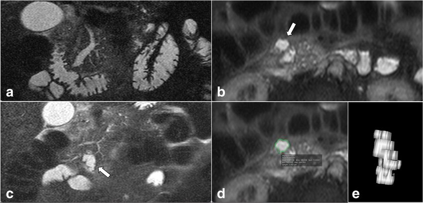
the obstructing effect of a solid mass on the MPD is very likely institutional MRI in the study were (1) magnetic fieldEur Radiol Fig. 1 Flow chart showing the selection of study population (MD-: main-duct type; BD- IPMN: branch-duct IPMN; PC: pancreatic cancer) strength ≥ 1.5 T; (2) availability of an axial and coronal Shot Turbo Spin Echo) or multi-shot PROPELLER tech- T2-weighted sequence acquired with the single-shot tech- nique; and (3) slice thickness and interslice gap not larger nique (HASTE, Single-Shot Fast Spin Echo or Single- than 6 mm and 20%, respectively. Fig. 2 Pancreatic MRI of an 80-year-old patient with weight loss and forming PC causes a stricture of the main pancreatic duct (MPD) with abdominal pain. The axial (a) and coronal (b) T2-weighted images upstream dilatation on coronal T2-weighted image (d). The patient was show a solid-mass-forming pancreatic cancer (PC) (open arrows) excluded from our cohort, as the dilation of the MPD upstream secondary originating from an adjacent IPMN (white arrows) located in the head to a solid mass may lead to overestimation of the positive yield of the of the pancreas. The pancreatic cancer is homogeneously hypointense in finding “dilated MPD” the T1-w axial image in the pancreatic arterial phase (c). The mass-
Eur Radiol

   All MR images were evaluated on a picture archiving                    Statistical analysis
and communication system (Sectra Workstation, IDS7 ver-
sion 23.1, Sectra AB) by two radiologists in consensus                    Normally and non-normally distributed variables were
reading (R.P.M. and N.K. with 15 and 12 years of post-                    expressed by means and medians, respectively. Since
residency experience in abdominal imaging, respectively).                 the EEG 2018 use a certain cut-off for MPD diameter
One cyst per patient was chosen for analysis (the largest or              and serum levels of CA 19-9 in their recommendations
the one with the most suspicion for malignancy appearance                 [4], categorical variables for MPD and CA 19-9 were
based on current guidelines at the time of surgery). The                  used for analysis. Wilcoxon rank sum test and chi-
collected imaging parameters are listed in Table 1.                       squared test were used to compare the outcome HGD/
                                                                          INV between two independent groups for numerical and
                                                                          categorical variables, respectively. Fisher’s exact test
Clinical features
                                                                          was applied when expected frequencies were less than
                                                                          5. Univariable logistic regression analysis was perform-
From each patient’s electronic medical record, the following
                                                                          ed to identify variables associated with the outcome
clinical parameters were collected: age at surgery, gender,
                                                                          HGD/INV. Odds ratios (OR) and 95% confidence inter-
presence of symptoms [such as jaundice, weight loss, abdom-
                                                                          vals (CI) were calculated. Variables that were shown to
inal pain, acute pancreatitis, recent (< 1 year) onset of diabetes
                                                                          be statistically significant at univariable logistic regres-
mellitus] or incidentally discovered IPMN, elevated serum
                                                                          sion were tested with multivariable logistic regression
levels of CA 19-9 (> 37 μmol/L), and presence of familial/
                                                                          (Enter Method), adjusted for age and gender. The pre-
genetic predisposition to PDAC.
                                                                          dicted probabilities for the outcome HGD/INV were cal-
                                                                          culated for hypothetical male patients at age ≥ 70 years
Histopathological features                                                old with and without the variables that were shown to
                                                                          be statistically significant in multivariable logistic re-
All the histopathological reports were examined, and the                  gression. The following diagnostic accuracy metrics of
grade of dysplasia for the resected specimen recorded. In cases           the solitary parameters were calculated: sensitivity, spec-
with an insufficient description of histopathological features            ificity, positive (PPV) and negative predictive values
[e.g., histotype, grade and location (i.e., cyst or MPD) of dys-          (NPV), and accuracy. A two-sided p value of < 0.05
plasia], a side-by-side revision of the pathological specimen             was considered statistically significant. The statistical
was performed by the pathologist (C.F.M.) in consensus with               analysis was performed with Stata16 (StataCorp. 2019,
one radiologist (R.P.M.). No further systematic radiologic-               Stata Statistical Software: Release 16, StataCorp LLC).
pathologic correlation was performed.

Table 1   Collected imaging parameters

Imaging parameters        Description

Diameter 1 (Diam1)        Maximum cyst diameter on axial T2-weighted sequence (mm)
Diameter 2 (Diam2)        Maximum craniocaudal cyst diameter on coronal T2-weighted sequence (mm)
Cyst maximum diameter     Either Diam1 or Diam2, depending on which was largest (mm)
Elongation value (EV)     Defined as [1 − (width/length)] according to previous publication [11], where length was represented by the maximum
                             diameter irrespective of the plane, and width as the maximum diameter perpendicular to length
Maximum MPD diameter      Expressed in mm
Mural nodules (MN)        Presence of contrast-enhancing mural nodules within the cyst
Cystic wall thickening    Present when cystic wall thickness ≥ 2 mm
Progress in size during   > 5 mm/year according to EEG 2018 [4]
  follow-up
Solitary/multifocal
  BD-IPMN
 Lesion localization      Head/uncinate process or body/tail
 Cyst volume (Vsegm)      Calculated on axial T2-w images after file export to a free DICOM medical imaging viewer (Horos v2.1.1). A region of
                            interest (ROI) was drawn along the edge of the BD-IPMN at multiple levels, using the tool “ROI volume” available in the
                            semi-automatic three-dimensional segmentation software implemented in the viewer. The common bile duct and the
                            MPD were excluded from the segmentation. Thereafter, the volume was automatically calculated by the software

MPD main pancreatic duct, EEG European evidence-based guidelines, BD-IPMN branch-duct IPMN
Eur Radiol
Results INV in the cohort’s patients depending on the number of pos-
itive risk factors. Figure 5 presents a case of a patient operated
The final study population comprised 106 patients (Fig. 1). Of on for suspected mixed-type IPMN with dilated MPD and
the 24 excluded patients (Fig. 1), 22 (92%) had a solid-mass- elevated CA 19-9, with final histology of HGD.
forming PC causing MPD stricture. Twenty-nine patients Interestingly, an MPD ≥ 5 mm represented the only surgi-
(27%) (operated on in the period 2008–2015) were part of cal indication in 15 patients, of whom three were diagnosed
the patient cohort in a previously published study [5] and 50 with HGD/INV (20%) (sensitivity 11.1% (95% CI: 2.3–29%;
(47%) (operated on in the period 2008–2017) of the patient n = 3/27) and positive predictive value (PPV) of 20% (95%
cohort in another study [6]. Patients’ characteristics are illus- CI: 4.3–48%; n = 3/15)). The diagnostic metrics for all resec-
trated in Table 2. In our series, one-fourth of patients had tion criteria taken alone are reported in Supplementary
HGD/INV (19/106 HGD and 8/106 invasive cancer). Material Table S1.
Among those eight patients with invasive cancer (one
microinvasive), no visible mass-forming PC was detected
pre-operatively. Fourteen patients had contrast-enhancing Discussion
MN (mean size/range: 12/4–32 mm): 3 with high-grade dys-
plasia and 4 with invasive cancer (Table 2). The MN size was Our results indicate that cystic volume as well as other imag-
not statistically significantly different among patients with ing features, such as the maximum cystic diameter, wall thick-
LGD and HGD/INV (p = 0.3). Seventy-eight patients (74%) ness and solitary/multifocal lesions, as well as progress in size
were diagnosed with mixed-type IPMNs at surgical histopa- (≥ 5 mm/year), failed in predicting HGD/INV in patients
thology. The gastric type was the most prevalent histotype operated on for BD- and mixed-type IPMN.
(70%) (Table 2). To the best of our knowledge, only one previous paper
Cyst volume was not statistically significantly different be- attempted to evaluate the role of tumoral volume in the pre-
tween patients with LGD and HGD/INV (p = 0.19). When diction of malignancy in patients with IPMN [10], showing
analyzed in logistic regression (both alone and in combination that an intraductal volume of ≥ 10 cm3 had a sensitivity and
with cystic EV), it was not associated with HGD/INV specificity of 70% and 73% in diagnosing malignant IPMN.
(Table 3). The mean EV was 0.36 (± SD 0.16), with a maxi- However, this paper included both CT and MRI scans without
mum value of 0.67 and an interquartile range of 0.25–0.5, stringent definitions of inclusion criteria. Moreover, the
showing that the majority of the segmented IPMN did not IPMN’s segmentation process was performed manually,
have a spheroid appearance (Table 2). At logistic regression, based on a “by-pen” tracing method on paper that eventually
EV showed a tendency for inverse association with the out- was scanned and digitalized, rendering this method not feasi-
come HDG/INV (OR = 0.38) although not statistically signif- ble for routine clinical practice. Our semi-automatic segmen-
icant (Table 3). The predicted probabilities for the outcome tation method is more practical, although not correlated to the
HGD/INV slightly decreased by increasing the elongation grade of dysplasia, and volumetry may potentially be perform-
value, although with broad confidence intervals (Fig. 3). ed with any segmentation tool available in any PACS system
At univariable logistic regression analysis, maximum cyst in a non-time-consuming fashion as proposed by Pandey P.
diameter, wall thickness, solitary or multiple lesions and prog- et al [12]. Furthermore, semi-automatic volumetric segmenta-
ress in size ( ≥ 5 mm/year) were not associated with HGD/INV tion has the important advantage of being independent of the
(Table 3). The only variables associated with HGD/INV at axis manually chosen by the radiologist and has very high
univariable logistic regression were the presence of contrast- intra- and interobserver reproducibility even in smaller lesions
enhancing MN, diameter of the MPD ≥ 5 mm and serum [12, 17]. Thus, it may potentially overcome the issue of non-
levels of CA 19-9 > 37 μmol/L. This strong association standardized manual measurements of cystic diameters, which
was also confirmed in a multivariable logistic regression are affected by intra- and inter-observer variability [17].
model adjusted for age and gender (Table 4). Histological We also hypothesized that the morphology of a BD-IPMN
cell subtypes did not correlate with cyst diameter, volume expressed by the EV might be associated with malignancy.
or EV (results not shown). For instance, a BD-IPMN with spheric appearance (i.e., EV
The predicted probabilities calculated for a hypothetical close to zero) might be characterized by a greater mucin se-
male patient with age ≥ 70 years old progressively increased cretion, depending on its MUC expression patterns and grade
by adding the risk factors contrast-enhancing MN, diameter of of dysplasia. Although the increase in EV appears slightly
the MPD ≥ 5 mm and serum levels of CA 19-9 > 37 (Fig. 4). inversely associated with the risk of HGD/INV (OR 0.38)
Namely, the predicted probability for the outcome HGD/INV per one-unit increase, this association was not statistically sig-
with none of the aforementioned risk factors was 0.08 and nificant. Thus, there is no sufficient evidence to support the
increased to 0.92 when all of the risk factors were present. hypothesis that the morphology of BD-IPMN may be associ-
Table 5 shows observed probabilities for the outcome HGD/ ated with HGD/INV.Eur Radiol
Table 2 Characteristics of patients with branch duct (BD)–intraductal papillary mucinous neoplasms (IPMN) and mixed-type IPMN
Number of patients 106 Low-grade dysplasia High-grade dysplasia/invasive cancer
Males 45/106 (42.4%) 31/45 (68.9%) 14/45 (31.1%)
Age (years) Mean 68.2, median 70 Mean 67.9, median 70 Mean 68.8, median 70
(min 43, max 86) (min 43, max 86) (min 48, max 86)
Individuals at risk 3/106 (2.8%) 3/3 (100%) 0
(2 familiarity; 1 Peutz-Jeghers)
Histology
Low-grade dysplasia 79/106 (74.5%)
High-grade dysplasia/invasive cancer 27/106 (25.5%) 8/106 inv.ca. (7.5%)
8/27 inv.ca. (29.6%)
Mixed-type IPMN 78/106 (73.6%) 53/79 (67.1%) 25/27 (92.6%)
BD-IPMN at pre-op MRI 25/78 (32%) 21/53 (39.6%) 4/25 (16%)
Mixed-type IPMN at pre-op MRI 53/78 (68%) 32/53 (60.4%) 21/25 (84%)
Histological cell subtypes
Gastric 75/106 (70.8%) 63/79 (79.8%) 12/27 (44.5%)
Pancreato-biliary (PB) 5/106 (4.7%) 2/79 (2.5%) 3/27 (11.1%)
PB + gastric 4/106 (3.8%) 3/79 (3.8%) 1/27 (3.7%)
Intestinal 7/106 (6.6%) 2/79 (2.5%) 5/27 (18.5%)
Intestinal + gastric 14/106 (13.2%) 8/79 (10.1%) 6/27 (22.2%)
PB + gastric + intestinal 1/106 (0.9%) 1/79 (1.3%) 0/27 (0%)
Symptomsa 32/106 (30.2%) 21/79 (26.6%) 11/27 (40.7%)
Jaundice 3/106 (2.8%) 1/79 (1.3%) 2/27 (7.4%)
Weight loss 3/106 (2.8%) 2/79 (2.5%) 1/27 (3.7%)
Abdominal pain 13/106 (12.3%) 9/79 (11.4%) 4/27 (14.8%)
Acute pancreatitis 15/106 (14.1%) 9/78 (11.4%) 6/27 (22.2%)
Diabetes (recent onset < 1 year) 0/53 (0%)
Serum CA 19-9 (μmol/L)b Median 11 (IQR 6–29) Median 8.8 (IQR 4.8–21) Median 29 (IQR 10–74)
min 0.3, max 30359 min 0.3, max 60 min 1, max 30359
CA 19–9 > 37 μmol/Lb 18/104 (17.3%) 9/77 (11.7%) 9/27 (33.3%)
IPMN localization
Head/uncinate process 59/106 (55.6%) 42/79 (53.2%) 17/27 (62.9%)
Imaging features IPMN
BD-IPMN at pre-op MRI 47/106 (44.3%) 41/79 (51.9%) 6/27 (22.2%)
Mixed-type IPMN at pre-op MRI 59/106 (55.7%) 38/79 (48.1%) 21/27 (77.8%)
Cyst max diameter (mm) Median 33 Median 32 Median 36
IQR 24–42; min 9, max 100 IQR 24–41; min 10, max 77 IQR 24–47; min 9, max 100
Diameter ≥ 30 mm 65/106 (61.3%) 47/79 (59.5%) 18/27 (66.6%)
Diameter ≥ 40 mm 37/106 (34.9%) 25/79 (31.6%) 12/27 (44.4%)
Elongation valuec Mean 0.36 ± 0.16 Mean 0.37 ± 0.16 Mean 0.34 ± 0.16
Volume (cm3) median 9.7 (IQR 4–19) median 9.4 (IQR 3–17) median 11.4 (IQR 5–22)
min 0.3, max 424.2 min 0.3, max 125.8) min 0.5, max 424.2
MPD max diameter (mm) Mean 5.8 ± 3.3 Mean 5.3 ± 2.9 Mean 7.2 ± 4.1
Median 5.1 (IQR 3.1–7.4) Median 4.9 (IQR 3–6.8) Median 6.6 (IQR 5.1–9.1)
min 1.5, max 19 min 2, max 15 min 1.5, max 19
MPD ≥ 5 mm 59/106 (55.7%) 38/79 (48.1%) 21/27 (77.8%)
MPD 5–9.9 mm 48/106 (45.3%) 32/79 (40.5%) 16/27 (59.3%)
MPD ≥ 10 mm 11/106 (10.4%) 6/79 (7.6%) 5/27 (18.5%)
Contrast-enhancing mural nodules 14/106 (13.2%) 7/79 (8.9%) 7/27 (25.9%)
Size mural nodules (mm) 12.1 ± 7.6 (min–max 4–32) 9.2 ± 3.9 (min–max 5.3–17) 15 ± 9.6 (min–max 4–32)
Wall thickness ≥ 2 mm 6/106 (5.6%) 3/79 (3.8%) 3/27 (11.1%)
Solitary lesion 39/106 (36.8%) 30/79 (37.9%) 9/27 (33.3%)Eur Radiol
Table 2 (continued)
Number of patients 106 Low-grade dysplasia High-grade dysplasia/invasive cancer
Progress in size ( > 5 mm/year) 29/106 (27.4%) 24/79 (30.4%) 5/27 (18.5%)
Pre-op pre-operative, MPD main pancreatic duct
a
Four patients had ≥ 2 symptoms
b
Preoperative CA 19-9 was not available in two patients
c
Elongation value calculated as [1 − (width/length)]
The presence of contrast-enhancing MN showed an asso- systematic review and meta-analysis published by
ciation with HGD/INV. Despite the low prevalence of this Marchegiani G. et al [18].
parameter in our cohort (14/106, 13%), seven patients with Interestingly, MPD dilatation was another imaging-related
contrast-enhancing MN were diagnosed with HDG/INV. This factor associated with a higher risk of malignancy in patients
is in line with other papers and, more recently, with the with IPMN without a solid-mass-forming PC. We decided to
Table 3 Univariable logistic
regression analysis for all clinical Patients’ features Nr. of observations Odds ratio 95% CI p value*
and imaging features
Demographic and clinical features
Age (years) 106 1.01 0.96–1.06 0.63
Age ≥ 70 (cohort’s median age) 106 1.05 0.44–2.51 0.91
Age < 70 106 0.95 0.39–2.28 0.91
Gender (male) 106 1.67 0.69–4.01 0.26
Localization (head/uncinate) 106 1.50 0.61–3.67 0.38
Mixed-type IPMN 106 6.13 1.34–27.89 0.02
Symptoms 106 1.90 0.76–4.74 0.17
Abdominal pain 106 1.35 0.38–4.81 0.64
Acute pancreatitis 106 2.22 0.71–6.97 0.17
Jaundicea 106 6.24 0.54–71.76 0.14
Weight loss 106 1.48 0.13–17.01 0.75
Serum CA 19-9 (μmol/L) 104 1.04 1.01–1.06 0.002
CA 19-9 > 37 μmol/L 104 3,77 1.30–10.9 0.014
Imaging-related features
Volume (cm3) 106 1.01 0.99–1.02 0.12
Cyst max diameter (mm) 106 1.02 0.99–1.04 0.18
Diameter ≥ 30 mm 106 1.36 0.54–3.4 0.51
Diameter ≥ 40 mm 106 1.72 0.7–4.22 0.23
Elongation value 106 0.38 0.02–5.93 0.49
MPD max diameter (mm) 106 1.17 1.02–1.33 0.02
MPD ≥ 5 mm 106 3.97 1.45–10.89 0.007
MPD 5–9.9 mm 106 2.13 0.87–5.19 0.09
MPD ≥ 10 mm 106 2.76 0.77–9.93 0.12
Mural nodules 106 3.6 1.13–11.47 0.03
Wall thickness ≥ 2mm 106 3.16 0.59–16.73 0.17
Solitary lesion 106 0.81 0.32–2.05 0.66
Multifocal lesions 106 1.23 0.49–3.07 0.66
Progress in size (≥ 5 mm/year)b 67 1.01 0.36–2.8 0.98
CI confidence intervals, CA carbohydrate antigen, MPD main pancreatic duct
*A p value < 0.002 was considered statistically significant (marked in bold)
a
No association was found between jaundice and elevated Ca19-9 (Fisher’s exact test, p = 0.56)
b
Calculated on 67 observations (39 subjects had no previous examinations)Eur Radiol
Fig. 3 Two-way plot showing
decreasing predicted probabilities
and their 95% CI (y-axis) for the
outcome high-grade dysplasia/
invasive cancer (HGD/INV) over
the elongation value (EV) (x-
axis). The lower the EV (i.e.,
spheroid cyst), the higher the
predicted probability of having
HGD/INV and vice versa,
although the variable did not
result statistically significant at
univariable logistic regression
evaluate the effect of the MPD dilatation in the absence of MPD 5–9.9 mm and ≥ 10 mm). It is also necessary to under-
detectable solid masses at preoperative MRI, which very like- line that this study cohort excluded all patients with main-duct
ly caused the obstruction of the MPD and its upstream dilata- IPMN since the main aim was the volumetric and morpholog-
tion. In this way, it was possible to analyze the real impact of ical analysis of BD-IPMNs.
MPD dilatation. Thus, as demonstrated by others [5, 6, 19], The only clinical feature correlated to a higher risk of
MPD dilatation appears to play an important role in terms of HGD/INV was the elevated serum level of CA 19-9. The
increased risk of HGD/INV in a surgical series, especially in presence of symptoms was not associated with HGD/INV
association with other imaging and/or clinical risk factors, in our cohort. Mucin-producing tumors such as IPMN may
such as contrast-enhancing MN and elevated serum levels of cause abdominal pain and/or acute pancreatitis, which
CA 19-9. However, when the indication for surgery was MPD were the most often encountered symptoms in our cohort,
dilatation alone (15 patients), the sensitivity and positive pre- and for these reasons, they are included among worrisome
dictive values were low (11% and 20%, respectively). This features and relative surgical indications in the current
might be explained by the fact that the MPD dilatation in guidelines [3, 4]. Possible reasons for symptoms not being
mixed-type IPMN is not exclusively related to diffuse malig- associated with malignant IPMN are the small sample size
nant epithelial changes but may result from passive distension and the fact that these symptoms are often secondary to
due to mucin secretion from solitary or multiple BD-IPMN. solid-mass-forming PC (an exclusion criterion in this
Thus, in patients with mixed-type IPMN, MPD dilatation as a study). The absence of pre-operatively detectable solid
sole resection criterion has to be carefully considered before masses may even explain the low prevalence of jaundice
the decision to proceed to surgery is taken. in our patient cohort (3/106, 2.8%).
Nonetheless, due to the small sample size, it was not pos- The main strength of our study is represented by the select-
sible to assess the impact of subclasses of MPD dilatation (i.e., ed population since, albeit small, it did not include patients
with a suspected solid tumor at preoperative MRI. On the one
hand, the presence of a solid mass associated with an IPMN
Table 4 Multivariable logistic regression analysis adjusted for age and
preoperatively is a major indication for surgery and potentially
gender
a very late stage of IPMN malignant transformation (thus,
Patients’ features Nr. of OR 95% CI p value* beyond the aim of preventive surgery). On the other hand,
observations pooling together subjects with IPMN and solid masses caus-
ing a MPD stricture with upstream dilatation (Fig. 2) may lead
Mural nodules 104 4.32 1.18 – 15.76 0.02
to overestimation of the yield of the risk factor “MPD dilata-
MPD ≥ 5 mm 104 4.2 1.34 – 13.1 0.01
tion”. In daily praxis, it is very common to encounter patients
CA19 - 9 > 37 μmol/L 104 6.72 1.89–23.89 0.003
with imaging-related risk factors (e.g., dilatation of the MPD,
Age at surgery (years) 104 1.01 0.95 – 1.07 0.61
enlarged BD-IPMN, MN, etc.) and no detectable solid mass.
Gender (male) 104 1.97 0.69 – 5.67 0.20
In these cases, the appropriate assessment of the risk-benefit of
*A p value < .05 was considered statistically significant surgery is mainly based on imaging and clinical featuresEur Radiol
Table 5 Observed probabilities for the outcome high-grade dysplasia/ enhancing mural nodules (MN), main pancreatic duct diameter equal to
invasive cancer (HGD/INV) versus low-grade dysplasia (LGD) in the or larger than 5 mm (MPD), and carbohydrate antigen 19-9 levels higher
cohort’s patients depending on the presence of risk factors contrast- than 37 μmol/L (CA 19-9)
LGD versus HGD/INV Sum of observed risk factors (MN, MPD, CA19-9)
0 1 2 3 Total
LGD 91.4% (32/35) 77.4% (41/53) 35.3% (6/17) 0 74.5% (79/106)
HGD/INV 8.6% (3/35) 22.6% (12/53) 64.7% (11/17) 100% (1) 25.5% (27/106)
according to previously published data, that if influenced by Our study has several limitations. The main ones are its
major suspected features (i.e., pancreatic mass causing MPD retrospective nature and the fact that it only included operated
obstruction), may possibly lead to wrong decisions. patients with a diagnosis of IPMN. This is an unavoidable and
Fig. 4 Two-way plot showing the
predicted probabilities and their
95% CI (y-axis) for the outcome
high-grade dysplasia/invasive
cancer (HGD/INV) over the
different combinations of risk
factors (x-axis) for a hypothetical
male patient with age ≥ 70 years
old. Predicted probabilities were
estimated by a multivariable
logistic regression model, as
described in the section
“Materials and methods.”
Abbreviations: MN: contrast-
enhancing mural nodules, MPD:
main pancreatic duct diameter
equal to or larger than 5 mm;
CA19-9: carbohydrate antigen
19-9 levels higher than 37
μ/μmol/L
Fig. 5 Pancreatic MR images of a 61-year-old man with recurrent IPMN was segmented using Horos v2.1.1 (d), and a volume of
episodes of acute pancreatitis. The main pancreatic duct (MPD) approximately 5 cm 3 was obtained (e). Due to the presence of
diameter is 9 mm in the head of the pancreas on coronal T2-weighted suspected IPMN–related acute pancreatitis, MPD diameter larger than
image (a), and a branch-duct intraductal papillary mucinous neoplasm 5 mm and elevated carbohydrate antigen 19-9 levels (80 μmol/L), the
(BD-IPMN; white arrows) is identified anteriorly in the uncinate patient underwent pancreaticoduodenectomy. The final histopathological
process on axial (b) and coronal (c) T2-weighted images. The BD- diagnosis was mixed-type IPMN with high-grade dysplasiaEur Radiol
well-known limitation for all studies investigating similar declare no relationships with any companies whose products or services
may be related to the subject matter of the article.
topics and affects the possibility of evaluating the real perfor-
mance of our findings on a “population-level” basis.
Statistics and Biometry Nicola Orsini, associate professor of Medical
Furthermore, the study cohort is small. However, we still had Statistics, head of the Biostatistics Team, Department of Global Public
statistically significant results. For a more precise estimation of Health, kindly provided statistical advice for this manuscript.
the role of new imaging-related risk factors in patients with
IPMN, larger samples are probably needed. Additionally, the Informed Consent Written informed consent was waived by the
Regional Review Board.
indications for surgery have varied during the span of the study,
which may have slightly affected our results. Furthermore, we
Ethical Approval Regional Review Board approval was obtained (EPN
did not correlate MN to the exact location of HGD/INV-focus. 2015/1544–31/4).
Another limitation is the use of T2-w axial images for the
segmentation and volumetry of BD-IPMN that may lead to less Study subjects or cohorts overlap The final population of our study
precise measurements. However, 3D-MRCP sequences were overlaps with two previously published papers.
Twenty-nine patients out of 106 (27%) (operated on in the period
not available in all patients, and, when available, the presence
2008–2015) were part of the patient cohort in a previously published
of artifacts oftentimes did not permit any measurements. study by our group [reference nr. 5: Ateeb Z, Valente R, Pozzi-Mucelli
Nonetheless, 3D-MRCP sequences are not considered neces- RM, et al (2019) Main pancreatic duct dilation greater than 6 mm is
sary for the assessment of resection criteria [4]. In case of arti- associated with an increased risk of high-grade dysplasia and cancer in
IPMN patients. Langenbecks Arch Surg].
facts, the more robust 2D-MRCP may be used, but it is not
Fifty out of 106 patients (47%) (operated on in the period 2008–2017)
suitable for computing volumetry. Additionally, the MR im- were part of the patient cohort in the study by Del Chiaro M. et al [refer-
ages were evaluated by two radiologists in consensus. For this ence nr. 6: del Chiaro M, Beckman R, Ateeb Z, et al (2019) Main duct
reason, it was not possible to assess the inter-observer agree- dilatation is the best predictor of high-grade dysplasia or invasion in
intraductal papillary mucinous neoplasms of the pancreas. Ann Surg].
ment regarding the measured parameters. Our study differs from the previous paper from Ateeb Z et al and del
Finally, we included MRI examinations from different ven- Chiaro et al as we only included patients with a preoperative pancreatic
dors and protocols. However, this increases the generalisability MRI, who had at least one histologically proven BD-IPMN detectable on
of our results. the T2-weighted images. Moreover, the main aim of our current study
was to assess the role of cystic volume, morphological features such as
In conclusion, our study shows that neither volumetry nor
elongation value and other well-established resection criteria after (1)
other novel imaging features of BD-IPMN can predict malig- excluding subjects with solid-mass-forming IPMN–associated or con-
nancy. The dilatation of MPD, especially in conjunction with comitant pancreatic cancer, and (2) patients with main-duct diameter ≥
contrast-enhancing MN and abnormally elevated serum levels 5 mm without a BD-IPMN clearly identifiable at preoperative MRI.
of CA 19-9, is associated with a higher risk of malignancy
Methodology
even when solid-mass-forming PC are excluded.
& retrospective
Supplementary Information The online version contains supplementary & cross-sectional diagnostic study
material available at https://doi.org/10.1007/s00330-022-08650-5. & performed at one institution
Acknowledgements The authors would like to thank Nicola Orsini, as-
sociate professor of Medical Statistics, head of the Biostatistics Team,
Department of Global Public Health. Open Access This article is licensed under a Creative Commons
Attribution 4.0 International License, which permits use, sharing, adap-
tation, distribution and reproduction in any medium or format, as long as
Funding Open access funding provided by Karolinska Institute. The
you give appropriate credit to the original author(s) and the source, pro-
study was supported by Region Stockholm (Nikolaos Kartalis, clinical
vide a link to the Creative Commons licence, and indicate if changes were
research appointment) and the Swedish Foundation for the Development
made. The images or other third party material in this article are included
of Pancreatology (SweSuP).
in the article's Creative Commons licence, unless indicated otherwise in a
credit line to the material. If material is not included in the article's
Declarations Creative Commons licence and your intended use is not permitted by
statutory regulation or exceeds the permitted use, you will need to obtain
Guarantor The scientific guarantor of this publication is associate pro- permission directly from the copyright holder. To view a copy of this
fessor Nikolaos Kartalis. licence, visit http://creativecommons.org/licenses/by/4.0/.
Conflict of Interest Lennart Blomqvist is co-founder and CMO at
Collective Minds Radiology. Nikolaos Papanikolaou is the owner of
MRIcons LTD company. Marco del Chiaro has been awarded an industry References
grant (Haemonetics, Inc) to conduct a multi-center study to evaluate the
prognostic implications of TEG in pancreas cancer. Further, he is a co-PI 1. Ren B, Liu X, Suriawinata AA (2019) Pancreatic ductal adenocar-
of a sponsored Boston Scientific study on the use of intra-operative cinoma and its precursor lesions: histopathology, cytopathology,
pancreatoscopy in IPMN patients. The other authors of this manuscript and molecular pathology. Am J Pathol 189:9–21. https://doi.org/
10.1016/j.ajpath.2018.10.004Eur Radiol
2. Maire F, Couvelard A, Palazzo L et al (2013) Pancreatic intraepi- 11. Aghaei Lasboo A, Rezai P, Yaghmai V (2010) Morphological anal-
thelial neoplasia in patients with intraductal papillary mucinous ysis of pancreatic cystic masses. Acad Radiol. https://doi.org/10.
neoplasms. Pancreas 42:1262–1266. https://doi.org/10.1097/ 1016/j.acra.2009.09.013
MPA.0b013e3182962723 12. Pandey P, Pandey A, Varzaneh FN et al (2018) Are pancreatic
3. Tanaka M, Fernandez-Del Castillo C, Kamisawa T et al (2017) IPMN volumes measured on MRI images more reproducible than
Revisions of international consensus Fukuoka guidelines for the diameters? An assessment in a large single-institution cohort.
management of IPMN of the pancreas. Pancreatology 17:738– European Radiology. https://doi.org/10.1007/s00330-017-5268-z
753. https://doi.org/10.1016/j.pan.2017.07.007 13. Paini M, Crippa S, Partelli S et al (2014) Molecular pathology of
4. The European Study Group on Cystic Tumours of the Pancreas intraductal papillary mucinous neoplasms of the pancreas. World J
(2018) European evidence-based guidelines on pancreatic cystic Gastroenterol 20:10008–10023. https://doi.org/10.3748/wjg.v20.
neoplasms. Gut 67:789–804. https://doi.org/10.1136/gutjnl-2018- i29.10008
316027 14. Jung W, Park T, Kim Y et al (2019) Validation of a nomogram to
5. Ateeb Z, Valente R, Pozzi-Mucelli RM et al (2019) Main pancreatic predict the risk of cancer in patients with intraductal papillary mu-
duct dilation greater than 6 mm is associated with an increased risk cinous neoplasm and main duct dilatation of 10 mm or less. Br J
of high-grade dysplasia and cancer in IPMN patients. Langenbecks Surg 106:1829–1836. https://doi.org/10.1002/bjs.11293
Arch Surg. https://doi.org/10.1007/s00423-018-1740-8 15. Tanaka M, Chari S, Adsay V et al (2006) International consensus
6. del Chiaro M, Beckman R, Ateeb Z et al (2019) Main duct dilata- guidelines for management of intraductal papillary mucinous neo-
tion is the best predictor of high-grade dysplasia or invasion in plasms and mucinous cystic neoplasms of the pancreas.
intraductal papillary mucinous neoplasms of the pancreas. Ann Pancreatology 6:17–32. https://doi.org/10.1159/000090023
Surg. https://doi.org/10.1097/sla.0000000000003174
16. del Chiaro M, Verbeke C, Salvia R et al (2013) European experts
7. Jang JY, Park T, Lee S et al (2017) Proposed nomogram predicting
consensus statement on cystic tumours of the pancreas. Dig Liver
the individual risk of malignancy in the patients with branch duct
Dis 45:703–711. https://doi.org/10.1016/j.dld.2013.01.010
type intraductal papillary mucinous neoplasms of the pancreas. Ann
Surg 266:1062–1068. https://doi.org/10.1097/SLA. 17. Dunn DP, Brook OR, Brook A et al (2016) Measurement of pancre-
0000000000001985 atic cystic lesions on magnetic resonance imaging: efficacy of stan-
8. Kwon W, Han Y, Byun Y et al (2020) Predictive features of ma- dards in reducing inter-observer variability. Abdom Radiol (NY) 41:
lignancy in branch duct type intraductal papillary mucinous neo- 500–507. https://doi.org/10.1007/s00261-015-0588-4
plasm of the pancreas: a meta-analysis. Cancers 12:1–18. https:// 18. Marchegiani G, Andrianello S, Borin A et al (2018) Systematic
doi.org/10.3390/CANCERS12092618 review, meta-analysis, and a high-volume center experience
9. Wong J, Weber J, Centeno BA et al (2013) High-grade dysplasia supporting the new role of mural nodules proposed by the updated
and adenocarcinoma are frequent in side-branch intraductal papil- 2017 international guidelines on IPMN of the pancreas. Surgery
lary mucinous neoplasm measuring less than 3 cm on endoscopic 163:1272–1279. https://doi.org/10.1016/j.surg.2018.01.009
ultrasound. J Gastrointest Surg 17:78–85. https://doi.org/10.1007/ 19. Marchegiani G, Andrianello S, Morbin G et al (2018) Importance of
s11605-012-2017-0 main pancreatic duct dilatation in IPMN undergoing surveillance.
10. Murayama S, Kimura W, Hirai I et al (2011) Volumetric and mor- Br J Surg 105:1825–1834. https://doi.org/10.1002/bjs.10948
phological analysis of intraductal papillary mucinous neoplasm of
the pancreas using computed tomography and magnetic resonance Publisher’s note Springer Nature remains neutral with regard to jurisdic-
imaging. Pancreas 40:876–882. https://doi.org/10.1097/MPA. tional claims in published maps and institutional affiliations.
0b013e31821fdcffEur Radiol
Affiliations
Raffaella M. Pozzi Mucelli 1,2 & Carlos Fernández Moro 3,4 & Marco Del Chiaro 5 & Roberto Valente 2,5,6 &
Lennart Blomqvist 7,8 & Nikolaos Papanikolaou 2,9,10,11 & Johannes-Matthias Löhr 2,12 & Nikolaos Kartalis 1,2
1
Department of Radiology Huddinge, Karolinska University Hospital, O-huset 42, 14186 Stockholm, Sweden
2 8
Department of Clinical Science, Intervention, and Technology, Department of Molecular Medicine and Surgery, Karolinska
Karolinska Institutet, O-huset 42, 14186 Stockholm, Sweden Institutet, L1:00, 17176 Stockholm, Sweden
3 9
Department of Clinical Pathology and Cancer Diagnostics, Computational Clinical Imaging Group, Centre for the Unknown,
Karolinska University Hospital, Huddinge, 141 Champalimaud Foundation, Av. Brasília, Doca de Pedrouços, 1400-
86 Stockholm, Sweden 038 Lisbon, Portugal
4 10
Division of Pathology, Department of Laboratory Medicine, Department of Radiology, Royal Marsden Hospital and The
Karolinska Institutet, Alfred Nobels Allé 8, 141 Institute of Cancer Research, London SM2 5NG, UK
52 Stockholm, Sweden 11
Computational Biomedicine Laboratory (CBML), Foundation for
5
Division of Surgical Oncology, Department of Surgery, University Research and Technology Hellas (FORTH),
of Colorado, Anschutz Medical Campus, 12631 E 17th Ave #6117, 70013 Heraklion, Greece
Aurora, CO 80045, USA 12
Department of Upper Abdominal Diseases, Karolinska
6
Department of Surgical and Perioperative Sciences, Umeå Comprehensive Cancer Center, Karolinska University Hospital,
University, Daniel Naezéns väg, 907 37 Umeå, Sweden Hälsovägen, 13, 141 57 Huddinge, Stockholm, Sweden
7
Department of Medical Radiation Physics and Nuclear Medicine,
Karolinska University Hospital, Solnavägen 1,
17177 Stockholm, SwedenYou can also read