British Art Studies
  February 2021
←
→
Page content transcription
If your browser does not render page correctly, please read the page content below
British Art Studies February 2021
British Art Studies
Issue 19, published 26 February 2021

Cover image: Victor Ehikhamenor, The King Returning from Holy Aruosa Cathedral
(detail), 2018, rosary beads, bronze statuettes, and thread on canvas, 116 x 71 in..
Digital image courtesy of Victor Ehikhamenor (all rights reserved).

PDF generated on 21 July 2021

Note: British Art Studies is a digital publication and intended to be experienced
online and referenced digitally. PDFs are provided for ease of reading offline. Please
do not reference the PDF in academic citations: we recommend the use of DOIs
(digital object identifiers) provided within the online article. These unique
alphanumeric strings identify content and provide a persistent link to a location on
the internet. A DOI is guaranteed never to change, so you can use it to link
permanently to electronic documents with confidence.

Published by:

Paul Mellon Centre
16 Bedford Square
London, WC1B 3JA
https://www.paul-mellon-centre.ac.uk

In partnership with:

Yale Center for British Art
1080 Chapel Street
New Haven, Connecticut
https://britishart.yale.edu

ISSN: 2058-5462
DOI: 10.17658/issn.2058-5462
URL: https://www.britishartstudies.ac.uk

Editorial team: https://www.britishartstudies.ac.uk/about/editorial-team
Advisory board: https://www.britishartstudies.ac.uk/about/advisory-board

Produced in the United Kingdom.

                                 A joint publication by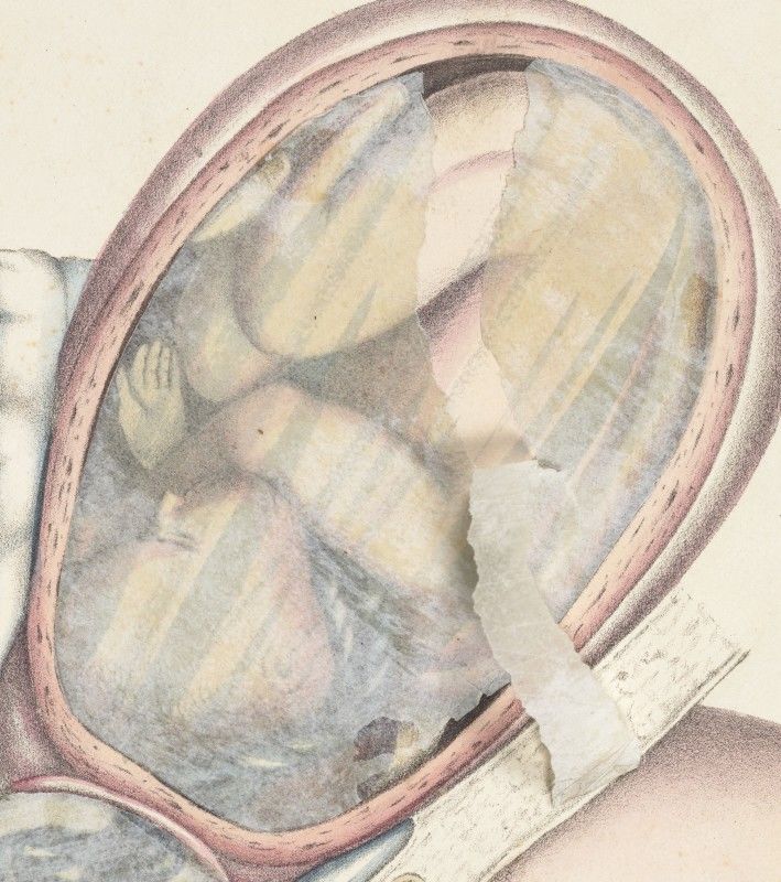
Contents
Spratt’s Flaps: Midwifery, Creativity, and Sexuality in Early Nineteenth-
Century Visual Culture, Rebecca Whiteley
Spratt’s Flaps: Midwifery, Creativity, and Sexuality
   in Early Nineteenth-Century Visual Culture
                                Rebecca Whiteley

    Abstract

    In 1833, an unusual book was published in London, Edinburgh, and Dublin.
    Written and illustrated by the surgeon and artist George Spratt, Obstetric
    Tables stood out among midwifery guides of the period for its coloured
    lithographic illustrations, mobilised by the construction of paper flaps. This
    article explores the way these flap prints contributed to medical pedagogy,
    but also looks much more widely at their cultural resonances. Through their
    interaction with wider visual cultures, Spratt’s tables engaged not only with
    medical literature, but also with social anxieties over nudity and sexuality,
    midwifery and propriety, and the power of popular print. By studying Spratt’s
    tables alongside comic and satirical mobile prints, obscene and pornographic
    prints, and “fine art” nudes, this article demonstrates how medical images
    can be addressed as rich and complex resources for histories that are
    medical, visual, and cultural.

    Authors

    Shreeve Fellow in the History of Medicine at the John Rylands Research
    Institute, University of Manchester

    Acknowledgements

    My thanks to Dr David Shreeve for generously funding my postdoctoral
    position and facilitating this research. My thanks also to the John Rylands
    Research Institute, which has hosted my fellowship, and to the librarians,
    archivists, photographers, and conservators at the University of Manchester
    Library for helping with my research and providing access to the library’s
    collections. In particular, this article would not be what it is without the
    expert help of Gwen Riley Jones in digitising the Spratt flap constructions,
    and Julie Ramwell in tracking down the Spratt family history.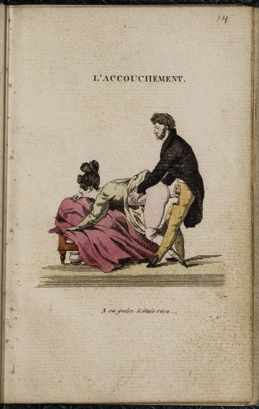
Cite as

Rebecca Whiteley, "Spratt’s Flaps: Midwifery, Creativity, and Sexuality in
Early Nineteenth-Century Visual Culture", British Art Studies, Issue 19,
https://dx.doi.org/10.17658/issn.2058-5462/issue-19/rwhiteley
Introduction

Under our eyes and hands, a woman conceives and grows pregnant; a foetus
turns and turns again, presenting different body parts for birth; forceps are
applied and an obstructed foetus is delivered; a vaginal examination is
conducted; a pregnant corpse is dissected layer by layer.

These are the actions users undertake when they view and handle George
Spratt’s Obstetric Tables: a slim, cloth-bound volume which presents a
summary of midwifery knowledge in hand-coloured lithographic plates, most
of which are mobilised and complicated by the construction of paper flaps
(Figs 1 and 2). First published in 1833, followed up with a supplement in
1835, and then regularly re-issued for roughly the next ten years, this
remarkable book had a short blaze of importance for British visual culture in
the last years of the Georgian and the first years of the Victorian eras. 1
Cultures of midwifery, of print, and of sexuality were all undergoing change
in these decades. Widespread social anxiety arose in response to: the
increasing dominance of male obstetric practice; the proliferation of cheap
and uncontrolled print; and the questions that both of these threw up about
propriety and sexual continence. This article explores what Spratt’s flap
constructions have to tell us not only about this period’s culture of medical
illustration and pedagogy, but also about its cultures of print, and of bodies,
much more widely.

View this illustration online

Figure 1.
George Spratt, Obstetric Tables: Comprising Graphic Illustrations, with
Descriptions and Practical Remarks; Exhibiting on Dissected Plates Many
Important Subjects in Midwifery. Second Edition, Considerably Enlarged and
Improved, volume 1. (London: The Author, 1835). Digital image courtesy of The
University of Manchester (All rights reserved).

View this illustration online

Figure 2.
George Spratt, Obstetric Tables: Comprising Graphic Illustrations, with
Descriptions and Practical Remarks; Exhibiting on Dissected Plates Many
Important Subjects in Midwifery. Second Edition, Considerably Enlarged and
Improved., volume 2. (London: The Author, 1835). Digital image courtesy of
The University of Manchester (All rights reserved).

Medical images can be interrogated as historic resources that chart the
progress of medical and biological discovery. But they are also parts of visual
culture, with complex relations to that which they represent, and when
looked at as evidence not of abstract truths but of the cultures that produced
them, they can speak in new ways. 2 Spratt’s tables, as this article will
demonstrate, can only be fully understood as images that worked both
earnestly within the culture of medical pedagogy, and as wider and more
subversive commentaries on that culture.

Looking at medical images as visual culture can be difficult—there is often
little textual evidence for their wider cultural significances, framed as they
were in the nineteenth century (and indeed today) by ideals of indexicality,
objectivity, and truthfulness. 3 But when we explore how such images
actually sort, interpret, and represent information, as well as how they
engage with the wider visual cultures and social preoccupations of the time,
they become historical sources in themselves, that work outside of the texts
that frame them. Artists, often not medically trained, turned an outsider’s
perspective on what they drew, even where they were directed by medical
authors. 4 They also produced outputs that were much more open to
interpretation, adaption, and misuse than medical texts. Exploring the
variety of influences on and interpretations of a medical image can,
therefore, give a much wider view of the place of medicine in a period’s
culture.

With Spratt’s tables, I aim to show that medical authors and artists often felt
an ambivalence about the official ambitions and approaches of their
profession, and that they were aware of the more critical and diverse ways in
which medicine was perceived by non-professionals. Indeed, Spratt makes a
particularly rewarding case study for this kind of history because he was both
the author and the artist of the Obstetric Tables. As a surgeon-accoucheur
and a lithographic artist, Spratt was both an insider and an outsider when it
came to the medical establishment and its rhetoric. His work as an artist led
him to comment with unusual frankness and criticality on the contemporary
culture of medical midwifery. It is this cultural commentary that this article
seeks to explore.

First establishing the context for Spratt’s work—its production history,
audience, and visual and material influences—I will then address a selection
of the tables. This selection demonstrates the diversity of Spratt’s output,
and moves through the different ways in which his flap productions engaged
with wider medical and body cultures: from anatomy and medical pedagogy;
to popular and comic mobile prints; to the complex and intertwined issues of
sex, nakedness, pornography, and the nude. Regarding these different
contexts, this article demonstrates how culturally complex a medical image
can be, and how it can be employed to knit together official medical
knowledge with wider cultures of medicine, the body, and its representation.

This article constitutes only the second scholarly work to address Spratt’s
output, and the first to focus on his medical illustrations. 5 Not known as a
pioneer in medicine or lithography, he barely features in histories of
obstetrics or print culture. Yet, the Obstetric Tables was popular in its time
and has much to tell us about visual cultures of medicine in the nineteenth
century. The last decades have seen increased interest among art historians
in the visual cultures of nineteenth-century medicine, but the field is still new
and by no means well covered. 6 Particularly, the focus within art history has
remained, in many cases, on famed practitioners and their interactions with
the fine arts. Book illustrations, which make up the core of the period’s
medical visual culture, are most often treated peripherally. This study turns
these trends inside out by placing the cheap, copied, “popular”, accessible
medical printed image at the centre of a visual culture study.

George Spratt

Spratt was both the author and artist of the Obstetric Tables, but this does
not mean that he worked in isolation. He drew heavily on existing sources for
both text and images, in a process that was common in the period, but which
has received little in-depth study since, either as a general phenomenon or
in specific instances. 7 Spratt borrowed images produced for the famed
midwifery authors of the eighteenth century, as well as popular
contemporary obstetric authors and others who produced flap anatomies. 8
The plates were printed by G.E. Madeley and Charles Hullmandel, the former
also undertaking some of the lithography. And while the drafting for the first
edition of 1833 was done largely by G. Spratt, the plates from the 1835
supplement, which were incorporated into all later editions, are signed not
only by G. but also by E., F., and W. Spratt. These other Spratts were most
likely his children, who included William Henry Williams baptised in 1811 and
Francis Edgar in 1814 in Blackmore, Essex. 9 The family were in the
printmaking line, with George’s wife Maria illustrating and publishing at least
one book in her own right, and their daughter Julia setting up an art school in
the 1840s. 10 George Spratt’s status as both a medical practitioner and a
professional artist, combined with his sharing of the work with other artists
who most likely had no medical training, allowed him to offer a particularly
broad and creative view of early nineteenth-century midwifery. 11

While Spratt claimed to be a “surgeon-accoucheur” on the title page to
Obstetric Tables, evidence suggests he was not a successful medical
practitioner. His simultaneous work as an artist points to this, as do the
listings of his bankruptcy in Northampton in 1824 12 and in Chelsea, London
in 1828. 13 Various scanty biographies of Spratt have suggested that he was
a member of the Royal College of Surgeons or a fellow of the Linnaean
Society, yet neither institution has any record of him. It is most likely, then,
that he achieved no particular prominence within medicine and was one of
that growing class of surgeons who struggled to make a living in what was
becoming an increasingly competitive marketplace. 14 He does seem to have
made some headway as an artist, however, producing illustrations for
several botanical compendia, toxicology wallcharts, and two series of popular
composite caricatures. 15

While many medical practitioners of the period could draw, learning as part
of a liberal education or training themselves in order to publish, Spratt’s
position is more unusual. Rather than being a doctor who drew, he made his
living on the boundaries between medicine and art, cobbling jobs together in
the simple pursuit of earning a living. As such, he was oriented not towards
pioneering new medical knowledge, but towards producing works that would
sell. His perspective on midwifery was both more representative of the
profession generally than that of the more rarefied medical elite, and
integrated an outsider’s perspective. As an artist—a caricaturist no less—he
was closely observant, critically witty, and experienced in understanding
what would appeal to a diverse audience. Because he was so embedded in
the cultures of popular printmaking, his flap constructions are both unusually
upfront about their wider cultural affiliations, and remarkably frank in their
commentary on the nature of medical practice and its visual representation.
Using existing texts and images, Spratt collected, combined, coloured, and
mobilised them into a work that was, as I will argue, popular, enticing, and
satirical as well as educational. As such, his work offers a remarkable case
study for the intersections between medical and wider visual cultures.

Readerships

Spratt’s book, while it would have been enticing and interesting to many
readers, was officially described as being intended for the “student in
obstetric science, and the more inexperienced accoucheur”. 16 Obstetric
Tables was meant to provide these male, professional readers with a simple
overview of the anatomy and practice of midwifery. The coloured and mobile
illustrations were valued by reviewers for the way they helped students to
decipher and remember medical knowledge. One reviewer for The London
Medical and Surgical Journal recommended the volume to both students and
practitioners, “for it instructs the former, and recals [sic] to the recollection
of the latter many most important circumstances, which it is impossible for
the memory to retain in vivid and fresh colours”. 17 The materially engaging
nature of the book was expressed by one American reviewer who told their
readers “It must be seen to be appreciated” and that “No single picture could
ever convey the same ideas, and enable the student to understand the
descriptions, but these dissected plates are almost equal to the manakin
itself.” 18
The book was characterised, therefore, as a useful revision guide, a
summation of the information and demonstrations on manikins provided in
midwifery lecture courses. It was often recommended to students, and
characterised as moderately priced, but it was clearly also appealing to more
established practitioners, and it certainly wasn’t as cheap as many other
contemporary student guides. The actual price seems to have
varied—between sellers and depending on whether or not the copy was
coloured—between £1 5s. and £2 5s. 19 Given that even the much more
comprehensive and heavily illustrated manual An Atlas of Plates by Francis
Ramsbotham sold for only 18 s., it seems likely that at least the less
financially secure among students and young practitioners would have
balked at purchasing Spratt’s book. 20 In fact, the list of subscribers printed
in each edition indicates that the actual core audience was more established
surgeons and general practitioners. 21 They likely saw the book as both a
serious medical study guide and more of a prestigious and beautiful
medically themed gift book. Such a visually and haptically attractive book
was likely intended to be both studied and shown off. Physicians and
surgeons may have made their copies available to colleagues, pupils,
midwives, and even patients.

Indeed, many of the same features likely made the work attractive to lay
readers. In the 1830s, as I will discuss further, professional and popular
medical texts were less distinct, and people also moved in and out of being
medical professionals in the search for sustaining employment. 22 Interested
lay readers with some money to spare might well have purchased such an
intriguing book, or browsed in it at a bookshop or a friend’s private library.
Indeed, a review in the literary magazine The Athenæum indicates that this,
as well as other medical books, were understood to be of interest to wider
and non-professional audiences. However, this particular review also
expresses a broader anxiety from the period: that books that were of value
to students for their accessibility might also be too attractive to lay readers
not trained to use and interpret them appropriately. So the reviewer noted of
the plates, with distinctly faint praise: “We hope they may repay the author
for the labour and pains he has evidently bestowed on them: their general
accuracy, together with their moderate price, will recommend them to those
who need their assistance.” 23 The reviewer clearly takes a dim view of those
who would need such a book, be they under-qualified practitioners or lay
people, suggesting that for the accomplished practitioner, the effort Spratt
put into creating the tables didn’t equal their worth.

Going forward, therefore, we will assume that most early nineteenth-century
viewers would have been excited and pleased, if not also puzzled and
troubled, to get their hands and eyes on a book with coloured and mobile
prints of naked women, female genitalia, and the mysterious unborn child.
Indeed, I argue that Spratt produced his tables specifically for such a diverse
audience—catering to the medical and the lay, the legitimate and the illicit
reader.

Medical Pedagogy

Turning to the tables themselves, I will begin with what we might call a
“textually sanctioned” reading of the tables as medically pedagogic, before
moving on to address wider audiences and more diverse interpretations.

Table 11 shows the dissected body of a pregnant cadaver (Fig. 3). It follows a
long-standing format for exposing the abdomen using a cruciform incision
and peeling back successive layers of tissue that was still, in this period,
heavily associated with the plates from William Hunter’s The Anatomy of the
Human Gravid Uterus Exhibited in Figures (1774). The flap format is lifted
from Edward William Tuson’s A Supplement to Myology (1828), but the twin
foetuses eventually uncovered are copied from Table 10 of William Smellie’s
A Sett of Anatomical Tables (1754).

                                        Watch Video

         Figure 3.
         E. Spratt (draftsman), Table 11, hand-coloured lithograph, 283 x 222 mm
         (page), from George Spratt, Obstetric Tables (London: The Author, 1835).
         Digital image courtesy of Courtesy of The University of Manchester (All rights
         reserved).
This is perhaps Spratt’s most conventional flap construction: it borrows from existing texts on midwifery, demonstrating Spratt’s comprehensive knowledge of the canonical literature and visual culture of the discipline. It also engages with the history of anatomical flap sheets. From the sixteenth century, printed anatomical flap sheets, sometimes called “fugitive sheets”, became popular. They are described by Andrea Carlino as translating “on to paper the whole concept of anatomical dissection, mimicking the progressive unveiling of the body, from skin to guts”. 24 These sheets provided anatomical information in a sanitised, bloodless, and reversible replication of the process of dissection. 25 The popularity of anatomical flap sheets waned from the late seventeenth century, but saw a resurgence in the nineteenth century. Some of the first of these were the large, intricate constructions produced for James Hogben in the 1810s and Edward William Tuson in the 1820s. Later in the century, this style of flap production was continued by Gustave-Joseph Witkowski and James Suydam Knox, among others. 26 These newly complex and delicate flaps describe the minutely detailed, austere anatomy of the nineteenth century. 27 While they were expensive, professional works, flap anatomies were also produced for wider popular audiences, by authors such as Achille Comte and Frederick Hollick. 28 Spratt’s flaps sit somewhere between the technical professional and the accessible lay publication. He saw in the paper technology of flaps a potential for playfulness that attracted viewers; a three-dimensionality that allowed for the description of bodies and operations on them; and a materiality that inclined the constructions to self-referentiality. In Table 11, what appears to be a quite straightforward paper reconstruction of uterine anatomy is also a meditation on materiality and representation. Moving through the uterine wall, the decidua, and the chorion, we reach the amnion, which is not represented by a hand-coloured lithograph on paper, but by hand-painted tissue paper. With this choice of material, the distance between object and representation is collapsed and the flap comes materially as well as visually to replicate the amnion—not only its translucency, but also its fragility. The care with which one has to handle this layer, sliding a nail under the tissue and gingerly lifting, brings home the fragility of the membrane, and the importance in midwifery and in anatomical dissection of not prematurely breaking it. The tissue also creates a confusion between the image and the material framing of the book pages, as the same kind of tissue is used to protect the plates. The material assonance pulls viewers out of the act of interpretation, to question whether we see something inside or out of the image—bodily membrane or tissue paper. This enforced attention to material also tells us something about the membrane represented: like tissue covering a bookplate, it is fundamentally
protective and yet in itself very fragile. All these material meanings in the
tissue layer direct the viewer to a kind of reverence, a carefulness born of
material and cultural knowledge, as we expose the foetus within.

Like Table 11, most medical flap prints in the nineteenth century and earlier
employ the technology to represent bodily layers as they are peeled back
during a dissection. But Spratt’s flaps are much more diverse: they are
remarkable not only for their self-conscious use of materials, but also for
their modes of representation, most often showing not the anatomical body
dissected, but the living body in movement or growth, or practised upon. The
editors of the American edition of Obstetric Tables noted this, claiming that
the book was “equivalent to a whole series of practical demonstrations” as
well as obviating “the necessity of continual post mortem examination”. 29
Essentially, the book was a surrogate for the demonstrations done on
working models and living women in a midwifery course, as well as for
anatomical dissection. It was a portable, re-useable, personal reconstruction
of the more physical and temporal aspects of midwifery training.

Handling Table 10, for instance, the viewer does not move through the body,
but cycles through different possibilities of foetal presentation (Fig. 4). Seeing
the foetus shift within the constant maternal body emphasises the variety of
possibilities, and the difficulty of establishing presentation, during an actual
labour. While in the first three flaps the membranes are broken and the
foetus is moving through the cervix, in the final flap, showing the “funis
presenting”, we find another tissue membrane. 30 Here, the layer is not a
flap—it is stuck down, permanently obscuring our view of the foetus, though
we are allowed to glimpse the umbilical cord in some copies. Unlike the
anatomical flap construction in Table 11, this “practitional” one emphasises
the mystery of the unborn child, its inaccessibility, as well as the
unpredictable dangers of an umbilical presentation.
Watch Video

Figure 4.
George Spratt (draftsman and printmaker), Table 10, hand-coloured lithograph,
283 x 222 mm (page), from George Spratt, Obstetric Tables (London: The
Author, 1835). Digital image courtesy of Courtesy of The University of
Manchester (All rights reserved).
Figure 5.
           E. Spratt (draftsman), Table 11, hand-coloured lithograph, 283 x 222 mm
           (page), from George Spratt, Obstetric Tables (London: The Author, 1835).
           Digital image courtesy of The University of Manchester (All rights
           reserved).

Trained by the images to search for flaps to lift and explore, when I first
encountered this image, I tried to slip a fingernail under the tissue layer and
lift it, before realising with a shock that I couldn’t and shouldn’t. Spratt
manipulates the viewer, encouraging us to lift and see, then denying us that
power. He balances abstract knowledge with the paucity of that knowledge
when attending an actual labour. There is something pointed about this
permanent frustration of the desire to see, particularly as the lithograph
underneath is detailed and coloured. The table becomes a physical
reconstruction of the body, and a physical manifestation of the nature and
emotional life of midwifery: the drive to know, the danger of exploring too
much, the ultimate mystery of the bodily interior. 31 This is perhaps shown
most starkly in the copies where an over-curious reader has torn the tissue
paper, leaving a permanent warning against rough handling and
unwarranted curiosity when it comes to both books and the body (Fig. 5).
Table 6 employs flaps in yet another way, describing the body changing over
time as the cervix dilates, is examined, and as the foetal head emerges (Fig.
6). In the final image on the paper ground, the uterine wall is cut away to
show the foetus. But from the third edition of 1838, an extra flap was added,
extending the image below the page as the paper ground was cut and
another image inserted beneath. This flap is different: materially, the paper
is finer and more fragile, the image too is sparser, more abstracted and
diagrammatic. It extends the image not just materially but temporally,
tracking the passage of the foetal head through the vagina. The folding is
also different here, not simply turning down but unfurling with a motion that
mimics the movement of the head it represents. The image of the foetus in
utero is copied from Table 12 of Smellie’s A Sett of Anatomical Tables, and
the extra flap comes from Plate 8 of Maygrier’s Nouvelles demonstrations
d’accouchemens. Spratt’s table brings these different visual knowledges
together to offer a fuller picture of the process of birth. The unfolding of the
flaps describes movement and change in a way that still images could not.
Watch Video
Figure 6.
George Spratt (draftsman), Table 6, hand-coloured lithograph, 283 x 222 mm
(page), from George Spratt, Obstetric Tables (London: The Author, 1838).
Digital image courtesy of Courtesy of The University of Manchester (All rights
reserved).By some measures, Spratt’s tables are derivative, simply taking existing illustrations, layering them and adding colour. Copying in itself, however, was not the problem in the early nineteenth century that it would be today. In fact, collecting and reproducing existing illustrations was a completely naturalised and ubiquitous technique employed by authors to consolidate their authority and guarantee the usefulness of their books. 32 On top of this, Spratt’s acts of colouring and mobilisation were innovative and creative in their own right. They added to the source images in multiple ways: making them more pedagogically engaging and communicative; making them enticing and appealing to diverse viewerships; and allowing them to speak about and subvert medical rhetoric in a variety of ways. It is in such processes of copying and adaption, as much as in original invention, that medical knowledge and medical cultures of the period were constructed. Comic Prints
Figure 7.
               M. Spratt (draftsman), The Crown Imperial, or Victoria Lily, ca.
               1837, hand-coloured lithograph, 26.3 x 20.5 cm. Collection of
               The British Museum (1902,1011.9680). Digital image courtesy
               of The Trustees of the British Museum (CC BY-NC-SA 4.0).

Spratt’s tables were created in dialogue with other anatomical flap prints, but
these were not the only mobile prints being produced in the early nineteenth
century. This period also saw a surge in interest in comic or satirical mobile
prints. Indeed, the Spratt family may have been directly involved in
producing popular mobile prints: The Crown Imperial, or Victoria Lily, in
which the head of the young Queen Victoria grows from a flower, is signed
“M. Spratt del.”—likely George’s wife Maria (Fig. 7). Such popular mobile
prints used flaps, tabs, and volvelles in order to produce visual jokes. Images
that could change allowed artists to set up situations and then subvert or
surprise the viewer’s expectations. In this period, according to Sileas Wood,
many such prints worked on the nuances of public and private life, and
family intimacy. 33 Spratt’s obstetric flaps work in similar ways, dealing with
issues of privacy and exposure, not of the domestic but of the bodily sphere.
Indeed, it is important to see Spratt’s obstetric tables, on one level, as visual
jokes. They often surprise or misdirect the viewer, working creatively with
medium as well as image to draw viewers in, and then to subvert
expectations or to pull the viewer back into an awareness of the process of
representation. In Table 6B, for instance, as we open the flaps, the
movements of a podalic version are enacted—the practitioner inserts a hand
into the uterus, finds the feet of a malpresenting foetus, turns it, and then
delivers it feet-first (Fig. 8). Opening the first flap, we see that, unusually, the
reverses are also printed. With each fold, the whole picture plane is
reformulated, extended downwards off the page. Again, the materiality
speaks to the content: as we turn the flaps, the foetus slips down and out of
the maternal body, and down and off the page. The table not only gives
details of podalic version, it also expresses the wider significance of
childbirth: the moving of the foetus from the secret inner world of the
maternal body to the world at large. As we manipulate the flaps, the foetus
seems to be repeatedly slipping out of our grasp, and we continually
recalculate the image with each turn to keep him in sight. There is a self-
consciousness about this construction, a joking that seems to come both
from the artist, and the foetus himself, who gives us a little wave as he
leaves.
Watch Video
Figure 8.
W. Spratt (draftsman and printmaker), Table 6B, hand-coloured lithograph, 283
x 222 mm (page), from George Spratt, Obstetric Tables (London: The Author,
1835). Digital image courtesy of The University of Manchester (All rights
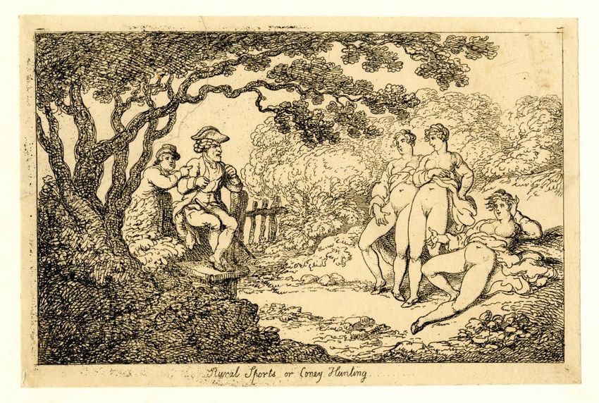
reserved).Midwifery and Obscenity Comic mobile prints were not, however, Spratt’s only point of interaction with wider print cultures. Print as a medium was becoming ever more pervasive, available, numerous, and diverse in its subject matter. Alongside its proliferation grew an anxiety about its uncontrollability and its potentially negative or corrupting influences. This anxiety was most manifest in discussions of what was coming, at this time, to be called pornography. 34 Prints with “obscene” content had a long history, but they underwent changes in both nature and number in the early nineteenth century. 35 They proliferated and they pervaded the spaces of the city—displayed in print shops and sold on street corners, they were seen as unavoidable, uncontrollable, and corrupting in a new way. No longer restricted to circles of wealthy men, obscene prints were not just cheap and available to everyone, they were also thrust on those who did not want to see them. 36 As Ian McCalman has shown, the older tradition of employing obscenity in political or satirical prints faded in this period, replaced by material that was more purely erotic. 37 These changes led to a middle-class moral panic about the corruption and sexualisation of women, children, and working people. Anti- vice societies sprang up to tackle the perceived problem, employing methods from entrapping potential buyers in the street to campaigning for stricter legislation. 38 This period saw the rise of the term “pornography” to describe this newly distinct and—according to its opponents—damaging form of print. And it was these very acts of definition and attempts at eradication that gave the genre its furtive, sinister, uncontrollable identity. 39 While the vast majority of this cheap printed visual material is now lost to us—neither kept by its original owners nor collected since—what remains indicates wider visual trends. Women who expose their own bodies by lifting skirts or loosing drapery, and who further expose their own genitalia with spread legs and forward-tilted hips, are familiar from the erotic prints of Thomas Rowlandson, which have survived in decent numbers thanks to the artist’s wider fame (Fig. 9). Illustrations have also survived in obscene books, where the same themes of female self-revelation, exposed genitals, and pubic hair are in evidence (Fig. 10). Some pornographic books also employed paper flaps, or played on the sexually suspect identity of the doctor (Fig. 11). 40 Erotic “art” prints and obscene books were joined, in the period, by cheap single sheet prints which no longer survive, but which were sold in vast numbers in shops and by street vendors. 41 This sheer availability, and the moral panic surrounding it, means that many people would have had at least a passing familiarity with such images. They would, in short, have been equipped to spot what I argue are intentional references in Spratt’s tables.
Figure 9.
Thomas Rowlandson (draftsman and printmaker), Rural Sports or Coney
Hunting, ca. 1790-1810, etching, 147 x 222 mm (trimmed). Collection of
The British Museum (1977,U.570). Digital image courtesy of The Trustees
of the British Museum (CC BY-NC-SA 4.0).
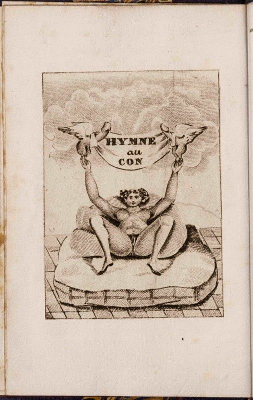
Figure 10.
Anon., Hymne au con, etching, measurements. From
Anon., Chansonnier du bordel, ou veillée d’un
fouteur (1833). Collection of the British Library (
P.C.31.a.30, p. [6]). Digital image courtesy of British
Library Board (All rights reserved).
Figure 11.
Anon., L’Accouchement, hand-coloured etching,
measurements. From Pierre Jean de Béranger,
Bérangiana. Mis en action ou choix de ses chansons
badines (1830). Collection of the British Library
(P.C.13.a.2, [facing p.26]). Digital image courtesy of
British Library Board (All rights reserved).
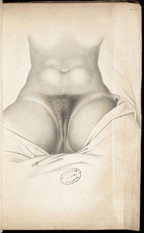
Two tables in particular from Spratt’s book play on these pornographic visual
tropes. Table 3 shows a woman’s body between belly and thighs, focusing on
her genitalia (Fig. 12). It includes two separate flaps, which represent the
body in different modes: the upper enacts an anatomical cut, opening up to
show the uterus and intestines beneath the skin; the lower describes the
body not under dissection but under medical examination with two flaps
representing the labia majora open like doors to expose the clitoris, labia
minora, and urethral and vaginal openings. These flaps do not represent a
cut into the body but a manipulable part of the body itself. As the illustration
to Hymne au con demonstrates, the view of the female genitalia, with pubic
hair, and framed by opened legs and the belly above was common to the
medical and the pornographic gaze (Fig. 10). While in the pornographic printthe depiction of the whole figure, the mattress on which she reclines, the
flying phalluses, and the title all encourage erotic looking, in Spratt’s table,
the cropped view and lack of context goes some way to directing away from
this kind of looking. Yet, the visual similarities cannot be completely denied,
in Spratt’s table we may say there is the potential for multiple kinds of
looking. To open the flaps in Spratt’s table might be interpreted as the
enacting of a medical examination, but it might also have been understood
as a sexual touching. Mary Hunter has noted the same potential for sexual
slippage in the medical image, describing the finger present in some medical
wax moulages of genitalia as both a masturbatory agent and a medical tool.
42
When we handle Spratt’s Table 3, we ourselves become this ambiguous
agent.
Watch Video
Figure 12.
F. Spratt (draftsman), Table 3, hand-coloured lithograph, 283 x 222 mm (page),
from George Spratt, Obstetric Tables (London: The Author, 1835). Digital image
courtesy of Courtesy of The University of Manchester (All rights reserved).
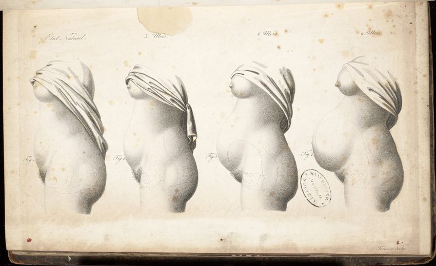
Table 4 shows a woman in profile from head to mid-thigh. She wears a lace
cap and holds up voluminous white drapery to expose her body (Fig. 13).
Lifting the flaps, we do not move inside her body, but rather observe her
exterior as her pregnancy develops, before finally getting a glimpse of the
fully gestated foetus in utero. This image is meant to describe for the
practitioner the external signs of pregnancy, focusing on the belly and the
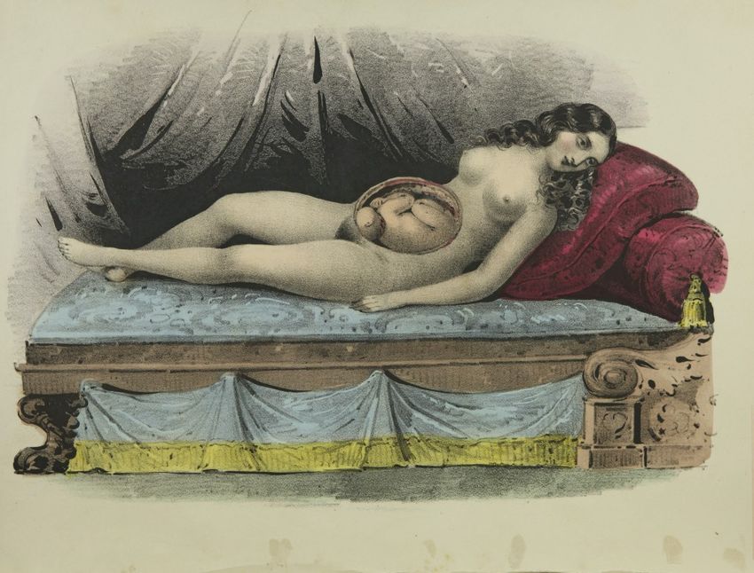
nipples. But, of course, this kind of close looking at the naked body can alsobe erotic, and the contemporary pornographic iconography of the self- exposing woman would have guided viewers towards such a reading (see Figs 9 and 10). Spratt’s text also seems to acknowledge the potential for erotic looking. He describes the figure in the first flap as a “virgin female”: 43 providing the “standard” body to compare with the pregnant one, but also offering a narrative of defloration. While the presence of pregnancy and the ability to look anatomically within the body might direct the viewer to a more medical interpretation, there is wider evidence that images of pregnancy could, in this period, also function as erotic images. 44 For instance, the illustrations in Wooster Beach’s An Improved System of Midwifery, published in New York in 1850, include copies of Spratt’s tables, as well as some other even more highly sexualised medical illustrations. 45 Incorporating, too, some of the woodcuts from Aristotle’s Masterpiece, the work is clearly in the vein of a midwifery guide/sex manual. 46 The frontispiece, for instance, shows a naked female figure lying on a luxurious couch, her expression somewhere between ecstasy and pain (Fig. 14). That she is presented as a sex object, despite her anatomised pregnant belly, is as clear here as it is in the many “anatomical Venuses” produced in the eighteenth and nineteenth centuries. 47
Watch Video

Figure 13.
G. Spratt (draftsman), Table 4, hand-coloured lithograph, 283 x 222 mm
(page), from George Spratt, Obstetric Tables (London: The Author, 1835).
Digital image courtesy of Courtesy of The University of Manchester (All rights
reserved).
Figure 14.
           Anon., Reclining Pregnant Woman, from Wooster Beach, An Improved
           System of Midwifery, 1850, hand-coloured lithograph, 29 cm (page
           height). Collection of The U.S. National Library of Medicine (WQ B365i
           1847). Digital image courtesy of The U.S. National Library of Medicine
           (Public Domain).

These visual associations invite the viewer to sexual looking, and force them
to consider the slippages between the medical and the pornographic. Indeed,
in their mobility, these tables forcibly implicate the viewer. Responding to the
demand for manipulation from the print itself, the viewer becomes an actor
in the medico-erotic realm of the image. It might be tempting to deny the
sexual in such images, to argue that they are purely medical, intended to be
looked at by a specialist trained in objective dispassion. But to do so would
be an act of anxiety-induced disengagement, particularly because, in the
nineteenth century, to say something was medical did very little to dispel its
sexual potential. The two were bound up with each other, and inextricable.

In the eighteenth century, debate had raged in both professional and public
print over the propriety of men attending women in labour. So-called “man-
midwives” were often looked on with deep suspicion not only as medically
incompetent but also as sexually predatory. This did not, however, stop the
spread of their practice. 48 By the late nineteenth century, man-midwives,
now called “obstetricians”, had established the propriety of their attendance
on women in childbirth; the necessity of physical and visual examinations;
and the legitimacy of their work as a medical specialism. Slowly,
obstetricians gained what Roy Porter has termed a “professional right of
entry”, allowing them to touch and look at the body in ways that, outside of
medical frameworks, were deeply inappropriate. 49 While, by late in the nineteenth century, this right had been established, it did not do away with all fears. Indeed, increased access and trust granted to doctors went hand in hand with an increased potential for abuse and raised the spectre of what McLaren calls the “murdering mad doctor”. 50 Earlier in the nineteenth century, while the “right of entry” was still being established, the acceptability of medical access to the female body was under active debate. Increased drives within medicine to closely examine patients were butting up against hardening notions of bodily modesty, propriety, and sexual continence, particularly for middle-class women. 51 Professional authors of the period sometimes expressed ambivalence over examination, wavering between an ideal of medical access and the reality of dealing with actual women. Francis Ramsbotham, for instance, argued both for the importance of manual intervention and visual examination, but also for not unduly exposing “the patient to the inconvenience of an ocular inspection”. 52 And while some practitioners had increasingly intolerant attitudes to their patients’ scruples, texts that denounced all male involvement in childbirth as “a disgrace to morality and Feminine Dignity” continued to be published. 53 An anonymous pamphlet published in 1826 explained that it was “unquestionably indelicate and unnatural for a surgeon to assist at a child-birth”, particularly because women’s modest “aversion to disclose”, combined with men’s “natural forwardness” meant that sexual misdemeanours could be easily committed and concealed by medical men. 54 These concerns were not restricted to a few vocal polemicists. The idea of the sexually suspect doctor appears in many aspects of culture, including contemporary pornography. In the illustration “L’Accouchement” (“Childbirth”) from Bérangiana (1830), the medical and sexual touch are explicitly linked, and the caption, which translates as “As he pleased, he was received”, points too to the easy slippage between kind and attentive doctor, and the wily seducer (Fig. 11). 55 While commentary and debate raged in many spheres of culture, what is clear is that by the 1830s most women who could afford it did call a male attendant when they were in labour. 56 But what is equally clear is that the access granted to such practitioners was tenuous, variable, and always subject to negotiation and worry. 57 In untangling these complex medico- social relations, medical images remain an under-used and often overlooked resource, yet as Elizabeth Stephens has demonstrated, they can be interrogated not simply as “medical representations of sexual bodies”, but also as expressions of the “sexualization inherent in the construction of medical knowledge itself”. 58 Spratt’s Tables 3 and 4, in the context of medical and public debates over access, and visual cultures that associated
doctors with philanderers or sexual predators, must have presented most
viewers with the troubling closeness of medical examination with sexual
looking and touching (Figs 12 and 13).

Spratt and Sexual Slippage

The entangling of medicine and sexuality was a public and important issue in
this period, as the proliferation of pornographic prints became associated
with a new genre of “popular” medical literature. 59 Higher literacy levels
and cheaper printing costs saw publishers finding new markets in pamphlets
and books aimed at educating a wider readership in all kinds of scientific
subjects, including medicine. Before the mid-century, the kinds of
information and images that went into these cheap and accessible works
were relatively unpoliced and often included information on sex and
generation. But, as with pornography, anti-vice societies and the medical
establishment felt moved to intervene. As demonstrated by the trials of
Frederick Hollick in America in the 1840s, doctors wanted to keep medical
expertise in-house, out of the hands of charlatan authors and the paying
public alike. 60 And anti-vice groups worried over the moral and social
implications of giving people information about sex and generation. By mid-
century, popular medical books were largely sanitised of such content and
professional works became increasingly inaccessible to lay readers. Popular
books that did contain prohibited information were characterised as obscene
and indeed were often sold alongside pornography. 61

A similar pattern has been traced by Sam Alberti and A.W. Bates with regards
to medical museums. Early in the century, they were popular and
widespread, providing a paying public with medical facts, frightening them
with gory specimens, and arousing them with supine and ecstatic female
anatomies. By the end of the century, such museums had largely been shut
down on the grounds of obscenity, or made private, accessible only to
medical students and professionals. 62 Those in power felt that the general
public could not cope with, or react appropriately to, the collections in these
museums. 63

Spratt’s Obstetric Tables has a specific place in this cultural moment: before
professional and public had more fully separated, and before medical and
legal institutions had settled on what constituted obscene content. His book
was able, in the 1830s and 1840s, to cater to professional surgeon-
accoucheurs; to students, apprentices, apothecaries, and midwives; to
curious lay people; and to those looking for either (or both) medical or sexual
content. The text uses technical language and references many canonical
obstetrical works, but is also brief and typographically accessible. The tables
too are less technical, detailed, and difficult to read than those produced in
many specifically “professional” works. I argue that the tables tread joyfully
all over the ambiguities of audience and propriety of medical content that
only existed in these decades between the 1820s and the 1850s.
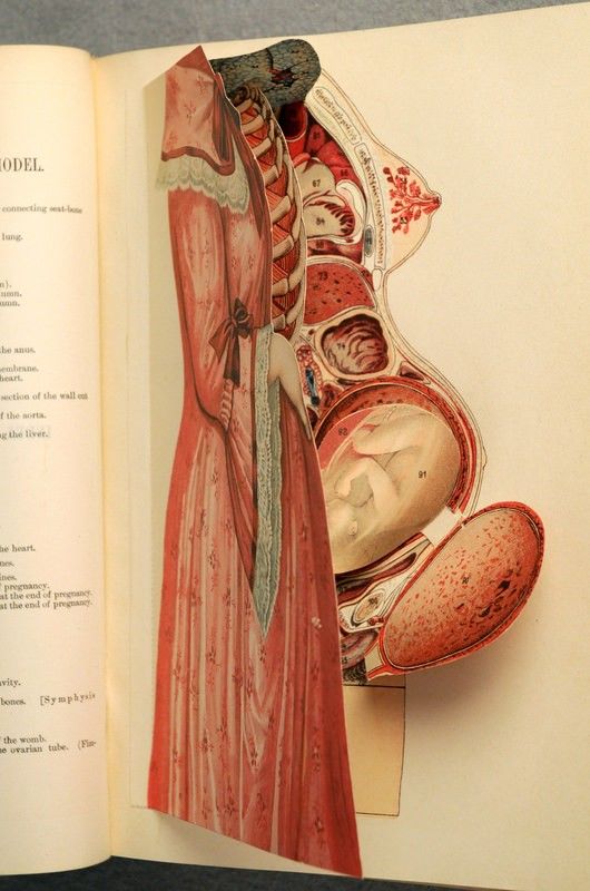
Figure 15.
Anon., Anatomical Female Model, from Frederick
Hollick, The Origin of Life (Philadelphia: David McKay,
1902), colour lithograph, 26 cm (page height).
Collection of David M. Rubenstein Rare Book &
Manuscript Library, Duke University (RC881.H73 1902
c.1). Digital image courtesy of David M. Rubenstein
Rare Book & Manuscript Library, Duke University (All
righs reserved).
That medical images at this time, and particularly Spratt’s tables, had the
capacity to accommodate multiple interpretations can be seen by comparing
Spratt’s Table 4 to a flap print that accompanied a 1902 edition of Hollick’s
The Origin of Life (Figs. 13 and 15). Where Spratt’s figure is irrevocably
naked, Hollick’s flaps move from full clothing straight to the bodily interior,
skipping the naked skin of the woman entirely. Hollick’s print provides only
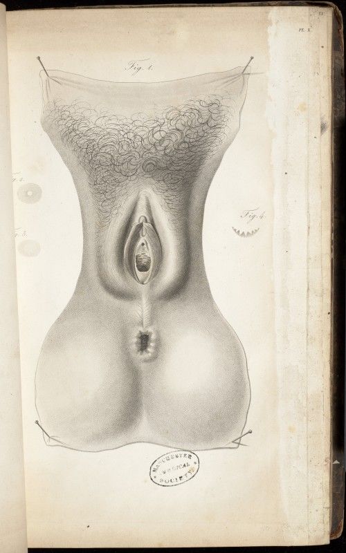
appropriate views of the female body, while Spratt’s forces us into anawareness of the potentially inappropriate nature of medical looking. Popular for a short period, I suspect that only a few decades after its first release, the Obstetric Tables had gone from a useful medical work to a collectible curiosity; and from one that was intriguingly risqué, to definitively obscene. In fact, Spratt’s work not only had its moment in this specific milieu but was also popular then because it commented knowingly on the ambiguities and anxieties of that culture. To modern eyes, it may seem confusing that a book ostensibly for medical professionals would also deal with the sexual problematics of their discipline, and even provide pornographic content. Yet this confusion arises partly from our own culture’s strict separation of professional medical content from wider cultures of sexuality. What Spratt’s book shows us is that in the decades before 1850 there was a market for this kind of multiplicity and ambiguity, indeed it is borne out in many illustrated books of the period. For contemporary medical viewers, it was acceptable, and even attractive, for a book to be medically pedagogic, culturally commentative, and potentially pornographic. All three functions were, after all, as interesting to doctors as to anyone else. Spratt was not alone in producing images that trod the line between medical and obscene, indeed, I argue that this tension infused all medical visual culture of the period. What does make Spratt different is his willingness to directly engage with the issue. His flap constructions do not defuse or deflect, they tackle the sexual in the medical and force us to examine it. This can best be seen by comparing Spratt’s images to one of his sources—the illustrations produced by Antoine Chazal for Maygrier’s Nouvelles demonstrations d’accouchemens. Table 3 in Obstetric Tables is an amalgamation of Maygrier’s Plates 9 and 10 (see Figs. 12, 16 and 17). With the flaps closed, Spratt’s table looks like a coloured copy of Maygrier’s Plate 9 (Fig. 16). In both, the sitting posture, the sheet covering the legs, and the fact that no anatomical cut has been made indicates that the woman is a living patient. While she is just as naked in Maygrier’s version as in Spratt’s, in the original, the labia majora are closed over the rest of the vulva, turning the female genitals into a neat cleft. This image, Maygrier declares, represents the female genitalia “in the natural state, and perfectly approximated”. 64 He associates the biologically ideal female body with a naturally modest, hidden vulva. Plate 10 shows the vulva under examination, after the labia majora have been “slightly separated” (Fig. 17). 65 But while Maygrier acknowledges that a separation of the labia majora is something a doctor might enact on a living patient, the image shows what is clearly an anatomical specimen. The pins stretching out the sectioned skin indicate that we are looking at a body part, separated not only from a cadaver but more broadly from the idea of a person. Visually, the social problematics of vaginal examination are denied, replaced by an abstracted, intellectualised body which is fully the property of the surgeon/dissector. 66
Figure 16.
Antoine Chazal (draftsman), Plate 9, from J. P.
Maygrier, Nouvelles demonstrations
d’accouchemens (Paris: Béchet,1822), 1822,
lithograph. Collection of The University of
Manchester Library. Digital image courtesy of The
University of Manchester Library (All righs
reserved).
Figure 17.
                   Antoine Chazal (draftsman), Plate 10, lithograph,
                   from J. P. Maygrier, Nouvelles demonstrations
                   d’accouchemens (Paris: Béchet,1822). Collection of
                   The University of Manchester Library. Digital image
                   courtesy of The University of Manchester Library
                   (All rights reserved).

Even the “naturally modest” image of the female genitalia in Maygrier’s Plate
9 seems to have been powerful and troubling. It has been removed from
many copies of the work for reasons, we may suppose, both erotic and
censorious. How much more confrontationally shocking did Spratt make his
version then, in relocating the entire vulva and the physical act of splaying
back onto the living body of the patient. Spratt makes explosive where
Maygrier and Chazal attempted to defuse. He doesn’t allow the world of
abstract medical knowledge to be separated from that of medical practice,
nor does he allow the specimen to be something different to the living body.
The turning of the closed labia in Maygrier’s Plate 9 into openable flaps
seems a direct challenge to Maygrier’s partial depiction—Spratt not only
exposes what is left hidden, he also forces the viewer to acknowledge the
action and consequences of exposure.
A similar process lies behind Table 4, which is modelled on Chazal and
Maygrier’s Plate 26 (see Figs. 13 and 18). In Chazal’s version, the body in
profile is repeated four times, growing increasingly pregnant. The crucial
difference in Spratt’s version is not so much the layering of these bodies, as
their re-personing. Spratt gives the woman a head, the cloth wound around
the shoulders and above the breasts of the original figures becomes a kind of
drapery, which she lifts to expose her body. She is even given a lace cap with
a bright blue ribbon, a standard item of domestic wear for married women. 67
This cap encourages the viewer to see the figure not in the abstract, but to
identify her as a real, specific late-Georgian woman of the middle classes—a
young wife. For women viewers, including patients, this adaption must have
encouraged identification with the figure, along with awareness (and
anxiety? or excitement?) of their own nakedness and vulnerability under the
medical gaze.
Figure 18.
Antoine Chazal (draftsman), Plate 26, lithograph, from J. P. Maygrier,
Nouvelles demonstrations d’accouchemens (Paris: Béchet,1822).
Collection of The University of Manchester Library. Digital image courtesy
of The University of Manchester Library (All righs reserved).
In Maygrier’s version, cloth is used to divorce the body from the face, and
from the idea of an actual patient, associating the figures instead with
anatomical specimens, which were often represented as partially draped.
The drapery in Spratt’s, while it lacks sleeves or tailoring that would make it
specifically identifiable as a chemise or nightgown, does evoke clothing in
the way it is worn, and lifted, by the figure. We are reminded by it not only ofthe small practicalities and negotiations of clothing and nakedness in medical examination but also of the much wider cultural significances of veiling. In textual and visual culture outside of medicine, veils were employed not simply to cover the body and ensure modesty, but also to create the possibility of exposure. Often translucent, they offered the form of modesty at the same time as a transgressive glimpse of the body beneath. As art and fashion historians have noted in many contexts, veiling heightens erotic potential. 68 The lifting of the cloth in Spratt’s Table 4 arguably adds a level of eroticism to the image that stark nakedness would not. But this is in itself complicated by the choice not to make the drapery a flap—while we can control the level of pregnancy of the figure, and even lift her skin to peer inside her uterus, we cannot clothe her. It is discomforting to be reminded of the act of unveiling but not given the capacity for re-veiling in an image that otherwise facilitates lifting and covering to such an unusual extent, and in which the foetus remains permanently veiled by the uterine membranes. Lynda Nead has discussed the association in the nineteenth century of sexuality and pornography with newly developing notions of the private and domestic realm. 69 Spratt’s image is one that addresses these problematics of privacy, sexuality, and medicine: the woman is an embodiment of the private realm that is destroyed by the examination of the doctor, and of the print’s viewer. Both medical and pornographic print exposed what should remain private, and in representing the woman’s garment as immovable, Spratt forces us into an awareness of our own discomfort with this exposure. Tables 3 and 4 force us to recognise the potential for sexual slippage in medical practice that troubled medical professionals and cultural commentators throughout the nineteenth century. Doctors themselves, reluctant to publicly acknowledge that some of their number could and did seduce and abuse their patients, often rechannelled their anxiety into concern over the power of their women patients to maliciously ruin their careers and reputations with inappropriate behaviour and false allegations. 70 This professional anxiety grew up alongside, and strengthened, the cultural enforcement of passivity on women’s bodies, both in terms of sexuality, and in childbearing. 71 A good woman was not a sexual woman, and she was a docile patient. These stories are well known: it was in the mid- century that some doctors claimed that women had no sex drive. While the view was never dominant, it did colour wider understandings of sexuality, and helped to disseminate the more accepted idea that women’s sexuality was less intense than men’s, and was often completely dormant. 72 In childbirth, increasing levels of male medical control rendered the woman’s body passive, at least rhetorically. This enforced passivity came from two sources: one had to do with the separation of medicine and physiological understanding from personhood. It was no longer women who laboured in childbirth, but their uteri, which did the job unconsciously. 73 The other
You can also read