Case Presentation: 50 year old state trooper with elevated liver function tests - Christian Yon MS3 Michael Baldwin, MD - UConn Health
←
→
Page content transcription
If your browser does not render page correctly, please read the page content below
Case Presentation: 50 year old
state trooper with elevated liver
          function tests
       Christian Yon MS3
       Michael Baldwin, MD
Initial Presentation to Family Medicine
50 year old man who works as a state trooper and has a past medical history significant for obesity,
gallstones, and kidney stones. He presented to the Family Medicine clinic for follow up on abnormal
liver function studies noted incidentally 1 month ago through a hematologic evaluation, which he
•
received for polycythemia.
•
Alkaline phosphatase 671
•
AST 197
•
ALT 335
Total bilirubin 1.4
•
Repeated liver function panel at the time of the Family Medicine visit demonstrated the following:
•
Alkaline phosphatase 1,016
•
AST 509
•
ALT 720
•
Total bilirubin 2.5
•
Direct bilirubin 1.6
•
Albumin 4.4
Total protein 6.7
Of note, the patient had gallstones incidentally noted on a CT in 2019 along with a dilated cystic duct
and thickening of the gallbladder wall.Family Med visit, cont.
• The patient denied abdominal pain and was overall
asymptomatic at this time. Minimal alcohol use, no drug
use, and no recent altercations or exposure to blood
products on the job as a state trooper.
• Physical exam:
109/72 72 97.9 F 99%
The patient is alert and well-appearing. No RUQ
tenderness, no hepatosplenomegaly.
• Additional labs: negative for Hep B, Hep C, HIV, CMV
• Patient counseled to report to ED if he develops pain or
feverHospital Presentation
2 days after the Family Med visit, the patient presented to the ED with the
following symptoms:
• RUQ abdominal pain
• Mild jaundice of the face
• Nausea
• Pruritus of the lower extremities at night
• Denies fevers, chills, chest pain, SOB, diarrhea, constipation
Admitted to medicine team; the following imaging was performed:
• RUQ ultrasound → demonstrated cholelithiasis and mild intrahepatic
biliary ductal dilatation
• CT abdomen pelvis with IV contrast
• MRI abdomen with and without IV contrast
• Magnetic resonance cholangiopancreatographyCT abdomen pelvis with IV contrast
CT abdomen pelvis with IV contrast

                                 Within the
                           gallbladder, there are
                            multiple foci of gas,
                              which are likely
                             contained within
                                gallstones.

                           Lack of evidence for
                              pericholecystic
                           inflammation or fluid
MRCP
MRCP

Thin-walled gallbladder is
filled with T2-hypointense
calculi.

No evidence of acute
cholecystitis.

A calculus is seen within
the neck of the
gallbladder and is
exerting a mass effect
upon the right lateral
aspect of the proximal
common bile duct.

Lack of filling defects
within the common bile
duct itself, so no evidence
of choledocholithiasis.
MRCP
MRCP

Axial view of the
calculus in the neck
of the gallbladder,
measured at 16 mm
MRCP
MRCP

Absence of focal
hepatic parenchymal
lesion

Mild to moderate
intrahepatic biliary
ductile dilatation
Differential Diagnosis
1.   Choledocholithiasis
     – Presents with fever, jaundice, RUQ pain (usually subacute/prolonged
        pain), ↑alk phos and ↑bilirubin
     – Would require at least 1 gallstone to be stuck in common bile duct
2.   Acute cholecystitis
     – Presents with sudden onset of RUQ pain, sonographic Murphy’s sign,
        and gallbladder wall thickening
     – Would not cause ↑alk phos or ↑bilirubin by itself; that would require a
        secondary process causing cholestasis
3.   Mirizzi syndrome
     – Presents with recurrent episodes of jaundice and cholangitis with biliary
        type RUQ pain
     – Dilated common bile duct on abdominal imaging
     – MRCP classically shows a large impacted gallstone in the gallbladder
        neck or cystic duct
4.   Cholangiocarcinoma
     – Presence of tumor(s) within bile ducts, either
        intrahepatic or extrahepatic
Diagnosis?
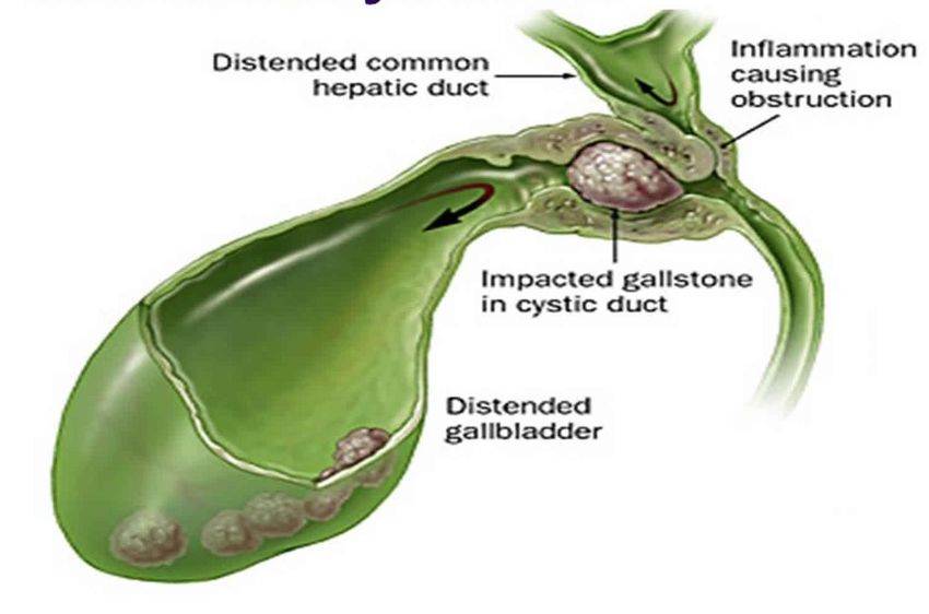
Diagnosis: Mirizzi Syndrome

stepwards.com
Discussion
•   Mirizzi Syndrome = extrinsic compression of an
    extrahepatic biliary duct caused by one or more calculi
    within the cystic duct or infundibulum of the
    gallbladder
     – Relatively rare
     – Can lead to acute cholecystitis, or eventually to
       fistulae between the gallbladder and common duct
        • Classified into types I-V depending on these
           complications and their severity
     – Associated with gallbladder cancer – likely due to
       recurrent inflammation and biliary stasis
     – Treatment of choice: cholecystectomy
Later Hospital and Surgical Course
• No surgical intervention anticipated at first
• HIDA scan 2 days later showed nonvisualization of the
gallbladder → now concerning for acute cholecystitis
• Went for laparoscopic cholecystectomy with
intraoperative cholangiogram
• Converted to open cholecystectomy and Roux-en-Y
hepaticojejunostomy for a chole-choledocho fistula,
discovered intraoperatively
• Further complications including likely abdominal
bleeding led to a transfer to the ICU and a long
hospital stay before his eventual recoverySources
1. Jones MW, Ferguson T. Mirizzi Syndrome. [Updated 2021 Feb 8].
StatPearls. Available from:
https://www.ncbi.nlm.nih.gov/books/NBK482491/
2. Vadera S, Weerakkody Y, et al. Radiopaedia. Available from:
https://radiopaedia.org/articles/mirizzi-syndrome?lang=us
3. Valderrama-Treviño AI, Granados-Romero JJ, Espejel-Deloiza M,
et al. Updates in Mirizzi syndrome. Hepatobiliary Surg Nutr.
2017;6(3):170-178. doi:10.21037/hbsn.2016.11.01
4. Umashanker R, Smink D. Mirizzi Syndrome. [Updated 2019 Feb
14]. Uptodate. Available from:
https://www.uptodate.com/contents/mirizzi-
syndrome#H2767415925
Illustration from: https://healthjade.net/mirizzi-syndrome/You can also read