Case Series Mycosis fungoides: A Chameleon of Dermatology
←
→
Page content transcription
If your browser does not render page correctly, please read the page content below
Case Series
Mycosis fungoides: A Chameleon of Dermatology
Swetha D1, D V S Pratap2
1
Assistant Professor,2Professor and Head,Department of Dermatology, Prathima Institute of Medical
Sciences,Karimnagar,Telangana,India.
Address for Correspondence: Dr.Swetha D,Assistant Professor,Department of Dermatology,Prathima Institute of Medical
Sciences,Karimnagar,Telanagana,India.
Email:swethambbs2@yahoo.com
ABSTRACT and serial biopsies may unravel the mystery and establish the
Mycosis fungoides (MF) is the most common type of cutaneous diagnosis of this great mimicker. We have encountered similar
T-cell lymphoma, characterized by skin-homing of clonal, situation in our study of 3 cases of MF.
mature malignant T-lymphocytes. MF develops slowly over CASE REPORT
several years and may have a variety of clinical presentations. Case 1 :
In addition to the classical clinical lesions-itchy patches, plaques
A young adult male aged 28 years old presenting with itchy,
or tumors, it may also present with atypical hypopigmented
erythematous, scaly patches over the trunk (Figure 1a, 1b)
lesions, dermatophytic lesions or psoriasiform lesions that may
visited several dermatologists and a diagnosis of
be confused with common benign conditions such as eczema
dermatophytosis was made. Topical and systemic anti-fungals
and psoriasis.
were prescribed repeatedly with no response. In View of
Clinical picture may pose a significant challenge and a chronicity and inadequate response to the above treatment
diagnostic dilemma to the dermatologist. Longterm follow up patient was subjected to thorough clinical examination which
and serial biopsies help to establish definitive diagnosis. revealed erythematous patches and plaques with mild scaling.
Keywords : Mycosis fungoides (MF), Cutaneous lymphoma, No induration was seen. Few patches were atrophic. No
Psoriasis, Vitiligo. lymphadenopathy was seen. Other systemic examination was
INTRODUCTION within normal limits . KOH examination of scales from the
lesions did not show any fungal hyphae. All biochemical
Mycosis fungoides (MF) is the commonest variant of primary
investigations were within normal limits. Histopathological
cutaneous T cell lymphoma, accounting for almost 50% of all
examination revealed mild spongiosis in epidermis and
primary cutaneous lymphomas.1, 2 It most commonly affects
perivascular lymphocytic infiltrate. Few lymphocytes showed
middle-aged and elderly adults of all races 3. Typically,
nuclear irregularity with halo around them.
neoplastic T cells localize to the skin and produce patches,
plaques, tumors or erythroderma4. It is characterized by a Case 2 :
relatively consistent constellation of clinical, histologic, A 52yr old male referred to the DVL department with six
immunophenotypic and molecular aberrations5. months history of scaly plaques with superficial erosions and
MF typically manifests as an indolent cutaneous eruption with crusted nodulo-ulcerative lesions all over the body. With these
erythematous scaly patches or plaques and may progress to symptoms he was misdiagnosed as plaque type psoriasis and
generalized erythroderma, cutaneous tumors or advised various mild and high potency topical corticosteroids
extracutaneous involvement. The initial skin lesions are often which did not offer any relief. Thorough skin examination
confined to sun protected areas. The rate of progression is revealed reddish brown ulcerated lesions and indurated
unpredictable. Tumors, however, develop only in a minority erythematous plaques with superficial erosions on trunk and
of patients from patches, plaques, or de novo. They more extremities (Figure 2a, 2b). His nails were intact without pitting
commonly arise on the face and body folds. Leonine facies or dystrophy. He had cervical and inguinal lymphadenopathy.
results from malignant T cell infiltration leading to extensive Auscultation of chest did not reveal any abnormality. No
thickening and skin fold accentuation. The palms and soles can organomegaly was seen. VDRL and HIV serology were negative.
develop hyperkeratosis and skin fissuring. These phases may Lesional biopsy revealed: atypical lymphocytes with
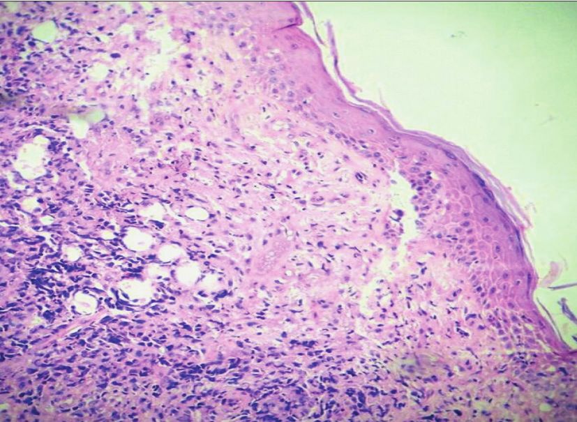
be distinct or overlapping at diagnosis6,7. cerebriform nuclei. Pautrier’s abscess was present in the
Several atypical forms resembling other dermatoses like epidermis (Figure 3).
vitiligo, dermatophytosis and psoriasis may pose a diagnostic
challenge to the dermatologist. Clinical diagnosis, follow up
18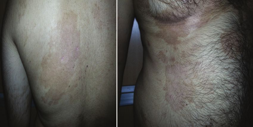
Swetha, et al

Case 3 :
A middle aged male came to DVL department with multiple
asymptomatic hypopigmented patches over the trunk and
proximal extremities. These hypopigmented patches first
appeared on the trunk and later on proximal extremities too.
The initial hypopigmented lesions gradually turned into
erythematous patches and increased in size as well. There was
no history of systemic symptoms such as fever, lassitude or
weight loss.
On examination there were asymptomatic, hypopigmented
and few vitiligo-like depigmented irregular atrophic macules
and patches on the trunk and extremities. These patches
showed a finely wrinkled slightly scaly surface (Figure4). They
were not indurated and there was no telangiectasia. No
lymphadenopathy or organomegaly seen. All other                     Figure 3: Section showing atypical lymphocytes with cerebri-
investigations were within normal limits.                           form nuclei. Pautrier’s abscess was present in the epidermis.
Histological examination of a vitiligo like lesion showed an
infiltrate of mononuclear cells at the interface with prominent
epidermotrophism. No atypical cells were seen.

 Figure 1: Photographs showing erythematous, scaly patches
 over the trunk.
                                                                    Figure 4: Photograph showing hypopigmented and few vitiligo-
                                                                    like depigmented irregular atrophic macules and patches on
                                                                    the trunk and extremities.

                                                                  DISCUSSION
                                                                  Mycosis fungoides is the most common type of cutaneous T-
                                                                  Cell lymphoma accounting for about fifty percent of all cases.
                                                                  It is an epidermotropic cutaneous T-Cell lymphoma caused by
                                                                  proliferation of small to medium sized lymphocytes with
                                                                  cerebriform nuclei. Clinically MF is divided into four main sub
                                                                  types:
                                                                  Patch stage, plaque stage, tumor stage & erythroderma. As
                                                                  the disease progresses, patches may evolve over a variable
                                                                  period of time, into infiltrated plaques with a more generalized
                                                                  distribution. Plaques may be followed by ulcerated and
 Figure 2: Photographs showing reddish brown ulcerated lesions
                                                                  exophytic tumors. Tumors develop only in a minority of
 and indurated erythematous plaques with superficial erosions
 on trunk and extremities.                                        patients, although it is common to have patch, plaque and
                                                                  tumor lesions simultaneously on different parts of the body.

     19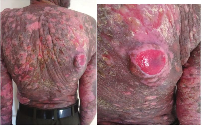
Swetha, et al

Some patients with patch stage MF never progress to other             other dermatoses and to ensure a proper diagnosis for better
forms of the disease. Tumors are the presenting sign in about         treatment. Thus the clinician must be vigilant and be prepared
10% of cases. Lymphadenopathy is usually a late occurrence.           to take a holistic view of the clinical, histological,
Visceral dissemination (lungs, spleen, liver, gastrointestinal        immunphenotypic and molecular genetic evidence to make a
tract) may develop subsequently. Extracutaneous                       proper diagnosis, accurate staging and classification.
dissemination is directly correlated to the extent of cutaneous       REFERENCES
disease7.                                                             1.     Whittaker SJ, Foss FM. Efficacy and tolerability of
 Median age at diagnosis is 55-60 years but MF may occur in                  currently available therapies for the mycosis fungoides
children and adolescents as well. Men are more commonly                      and Sezary syndrome variants of cutaneous T-cell
affected than women. It mimics several dermatoses. In addition               lymphoma. Cancer Treat Rev. 2007;33:146–160.
to the classical presentation, several atypical forms                 2.     Scarisbrick JJ. Staging and management of cutaneous
hypopigmented lesions, psoriasiform lesions, palmo plantar                   T-cell lymphoma. Clin Exp Dermatol. 2006;31:181–186.
form and dermotophyte like lesions have been reported. Early
                                                                      3.     Hwang ST, Janik JE, Jaffe ES, Wilson WH. Mycosis
lesions of MF may pose a significant diagnostic challenge to
                                                                             fungoides and Sézary syndrome. Lancet. 2008;371:945–
clinicians and dermoto-pathologists. Many such patients need
                                                                             957.
long term follow up and serial biopsies to make definitive
diagnosis.                                                            4.     Nashan D, Faulhaber D, Ständer S, Luger TA, Stadler R.
                                                                             Mycosis fungoides: a dermatological masquerader. Br J
The descriptive term “Mycosis fungoides” chosen in 1806 by
                                                                             Dermatol. 2007;156:1–10.
Alibert, suggests that first differential diagnosis of tinea
corporis for a typical MF lesion.Here the patient presented           5.     Smoller BR. Mycosis fungoides: what do/do not we
with scaly annular plaques all over the body. He was diagnosed               know? J Cutan Pathol. 2008;35(suppl 2):35–39.
to be a case of dermatophytosis and was given several                 6.     Kim-James HY, Heffernan MP. The diagnosis, evaluation,
antifungals but without any clinical relief. Then a biopsy was               and treatment of cutaneous T-cell lymphoma. Curr
done and it revealed atrophy of the epidermis; a superficial,                Probl Dermatol. 2001;13:307–340.
perivascular, and interstitial lymphocytic infiltrate with            7.     Zinzani PL, Ferreri AJ, Cerroni L. Mycosis fungoides. Crit
numerous atypical lymphocytes; and exocytosis of atypical                    Rev Oncol Hematol. 2008;65:172–182.
lymphocytes into the epidermis with formation of
                                                                      8.     Ryan EA,Sanderson KV, Bartak P,Samman PD.Can
microabscesses-findings consistent with the diagnosis of
                                                                             mycosis fungoides begin in the epidermis?A
mycosis fungoides. Treatment with Nb UVB led to long-term
                                                                             hypothesis.Br J Dermatol 1973;88:419-29.
remission of the mycosis fungoides.
Recalcitrant tinea corporis has been reported as the presenting        Please cite this article as: Swetha D,Pratap D V S. Mycosis
manifestation of patch-stage MF in several studies. This may           fungoides: A Chameleon of Dermatology.Perspectives in
be due to the immunosupression by the underlying condition.            medical research 2015;3:2:18-20.
In early stages of MF the characteristic lesion consists of            Sources of Support: Nil,Conflict of interest:None declared
erythematous macules or papules. Often some degree of
scaling is observed, similar to psoriasis. The edges of the lesions
may exhibit increased scaling, corresponding to a growing
infiltrate but the histopathological picture differs from that of
psoriasis.
The term hypopigmented MF was mentioned for the first time
by Ryan et al in 19738. This variant of MF is more commonly
seen in children and dark skinned individuals; especially Asians
and occurs at a much earlier age as compared to the classical
MF. Hypopigmented MF lesions were considered to be the
earliest and benign lesions and often clinically mistaken for
other hypopigmented lesions like leprosy, vitiligo etc.
Histopathology and immunohistochemistry are helpful to make
accurate diagnosis. The histopathological lesions shows
scattered lymphocytes in basal epidermis with or without
Pautrier’s microabscesses. The main purpose of presenting
these case reports is to show the clinical similarity of MF with

     20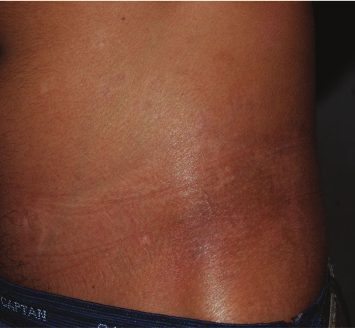

You can also read