Annals of Clinical and Medical Case Reports
←
→
Page content transcription
If your browser does not render page correctly, please read the page content below
Annals of Clinical and Medical
                                         Case Reports

Case Report                                                                                                       ISSN 2639-8109 Volume 6

Primary Cardiac Angiosarcoma Diagnosed by 3D Transesophageal Echocardiography
Guided Endomyocardial Biopsy – Case Report
Ozieranski K1, Szczerba E1*, Tyminska A1, Marchel M1, Wojnicz R2, Piatkowski R1, Opolski G1 and Grabowski M1
1
  First Department of Cardiology, Medical University of Warsaw, Poland
2
  Department of Histology and Cell Pathology in Zabrze, School of Medicine with the Division of Dentistry, Medical University of
Silesia in Katowice, Poland
 *
  Corresponding author:                                  Received: 22 May 2021           Copyright:
Ewa Szczerba,                                            Accepted: 11 May 2021           ©2021 Szczerba E. This is an open access article dis-
First Department of Cardiology, Medical University       Published: 16 Jun 2021          tributed under the terms of the Creative Commons Attri-
of Warsaw, ul. Banacha 1a, 02-097 Warszawa,                                              bution License, which permits unrestricted use, distribu-
Poland, Tel. +48 22 599 29 58,                                                           tion, and build upon your work non-commercially.
E-mail: ewa_szczerba@poczta.onet.pl
                                                                                         Citation:
                                                                                         Szczerba E, Primary Cardiac Angiosarcoma Diagnosed
Keywords:                                                                                by 3D Transesophageal Echocardiography Guided En-
Cardiac tumor; Cardio-oncology; Heart failure; Histo-                                    domyocardial Biopsy – Case Report. Ann Clin Med Case
pathology                                                                                Rep. 2021; V6(18): 1-7

1. Abstract                                                               tochemical stainings, electron microscopy) allows for quick, safe
Cardiac angiosarcomas are uncommon, primary malignant car-                and definite diagnosis of patients with cardiac tumors of unknown
diac tumors, characterized by an aggressive local growth within           origin facilitating further management. EMB with 3D-TEE guid-
the myocardial structures. The majority occur in the right atrium,        ance is technically feasible and increases the accuracy and safety
and at the moment of diagnosis usually infiltrate into neighboring        of the diagnosis of intracardiac tumors.
structures. Due to the lack of typical features in clinical presenta-     2. Introduction
tion, the diagnosis of cardiac angiosarcoma is challenging. Initial       Primary malignant cardiac tumors are extremely uncommon
clinical workup includes different imaging modalities [transtho-          (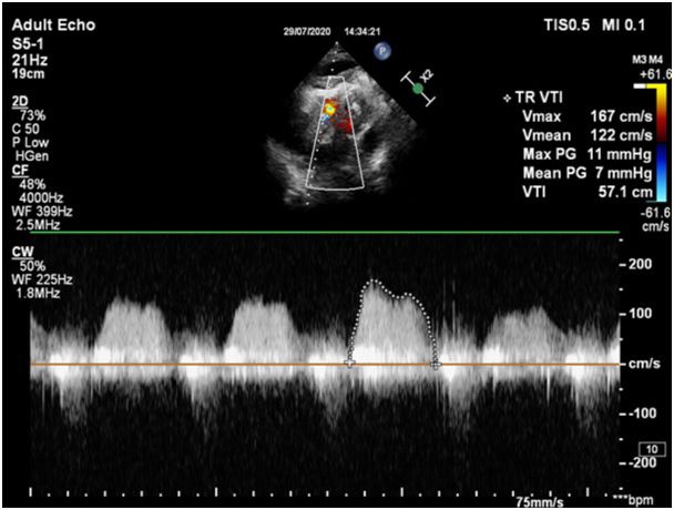
Volume 6 Issue 18 -2021                                                                                                                Case Report

She denied chest pain, palpitations or other relevant symptoms.           A TEE confirmed a large polycyclic tumor (80 x 66 mm) with
On physical examination, her vital signs were as follows: hemo-           heterogeneous echogenicity, attached to the wall of the right atri-
globin saturation, 94% (Fraction of Inspired Oxygen (FiO2) 0.21);         um and infiltrating the right atrium wall, superior vena cava and
heart rate, 90 beats/min; blood pressure, 95/78 mmHg; and no              visceral pericardium. The tumor occupied the entire right atrium
fever. Systolic-diastolic murmur on cardiac auscultation; general         causing functional tricuspid stenosis, but without invading the tri-
peripheral oedema; signs of bilateral pleural effusion and liver en-      cuspid valve and inferior vena cava (Figure 4).
largement were present. Laboratory studies showed the following           Chest, abdomen, and pelvis contrast-enhanced computed tomog-
abnormalities suggesting acute kidney injury and decompensation           raphy revealed a large polycyclic tumor (88 x 67 x 74 mm) with
of HF: serum creatinine, 1.94 mg/dl; estimated glomerular filtra-         heterogeneous densities (moderately enhanced by contrast, most
tion rate (eGFR), 25 ml/min/1,73 m2; urea, 110 mg/dl; N-terminal          likely containing necrotic areas) covering most of the right atrium,
pro-brain natriuretic peptide, 2392 pg/ml (N:
Volume 6 Issue 18 -2021                                                                                                                       Case Report

Figure 2: A large tumor with heterogeneous echogenicity (shown with a white arrow) infiltrating the wall of the right atrium and filling its cavity almost
completely, visible from the substernal view. LA-left atrium, LV-left ventricle, RA-right ventricle.

Figure 3: Maximal and mean gradient through the right atrium and the tricuspid valve assessed by continuous Doppler ultrasound measurements indi-
cating severe obstruction of blood flow into the right ventricle.

http://www.acmcasereport.com/                                                                                                                           3
Volume 6 Issue 18 -2021 Case Report
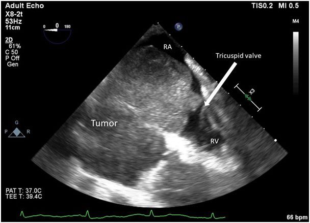
Figure 4: A modified mid-esophageal four-chamber view demonstrating a large hypoechogenic tumor infiltrating the wall of the right atrium and filling
its cavity almost completely. RA-right atrium, RV-right ventricle.
Figure 5: Tumor biopsy controlled by transesophageal echocardiography. RA-right atrium.
http://www.acmcasereport.com/ 4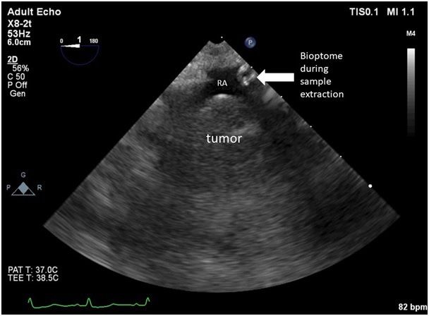
Volume 6 Issue 18 -2021 Case Report
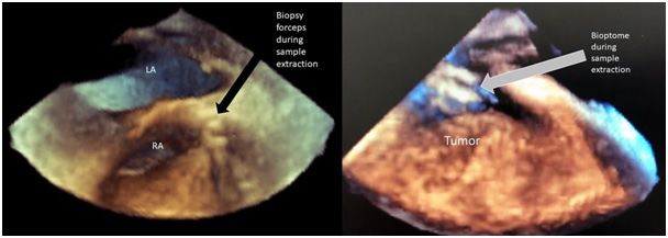
Figure 6: Tumor biopsy controlled by transesophageal echocardiography – a 3D view. LA-left atrium, RA-right atrium.
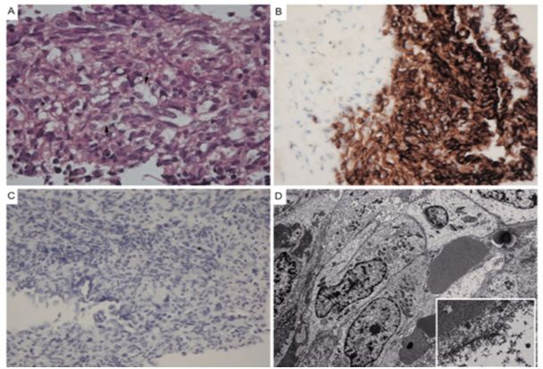
Figure 7: Primary cardiac angiosarcoma. A: Anaplastic cells with poorly formed vascular channels (arrows) (H&E); B: Strong immunohistochemical
staining for CD31 marker (brown color); C: Negative immunohistochemical staining for cytokeratin filaments AE1/AE3 (brown color); D: Electron
micrographs showing immature endothelial cells.
During hospitalization, she was initially treated with furosemide Therefore, an early diagnosis of angiosarcoma is crucial for thera-
and i.v. fluids (under blood pressure and diuresis monitoring) pre- peutic options and the patient’s prognosis. Comprehensive clinical
senting slight clinical improvement. However, three days after the and multimodality imaging (TTE and TEE, including 3D, contrast
EMB, she developed cardiogenic shock and subsequent sudden or intracardiac echocardiographic imaging; cardiac magnetic reso-
cardiac arrest. The patient died despite cardiopulmonary resusci- nance (CMR); positron emission tomography (PET)) evaluation of
tation. cardiac tumors is fundamental to obtain a proper initial differential
4. Discussion diagnosis [4,5,6,7]. In our case, an initial diagnosis of a malignant
cardiac tumor was made by use of echocardiography, both TTE
Cardiac angiosarcomas are characterized by aggressive growth and
and TEE, followed by contrast-enhanced computed tomography,
early metastases. The diagnostic and therapeutic approach is very
which revealed a huge, dense mass with areas of necrosis in the
demanding because of the intracardiac localization of the tumor.
right atrium. Angiosarcomas are mostly immobile and broad-
Clinical presentation of cardiac angiosarcomas depends on their
based with endocardial to myocardial growth [1]. CMR could
size, location, status of local infiltration and/or distant metastases,
be useful but in our patient a CMR was not performed because
relation with other cardiac structures and potential signs of hemo-
of kidney insufficiency. CMR findings in angiosarcomas include
dynamic compromise. In the majority of cases, angiosarcomas are
heterogeneous T1 and T2-weighted signal intensity and a hetero-
diagnosed when the disease is already advanced with evidence of
geneous contrast enhancement pattern [1]. A PET scan with the
metastases, and often with atypical signs of HF [3].
use of 18F-2-fluoro-2-deoxy-D-glucose (FDG) can reveal areas
http://www.acmcasereport.com/ 5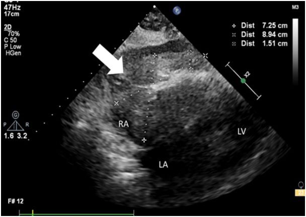
Volume 6 Issue 18 -2021                                                                                                               Case Report

of high FDG uptake within the mass and evidence of metastatic           5. Ethical Statement
disease [8,9].                                                          The authors are accountable for all aspects of the work and ensur-
Histopathology is necessary to reach the final diagnosis and plan       ing that questions related to the accuracy or integrity of any part
subsequent clinical management in some types of cardiac masses          of the work are appropriately investigated and resolved. All pro-
[10]. The histopathological features of angiosarcomas are: a highly     cedures performed in studies involving human participants were
vascularized mass with myocardial infiltration and signs of pleo-       in accordance with the ethical standards of the institutional and/or
morphism, necrosis and mitosis, clearly indicating diagnosis [1].       national research committee(s) and with the Helsinki Declaration
EMB is still the only method allowing for a definite diagnosis,         (as revised in 2013). Written informed consent was not obtained
but it is not commonly performed, despite a very low complica-          from the patient. Presented data do not allow for the subject to be
tion rate (
Volume 6 Issue 18 -2021                                                     Case Report

     myocardial biopsy in the management of cardiovascular disease: a
     scientific statement from the American Heart Association, the Amer-
     ican College of Cardiology, and the European Society of Cardiology.
     Endorsed by the Heart Failure Society of America and the Heart Fail-
     ure Association of the European Society of Cardiology. J Am Coll
     Cardiol 2007; 50: 1914-1931.
13. Aghdassi A, Gupta A. Endomyocardial Biopsy: Examining Indica-
    tions and Trends in Use – More Native Heart Biopsies to Come?
    Cardiology 2019; 142: 102-104. doi: 10.1159/000494612
14. Toscano G, Gambino A, Bagozzi L, et al. Endomyocardial biopsy
    under echocardiographic monitoring. Multimed Man Cardiothorac
    Surg. 2016; 2016: mmw006.
15. Zanobini M, Dello Russo A, Saccocci M, et al. Endomyocardial bi-
    opsy guided by intracardiac echocardiography as a key step in intrac-
    ardiac mass diagnosis. BMC Cardiovasc Disord. 2018; 18: 15.
16. Park KI, Kim MJ, Oh JK, et al. Intracardiac echocardiography to
    guide biopsy for two cases of intracardiac masses. Korean Circ J.
    2015; 4(2): 165-168. doi:10.4070/kcj.2015.45.2.165

http://www.acmcasereport.com/                                                        7
You can also read