Causes of death in Prader-Willi syndrome: Prader-Willi Syndrome Association (USA) 40-year mortality survey - Nature
←
→
Page content transcription
If your browser does not render page correctly, please read the page content below
© American College of Medical Genetics and Genomics Original Research Article
Causes of death in Prader-Willi syndrome: Prader-Willi
Syndrome Association (USA) 40-year mortality survey
Merlin G. Butler, MD, PhD1,2, Ann M. Manzardo, PhD1, Janalee Heinemann, MSW3, Carolyn Loker3
and James Loker, MD4
Background: Prader-Willi syndrome (PWS) is a rare, complex, neu- was the most common cause, accounting for 31% of all deaths. Males
rodevelopmental genetic disorder that is associated with hyperphagia were at increased risk for presumed hyperphagia-related accidents/
and morbid obesity in humans and leads to a shortened life expec- injuries and cardiopulmonary factors compared to females. PWS
tancy. This report summarizes the primary causes of death and evalu- maternal disomy 15 genetic subtype showed an increased risk of
ates mortality trends in a large cohort of individuals with PWS. death from cardiopulmonary factors compared to the deletion sub-
type.
Methods: The US Prader-Willi Syndrome Association (PWSA
(USA)) syndrome-specific database of death reports was collected Conclusions: These findings highlight the heightened vulner-
through a cursory bereavement program for PWSA (USA) families ability to obesity and hyperphagia-related mortality in PWS. Future
using a brief survey created in 1999. Causes of death were descrip- research is needed to address critical vulnerabilities such as gender
tively characterized and statistically examined using Cox propor- and genetic subtype in the cause of death in PWS.
tional hazards.
Genet Med advance online publication 17 November 2016
Results: A total of 486 deaths were reported (263 males, 217 females,
6 unknown) between 1973 and 2015, with mean age of 29.5 ± 16 years Key Words: cardiac and respiratory failure; GI problems; genetic
(2 months–67 years); 70% occurred in adulthood. Respiratory failure subtypes; obesity; thromboembolism
INTRODUCTION background are typical, and behavioral problems—including
Prader-Willi syndrome (PWS) is a rare, complex, neurode- tantrums, stubbornness, obsessive compulsions and skin pick-
velopmental genetic disorder with multiple cognitive, behav- ing—frequently occur in childhood and continue into adoles-
ioral, and endocrine abnormalities and recognizable physical cence and adulthood .1–3,7
changes. The disorder is characterized by a narrow bifron- PWS is the most common known cause of morbid obe-
tal diameter, almond-shaped eyes, short upturned nose, and sity in humans, with a prevalence of 1 in 10,000–30,000 live
downturned corners of a dry mouth, with sticky saliva, enamel births.1–3,7,8 The annual mortality rate is estimated at 1–4%;
hypoplasia, and dental caries. Severe central hypotonia, poor the shortened life expectancy is greater than anticipated by
sucking, and feeding difficulties are cardinal features during patients’ level of intellectual disability and is due primarily to
infancy. Hypogonadism and hypogenitalism have been noted complications of hyperphagia and obesity-related causes.8 The
in both males and females, with cryptorchidism and a micrope- role of obesity as a cause of death in PWS is often discussed as
nis in males. Growth hormone deficiency leads to short stature a consequence of cardiorespiratory failure.9 The characteristic
and small hands with a flattened ulnar border and small feet. food-seeking behavior increases the risk for mortality because
If uncontrolled, hyperphagia (the unrelenting pathologic urge of the potential for gastrointestinal perforation, aspiration/
to consume food and unremitting hunger) can lead to dan- choking due to rapid consumption, and hypotonia-related
gerous food-seeking behavior with life-threatening obesity.1–5 swallowing difficulties. Accidental deaths (e.g., traffic fatali-
Decreased muscle mass and increased fat mass are accompa- ties, hypothermia) and other physiological and genetic differ-
nied by a low metabolic rate—approximately 60% of normal.1,2,6 ences unique to this syndrome further increase the mortality
Limited treatment options are available for the intractable obe- rate of PWS.2
sity and hyperphagia, which continues throughout adulthood PWS is caused by errors in genomic imprinting from lost
and diminishes the quality of life for those with this disorder expression of paternal genes in the chromosome 15q11–q13
as well as their family members. Delayed developmental mile- region, most commonly due to a de novo deletion (approxi-
stones and mental deficiency (average IQ = 65) for the family mately 70% of cases) followed by maternal disomy 15 (UPD15)
1
Department of Psychiatry and Behavioral Sciences, University of Kansas Medical Center, Kansas City, Kansas, USA; 2Department of Pediatrics, University of Kansas Medical Center,
Kansas City, Kansas, USA; 3Prader-Willi Syndrome Association (USA), Sarasota, Florida, USA; 4Bronson Children’s Hospital, Kalamazoo, Michigan USA.
Correspondence: Merlin G. Butler (mbutler4@kumc.edu)
Submitted 26 July 2016; accepted 21 September 2016; advance online publication 17 November 2016. doi:10.1038/gim.2016.178
Genetics in meDicine | Volume 19 | Number 6 | June 2017 635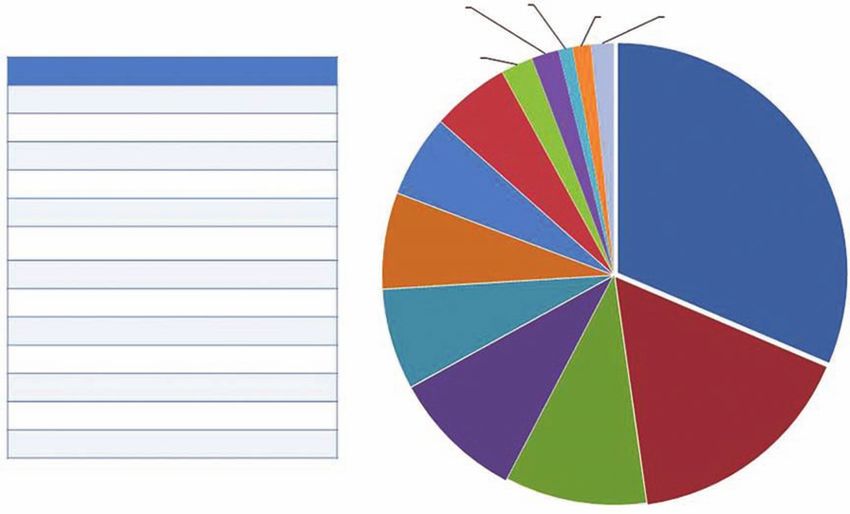
Original Research Article                                                                    BUTLER et al | Causes of death in Prader-Willi syndrome

or both 15s from the mother (approximately 25% of cases), with      time of death. The questionnaires were disseminated to families
the remaining cases having imprinting defects.2–5 Imprinting        known to have experienced the death of a relative with PWS.
defects may be inherited, with the potential of subsequent          The collected data were reviewed by the same PWS experts.
children having a PWS risk of 50%. The PWS genetic sub-             Inconsistencies in the data were reviewed and results clarified
types include the most frequent paternally derived 15q11–q13        and confirmed by consulting with the reporting family mem-
deletion consisting of the larger typical type I deletion or the    ber. The reported causes and contributors to each death and
smaller type II deletion involving two different proximal           autopsy reports were evaluated by a clinically licensed cardiolo-
15q11–q13 breakpoints or, rarely, atypically sized cytogenetic      gist, parent, and expert in the care of PWS, who assessed the
deletions.2,5,10–12 Clinical and behavioral differences have been   primary cause of death for each individual. The defined causes
reported in those with the two typical type I and type II dele-     of death were then classified into major categories.
tions and those with maternal disomy 15. Generally, those with
PWS and the larger type I deletion have more behavioral prob-       Statistical analysis
lems, obsessions, and self-injury than those with the smaller       Causes of death for PWS fell into 13 categories that were descrip-
type II deletion or maternal disomy 15.2,4,13–15 Reduced pain       tively characterized (e.g., frequencies and means with standard
threshold further increases propensity toward self-injury and       deviations) by gender, age, and body mass index (BMI) at death
infection, possibly related to disturbances in beta endorphin       and PWS genetic subtype. The frequencies of the 13 causes were
and substance P levels.16 Differential behavioral and physi-        summarized and reported for four age groups: infants (new-
ological profiles and vulnerabilities may also be associated with   born to
Causes of death in Prader-Willi syndrome | BUTLER et al                                    Original Research Article
Table 1 PWSA(USA) 40-year mortality survey syndrome-specific database summary
 Characteristic, total N = 486                            Value
 Gender, N = 480 (99%)                                    N = 263 (54%) Male
                                                          N = 217 (45%) Female
 Age of death, N = 425 (87%)                              Mean (±SD) = 29.5 (±15) years (range 2 months − 67 years)
 Male, N = 224 (53%)                                      Mean (±SD) = 27.8 (±16) years (range 1 months − 61 years)
 Female, N = 199 (47%)                                    Mean (±SD) = 31.7 (±15) years (range 2 months − 67 years) (F = 6.5 P < 0.011 (gender))
 Age range                                                Infant (90th percentile).
gender or age at death. Considering only those with a known
cause of death, the most common single cause of death was                     PWS genetic subtype
respiratory failure, reported for 98 individuals (31%), followed              Investigation of the relationship between PWS genetic sub-
by cardiac disease/failure (n = 51; 16%), gastrointestinal (GI)               type and cause of death was hindered by a lack of necessary
problems such as perforation, distension, or obstruction (n =                 genetic characterization in the selected sample, particularly for
31; 10%), and infections (n = 29; 9%). Obesity was listed as a                the older individuals who may not have had access to accurate

Genetics in meDicine | Volume 19 | Number 6 | June 2017                                                                                            637
Original Research Article BUTLER et al | Causes of death in Prader-Willi syndrome
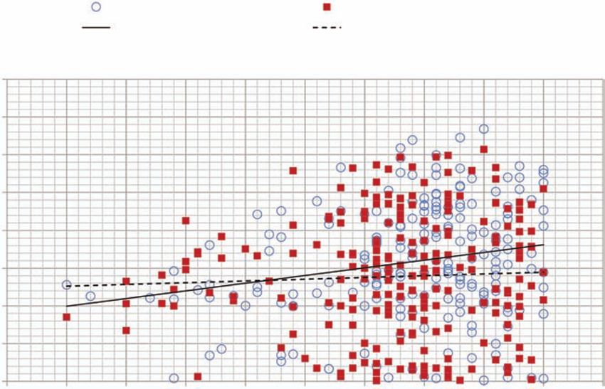
Females, n = 199 Males, n = 224
Linear (females, n = 199) Linear (males, n = 224)
y = 0.4072x – 784.26 y = 0.093x – 158.38
R 2 = 0.0428 R 2 = 0.0022
80.0
70.0
60.0
50.0
Age of death
40.0
30.0
20.0
10.0
0.0
1970 1975 1980 1985 1990 1995 2000 2005 2010 2015 2020
Paternal origin of FISH and methylation Microarray developed
38 Year of death
34 36,37
deletion discovered PCR developed
Chromosome 15 Uniparental maternal Growth hormone
33 35
deletion discovered disomy 15 discovered treatment approved
Figure 1 Relationship between the reported age and year of death for Prader-Willi syndrome. Scatter plot of the age at death by the year of death
for males (red squares) and females (blue circles) with Prader-Willi syndrome (PWS) with respect to historical benchmarks in genomic discovery and treatment
advances in PWS. Trend lines show the correlation coefficient for females (solid line) and males (dotted line).
genetic testing. DNA methylation testing, which is 99% accu- male with an imprinting defect died from GI perforation at 13
rate in confirming the diagnosis of PWS but does not deter- years of age.
mine the specific genetic subtype (chromosome 15q11–q13
deletion, maternal disomy 15, or imprinting defects), did not Survival analyses
become readily available until the mid- to late 1990s.21,22 The Cox regression analysis of our sample of 425 individuals with
deletion subtype was approximately equally divided among PWS and a known age at death identified quartile point esti-
male (n = 14; 45%) and female (n = 17; 55%) patients, with mates of 25% mortality for those 20 years of age (95% CI 18–21
a mean age of 34.8 ± 16 years at death, which was not related years); 50% mortality for those 29 years of age (95% CI 27–32
to the year in which the death occurred. However, the age of years); and 75% mortality for those 42 years of age (95% CI 39–
death for individuals with the deletion subtype was signifi- 44 years). A 99% mortality rate was achieved for those 60 years
cantly higher among females (41.0 ± 13.3 years, range 14.7– of age. A primary sex difference in mortality risk was identi-
55.3 years) than among males (27.2 ± 16 years, range 0.97–59 fied for PWS, with males displaying a significantly higher risk
years; t = 2.0; P < 0.05). The leading causes of death associated of early mortality compared with females (χ2 = 5.0; P < 0.025;
with the deletion subtype were cardiac problems (n = 9; 30%), hazard ratio = 1.2; 95% CI 1.0–1.5; Figure 3a). Furthermore,
respiratory failure (n = 8; 27%) and infections (n = 5; 17%), sex significantly interacted with the primary cause of death
followed by GI problems (n = 4; 13%), pulmonary embolism (cardiopulmonary versus hyperphagic), with males showing a
(n = 2; 7%), choking (n = 1; 3%), and hypothermia (n = 1; 3%); significantly higher risk of death due to presumed hyperpha-
females comprised seven of the nine cases of cardiac problems gia-related causes (accidents, choking, GI problems) relative to
and five of eight cases of respiratory failure in the deletion sub- females (Figure 3b; Supplementary Table S1 online). There
sample. The limited data from uniparental maternal disomy was a nearly significant trend toward increased risk for cardio-
15 showed that 8 of the 12 patients (9 males; 3 females) died pulmonary-related versus hyperphagia-related deaths among
from respiratory failure and the remaining 4 patients (3 males; females. PWS genetic subtype also significantly impacted mor-
1 female) died from GI problems, cancer, infections, and renal tality risk over the life span, with individuals possessing the
complications, respectively, at a mean age of 22.2 ± 18 years maternal disomy 15 subtype showing a significantly higher risk
(range 1.2 to 49 years), which did not differ by gender. A single (HR = 2.0; 95% CI 1.0–3.9; χ2 = 4.1; P < 0.04) of death due
638 Volume 19 | Number 6 | June 2017 | Genetics in meDicineCauses of death in Prader-Willi syndrome | BUTLER et al Original Research Article
Hypothermia
1% Drug reaction
Neurologic 1%
2% Cancer
2%
Renal failure
Category Mean age (SD) 2%
Respiratory failure, n = 94 24.6 (16) years
Accident
Cardiac, n = 50 32.1 (14) years
6%
GI, n = 30 32.4 (16) years
Choking
Infection, n = 29 35.7 (16) years Respiratory failure
6%
Obesity, n = 22 30.7 (12) years 31%
Pulmonary embolism, n = 19 34.1 (12) years Pulmonary
embolism
Choking, n = 18 30.1 (17) years 7%
Accident, n = 17 25.0 (16) years
Obesity
Renal failure, n = 7 34.2 (11) years
7%
Neurologic, n = 6 18.0 (21) years
Hypothermia, n = 3 30.8 (14) years Cardiac
Infection
16%
Drug reaction, n = 3 25.1 (9) years 9%
Cancer, n = 4 39.7 (27) years GI
10%
Figure 2 Causes of death among 312 individuals with Prader-Willi syndrome divided into 13 major categories.
to cardiopulmonary versus hyperphagic causes compared with maternal disomy 15 compared with deletion genetic subtypes,
the deletion subtype (Figure 4). which may reflect differential vulnerability due to amplified
effects of homozygous mutations from duplicated maternal
DISCUSSION contributions on chromosome 15.
We report a descriptive analysis of mortality data collected Despite the report of causes of death in PWS in individual
from individuals with PWS with reported deaths between case reports, case series, population surveys, and syndrome-
1973 and 2015 as part of a supportive bereavement program specific registry databases for both children and adults, there
for PWS families. Respiratory failure was the most common is a paucity of data for systematic collection and analysis of
overall cause of death reported in our database of adults and cause of death. For example, the Cambridge PWS study in 2004
children with PWS, whereas cardiac disease and failure with reported on fewer than 100 individuals with confirmed PWS
pulmonary thromboembolism were more commonly found in and found a mortality rate of 3% per year after the age of 5 years.
adulthood in combination with obesity-related morbidity. This More recently, in 2012, Lionti et al.23 reviewed an Australian
is the first report to characterize and quantify deaths attribut- registry of 163 individuals with PWS from ages 3 weeks to 60
able to pulmonary embolism in PWS; it accounted for 7% of years; 15 deaths were recorded, corresponding to an 87% prob-
all deaths. Cardiopulmonary and BMI-related mortality factors ability of survival to 35 years of age, which equates to a sur-
predominated among females, whereas males were more likely vival rate reported by an Italian survey of 80% at 40 years of
to experience accidents, choking, and infection at a young age. age for 425 individuals with PWS. The most common causes
Accidents, aspiration, sepsis, and choking were the most com- of death were the following, in no particular order: pulmonary
mon causes of death in children and adolescents, accounting thromboembolism, sepsis, accidents, diabetes, cardiac disease
for approximately half of all deaths in childhood and one-third and problems, choking, aspiration, gastric rupture, respiratory
of adult deaths. A progressive increase in the life span (mea- failure, and obesity-related complications.23–30
sured as age at mortality) was observed over time, particularly The present summary of mortality in PWS is consistent with
for deaths due to cardiac problems in females, possibly due to earlier reports indicating the young age of death and high rates
protective effects associated with earlier diagnosis, treatment of mortality due to cardiac and respiratory failure. Deaths
intervention and monitoring (e.g., growth hormones), and attributable to obesity-related and cardiac diseases can be antic-
weight management of recent cohorts. This was not observed ipated for this highly vulnerable group. Our data suggest that
among males, possibly owing to an increased rate of hyper- early interventions have impacted disease trajectory, resulting
phagia-related rather than obesity-driven cardiopulmonary in delayed mortality (i.e., older age of death) among females.
deaths among males with PWS. A significantly increased risk However, the underlying pathology for respiratory failure—the
of mortality due to cardiopulmonary factors was observed for leading cause of death for adults and children—remains elusive
Genetics in meDicine | Volume 19 | Number 6 | June 2017 639Original Research Article BUTLER et al | Causes of death in Prader-Willi syndrome
a Product-limit survival estimates
With number of subjects at risk and 95% Hall-Wellner bands
1.0
Logrank P = 0.0245
0.8 Females
Survival probability
0.6 Males
0.4
0.2
0.0
F 199 176 161 100 65 24 4 0
M 224 187 155 103 54 17 1 0
0 20 40 60
Age death
Sex F M
b Product-limit survival estimates
With number of subjects at risk
1.0
1
0.8
2
Survival probability
3
0.6
0.4
4
0.2
0.0
1 77 65 57 33 22 8 1 0
2 26 24 24 18 13 6 0
3 85 69 59 39 17 6 1 0
4 39 31 22 14 7 2 0
0 20 40 60
Age death
1: Sex=F cause=Cardiopulmonary 2: Sex=F cause=Hyperphagia
3: Sex=M cause=Cardiopulmonary 4: Sex=M cause=Hyperphagia
Figure 3 Survival analysis illustrates the effects of gender and cause of death on age of death for Prader-Willi syndrome. Kaplan-Meir plot
of survival probability is shown as a function of age at death for a) males versus females with 95% Hall-Wellner bands and for b) cardiopulmonary versus
hyperphagia-related causes by gender. Numbers of uncensored participants by group are listed at the bottom of each figure.
and does not appear to be impacted by recent advancements in or hyperphagia-related deaths appear to disproportionately
treatment modalities. Reported deaths due to respiratory failure impact younger males, possibly owing to increased activity
could also be secondary to undiagnosed aspiration, pulmonary and/or impulsive characteristics.
embolism, or some unspecified neurological disturbance and Early diagnosis of PWS and prevention of overweight are key
may vary by gender and/or age. Future targeted research should factors in preventing early causes of death in individuals. These
seek to characterize this phenomenon to advance treatment include close monitoring and supervision of food access and
development. Similarly, individuals with PWS have high rates quantity to avoid choking caused by eating quickly and gastric
of choking, accidents, and GI perforation, presumably related rupture resulting from excessive food consumption The risks
to uncontrolled hyperphagia and food-seeking behaviors, con- associated with choking are greater in the PWS population,27
tributing to approximately one-third of all reported deaths and with potential causes of increased choking related to poor oral/
approximately half of all deaths in childhood. Accidental and/ motor coordination, poor sensation, hypotonia, hyperphagia,
640 Volume 19 | Number 6 | June 2017 | Genetics in meDicineCauses of death in Prader-Willi syndrome | BUTLER et al Original Research Article
Product-limit survival estimates composition and stature in PWS, with a positive impact on the
With number of subjects at risk
1.0 control of obesity.
In summary, causes of death seen in infancy or young child-
0.8 hood in PWS are more likely to be related to respiratory failure,
aspiration, infection, and choking than to obesity-related fac-
Survival probability
0.6 tors, whereas cardiac disease and failure, pulmonary thrombo-
embolism, accidents, sepsis, and obesity-related complications
0.4 are more commonly found in adolescents and adulthood.
Deaths from cardiac disease typically reflect right heart fail-
0.2 ure rather than atherosclerotic disease. Males may be more
likely to engage in aggressive or risky food-seeking behav-
0.0 iors than females, particularly in childhood, whereas females
Del 24 23 21 14 12 5 0
UPD 9 5 5 4 1 0 may be more likely to experience obesity-related morbidity.
0 10 20 30 40 50 60 Standardized syndrome-specific growth charts developed for
Age death
growth hormone–treated individuals with PWS appear to show
Subtype Del UPD differences in the degree of obesity (weight) between males and
females. At age 18 years, females with PWS were heavier than
Figure 4 Survival analysis of deaths due to cardiopulmonary causes as
males of the same age with PWS.32
a function of Prader-Willi syndrome (PWS) genetic subtype. Kaplan-Meir
plot of survival probability is shown for deaths attributable to cardiopulmonary Our analyses and interpretation of data trends are limited by
versus hyperphagia-related causes for individuals with 15q11-q13 deletion the reliability of death reporting based on the availability and
(Del) versus maternal uniparental disomy 15 (UPD). Numbers of uncensored knowledge of family members and access to confirmed genetic
participants by PWS genetic subtype are listed at the bottom of the figure. status and autopsy reports. Older individuals without living
relatives to report the death may have been underreported.
a dry mouth, decreased mastication, and voracious feeding Similarly, deaths of infants and children with PWS, prior to
habits. Therefore, implementation of preventive measures and better awareness with recognition of PWS and the availability
education, with better awareness of group home-care providers, of advanced genetic testing, may have escaped diagnosis prior
are recommended, including training of the Heimlich maneu- to death. Statistical corrections for age and time effects have
ver, supervised meals, food security, food preparation, and diet been incorporated to minimize the influences of these factors
modification. One operational strategy is sipping fluid between on our conclusions, but the sample size is small. Gender differ-
bites of food to help clear the esophagus to lessen the risk of ences in PWS genetic subtype may have influenced the result.
choking. Pain in the upper abdomen and/or vomiting should Nevertheless, this study is the largest and most extensive exam-
be taken as a possible sign of an emergent event such as acute ination of mortality due to PWS to date. The results support
serious gastric/intestinal dilatation with risk for rupture. An the current understanding of disease pathology and mortality
established algorithm for evaluating individuals with PWS and for PWS and provide useful insight into risk factors, mortal-
GI symptoms (available at http://www.pwsausa.org) should be ity trajectory over time, and areas of need. Family members,
followed, along with intravenous fluid support. care providers, and health-care professionals involved with the
In adults, weight reduction is complicated by daytime sleepi- immediate and long-term care of individuals with PWS should
ness due to apnea. Adults with PWS and obesity can be expected be made aware of these risk factors and causes of death to
to have the same life-threatening complications as obese indi- improve the longevity and quality of life of these individuals (at
viduals without PWS, including complications of cardiopulmo- all ages) and their family members.
nary problems, hypertension, diabetes, and skin infections.31
Weight reduction lowers the risk of obesity-related, possibly SUPPLEMENTARY MATERIAL
life-threatening complications. Supplementary material is linked to the online version of the paper
Because hyperphagia and subsequent obesity are cardinal fea- at http://www.nature.com/gim
tures of this rare genetic disorder, excessive overeating, stomach
necrosis and rupture, cardiovascular disease, respiratory fail- ACKNOWLEDGMENTS
ure, sleep apnea, diabetes mellitus, and related comorbidities We acknowledge the support of the Prader-Willi Syndrome Asso-
can be life-threatening. Early diagnosis, dietary intervention ciation (USA) and families as well as the National Institute of Child
with restricted caloric intake, and significant controls of access Health and Human Development (grant HD02528). Partial fund-
to food (e.g., locking cabinets) along with exercise programs ing was also received through an unrestricted grant from Zafgen,
developed for those with PWS have led to successful weight Inc.
loss and BMI reduction; active management of hyperphagia
prolongs life. Furthermore, the use of growth hormones and DISCLOSURE
other hormone replacements have helped to normalize body The authors declare no conflict of interest.
Genetics in meDicine | Volume 19 | Number 6 | June 2017 641Original Research Article BUTLER et al | Causes of death in Prader-Willi syndrome
References 20. Schrander-Stumpel C, Sijstermans H, Curfs L, Fryns JP. Sudden death in children
1. Butler MG. Prader-Willi syndrome: current understanding of cause and with Prader-Willy syndrome: a call for collaboration. Genet Couns 1998;9:231–2.
diagnosis. Am J Med Genet 1990;35:319–332. 21. Muralidhar B, Butler MG. Methylation PCR analysis of Prader-Willi syndrome,
2. Butler MG, Lee PDK, Whitman BY (eds). Management of Prader–Willi Angelman syndrome, and control subjects. Am J Med Genet 1998;80:263–265.
Syndrome, 3rd edn. Springer: New York, 2006. 22. Kubota T, Sutcliffe JS, Aradhya S, et al. Validation studies of SNRPN methylation
3. Cassidy SB, Schwartz S, Miller JL, Driscoll DJ. Prader-Willi syndrome. Genet Med as a diagnostic test for Prader-Willi syndrome. Am J Med Genet 1966;66:77–80.
2012;14:10–26. 23. Lionti T, Reid SM, Rowell MM. Prader-Willi syndrome in Victoria: mortality and
4. Angulo MA, Butler MG, Cataletto ME. Prader-Willi syndrome: a review causes of death. J Paediatr Child Health 2012; 48:506–511.
of clinical, genetic, and endocrine findings. J Endocrinol Invest 2015; 38: 24. Schrander-Stumpel CT, Curfs LM, Sastrowijoto P, Cassidy SB, Schrander JJ,
1249–1263. Fryns JP. Prader-Willi syndrome: causes of death in an international series of 27
5. Butler MG. Single gene and syndromic causes of obesity: Illustrative examples. cases. Am J Med Genet 2004;A.124A:333–338.
Prog Mol Biol Transl Sci 2016;140:1–45. 25. Van Vliet G, Deal CL, Crock PA, Robitaille Y, Oligny LL. Sudden death in growth
6. Hill JO, Kaler M, Spetalnick B, Reed G, Butler MG. Resting metabolic rate in hormone-treated children with Prader-Willi syndrome. J Pediatr 2004;144:
Prader-Willi syndrome. Dysmorphol Clin Genet 1990;4:27–32. 129–131.
7. Hoybye C (ed). Prader-Will Syndrome. Nova Science Publishers: New York, 26. Eiholzer U. Deaths in children with Prader-Willi syndrome. Horm Res
2013. 2005;63:33–39.
8. Butler JV, Whittington JE, Holland AJ, Boer H, Clarke D, Webb T. Prevalence of, 27. Stevenson DA, Heinemann J, Angulo M, et al. Deaths due to choking in Prader-
and risk factors for, physical ill-health in people with Prader-Willi syndrome: a Willi syndrome. Am J Med Genet 2007;A.143A:484–487.
population-based study. Dev Med Child Neurol 2002;44:248–255. 28. Stevenson DA, Heinemann J, Angulo M, et al. Gastric rupture and necrosis in
9. Hertz G, Cataletto M, Feinsilver SH, Angulo M. Sleep and breathing patterns Prader-Willi syndrome. J Pediatr Gastroenterol Nutr 2007;45:272–274.
in patients with Prader Willi syndrome (PWS): effects of age and gender. Sleep 29. Nagai T, Obata K, Tonoki H, et al. Cause of sudden, unexpected death of Prader-
1993;16:366–371. Willi syndrome patients with or without growth hormone treatment. Am J Med
10. Bittel DC, Butler MG. Prader-Willi syndrome: clinical genetics, cytogenetics and Genet 2005;136A:45–48.
molecular biology. Expert Rev Mol Med 2005;7:1–20. 30. Tauber M, Diene G, Molinas C, Hébert M. Review of 64 cases of death in
11. Butler MG. Prader-Willi syndrome: obesity due to genomic imprinting. Curr children with Prader-Willi syndrome (PWS). Am J Med Genet 2008;A.14
Genomics 2011;12:204–15. 6A:881–887.
12. Kim SJ, Miller JL, Kuipers PJ, et al. Unique and atypical deletions in Prader- 31. Whittington JE, Holland AJ, Webb T, Butler J, Clarke D, Boer H. Population
Willi syndrome reveal distinct phenotypes. Eur J Hum Genet 2011;20: prevalence and estimated birth incidence and mortality rate for people with
283–290. Prader-Willi syndrome in one UK Health Region. J Med Genet 2001;38:792–798.
13. Butler MG, Bittel DC, Kibiryeva N, Talebizadeh Z, Thompson T. Behavioral 32. Butler MG, Lee J, Cox DM, et al. Growth charts for Prader-Willi syndrome during
differences among subjects with Prader-Willi syndrome and type I or type II growth hormone treatment. Clin Pediatr (Phila) 2016;55:957–974.
deletion and maternal disomy. Pediatrics 2004;113(3 Pt 1):565–573. 33. Ledbetter DH, Riccardi VM, Airhart SD, Strobel RJ, Keenan BS, Crawford JD.
14. Zarcone J, Napolitano D, Peterson C, et al. The relationship between compulsive Deletions of chromosome 15 as a cause of the Prader-Willi syndrome. N Engl J
behaviour and academic achievement across the three genetic subtypes of Med 1981;304:325–329.
Prader-Willi syndrome. J Intellect Disabil Res 2007; 51(Pt. 6):478–487. 34. Butler MG, Palmer CG. Parental origin of chromosome 15 deletion in Prader-
15. Hartley SL, Maclean WE Jr, Butler MG, Zarcone J, Thompson T. Maladaptive Willi syndrome. Lancet 1983;1:1285–1286.
behaviors and risk factors among the genetic subtypes of Prader-Willi syndrome. 35. Nicholls RD, Knoll JH, Butler MG, Karam S, Lalande M. Genetic imprinting
Am J Med Genet A 2005; 136:140–145. suggested by maternal heterodisomy in nondeletion Prader-Willi syndrome.
16. Butler MG, Nelson TA, Driscoll DJ, Manzardo AM. Evaluation of plasma Nature 1989;342:281–285.
substance P and beta-endorphin levels in children with Prader–Willi syndrome J 36. Kuwano A, Mutirangura A, Dittrich B, et al. Molecular dissection of the Prader-
Rare Disord 2015; 3:1–9. Willi/Angelman syndrome region (15q11-13) by YAC cloning and FISH analysis.
17. Swaab DF. Prader-Willi syndrome and the hypothalamus. Acta Paediatr Suppl Hum Mol Genet 1992;1:417–425.
1997;423:50–54. 37. Driscoll DJ, Waters MF, Williams CA, et al. DNA methylation imprint, determined
18. DiMario FJ Jr, Burleson JA. Cutaneous blood flow and thermoregulation in by the sex of the parent, distinguishes the Angelman and Prader-Willi
Prader-Willi syndrome patients. Pediatr Neurol 2002;26:130–133. syndromes. Genomics 1992;13:917–924.
19. Hayashi M, Itoh M, Kabasawa Y, Hayashi H, Satoh J, Morimatsu Y. A 38. Wang NJ, Liu D, Parokonny AS, Schanen NC. High-resolution molecular
neuropathological study of a case of the Prader-Willi syndrome with an characterization of 15q11-q13 rearrangements by array comparative genomic
interstitial deletion of the proximal long arm of chromosome 15. Brain Dev hybridization (array CGH) with detection of gene dosage. Am J Hum Genet
1992;14:58–62. 2004;75:267–281.
642 Volume 19 | Number 6 | June 2017 | Genetics in meDicineYou can also read