Cholesterol crystal embolization following plaque rupture: a systemic disease with unusual features
←
→
Page content transcription
If your browser does not render page correctly, please read the page content below
Available online at www.jbr-pub.org
                                                Open Access at PubMed Central

                                   The Journal of Biomedical Research, 2017 31(2): 82–94

                                                              Review Article

Cholesterol crystal embolization following plaque rupture: a
systemic disease with unusual features
Firas Ghanem1,2, Deepthi Vodnala3, Jagadeesh K. Kalavakunta1,4, Sridevi Durga1, Noah Thormeier5,
Prem Subramaniyam1, Scott Abela5, George S. Abela1,6, ✉
1
  Department of Medicine, Division of Cardiology, College of Human Medicine, Michigan State University, East Lansing,
  MI 48824, USA;
2
  Wheaton Franciscan Health, Brookfield, WI 53005, USA;
3
  University of Missouri, St. Luke's Health System, Kansas City, MO 64111, USA;
4
  Borgess Hospital, Kalamazoo, MI 49048, USA;
5
  College of Osteopathic Medicine, Michigan State University, East Lansing, MI 48824, USA;
6
  Department of Physiology, Division of Pathology, College of Human Medicine, Michigan State University, East Lansing,
  MI 48824, USA.

Abstract
   Cholesterol crystal embolic (CCE) syndrome is often a clinically challenging condition that has a poor prognostic
implication. It is a result of plaque rupture with release of cholesterol crystals into the circulation that embolize into
various tissue organs. Plaque rupture seems to be triggered by an expanding necrotic core during cholesterol
crystallization forming sharp tipped crystals that perforate and tear the fibrous cap. Embolizing cholesterol crystals
then initiate both local and systemic inflammation that eventually lead to vascular fibrosis and obstruction causing
symptoms that can mimic other vasculitic conditions. In fact, animal studies have demonstrated that cholesterol
crystals can trigger an inflammatory response via NLRP3 inflammasome similar to that seen with gout. The diagnosis
of CCE syndrome often requires a high suspicion of the condition. Serum inflammation biomarkers including
elevated sedimentation rate, abnormal renal function tests and eosinophilia are useful but non-specific. Common
target organ involvement includes the skin, kidney, and brain. Various testing including fundoscopic eye examination
and other non-invasive procedures such as trans-esophageal echocardiography and magnetic resonance imaging may
be helpful in identifying the embolic source. Treatment includes aspirin and clopidogrel, high dose statin and possibly
steroids. In rare cases, mechanical intervention using covered stents may help isolate the ruptured plaque.
Anticoagulation with warfarin is not recommended and might even be harmful. Overall, CCE syndrome is usually a
harbinger of extensive and unstable atherosclerotic disease that is often associated with acute cardiovascular events.
Keywords: cholesterol crystal embolic syndrome, plaque rupture, cholesterol crystal pathogenesis, clinical
presentation, diagnosis

Introduction                                                           atherosclerotic plaques lining the walls of the aorta or
                                                                       major inflow arteries that showered particulates down-
  Cholesterol crystal embolism (CCE) refers to the                     stream via smaller arteries and arterioles to end organs.
embolization of cholesterol crystals (CCs) released from               This leads to a variety of clinical manifestations that
✉
  Corresponding author: George S. Abela, M.D., F.A.C.C., Michi-        Received 08 August 2016, Revised 07 September 2016, Accepted 26
gan State University, B208 Clinical Center, East Lansing, MI 48824,    September 2016, Epub 20 November 2016
USA. Tel: 1- (517) 353-1754, Fax: 1- (517) 353-4978, Email:            CLC number: R587.1, Document code: A
george.abela@ht.msu.edu.
© 2017 by the Journal of Biomedical Research. All rights reserved.                                        doi:10.7555/JBR.31.20160100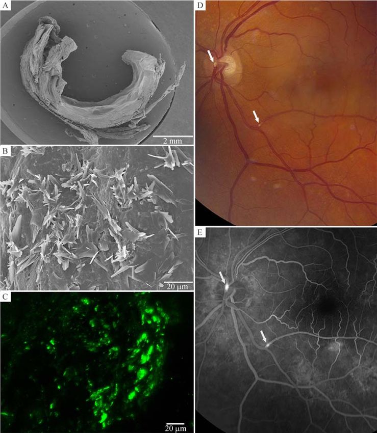
Cholesterol crystal induced vasculitis                                     83

depend on the specific organ affected by emboli as well       incidence of pathologic CCE may be higher due to
as systemic symptoms[1]. Cholesterol crystal emboliza-       high rate of aortic debris retrieved with placement of
tion is an under-recognized disease that can occur           guiding catheters during percutaneous interventions.
spontaneously or accompany a myriad of vascular and          This, however, did not seem to result in greater in-
endovascular procedures[2]. The first single autopsy          hospital ischemic events[13].
findings was described in 1862 by the Danish                     Among patients undergoing coronary artery bypass
pathophysiologist Peter Ludvig Panum[3]. Eighty years        grafting (CABG) within 1 month of an acute myocardial
elapsed before the first autopsy series by Curtis Flory       infarction, muscle and skin biopsies done at the site of
who provided support to the theory of distal emboliza-       vein harvest demonstrated a 12% incidence of CCE.
tion of CCs from eroded aortic plaques as a cause of         There was no significant difference between those who
arterial occlusions rather that the theory of in situ        received thrombolytic therapy and those who did not;
formation of CCs[4]. The most commonly involved              most of the cases were subclinical[14]. Overall, retro-
organs are the skin and the kidneys with a plethora of       spective autopsy or biopsy studies may detect sub-
nonspecific signs and symptoms that include fever,            clinical disease and thus exaggerate the frequency of
weight loss, myalgias and headache[1-2]. This can result     CCE; whereas the incidence of CCE in clinical reports
in presentations mimicking various other diseases (i.e.      is considerably less and can be affected by sample
polymyositis) leading to difficulties and delays in           characteristics and the duration of follow up. In
diagnosis, thus making this CCE a "great masquerader".       addition, the inherent selection bias of autopsy studies
                                                             may explain the discrepancy between pathology and
                                                             clinical data. The incidence of CCE may be expected to
Epidemiology
                                                             increase with a rise in the aging population and the
   CCE has been noted to predominantly affect males          number of vascular procedures performed. The current
(age over 60 years) often with clinical evidence of          estimates of incidence are probably greatly under-
atherosclerotic cardiovascular disease[1-2,4-5]. The true    estimated because tissue preparation methods using
incidence of CCE varies widely due to inconsistency in       ethanol dehydration dissolves CCs[15]. This could
diagnostic approaches based on the reporting used i.e.       greatly skew the data in favor of a lower incidence of
clinical (mainly dermal manifestations or elevation in       CCE when using standard histological methods.
serum creatinine) or histopathological criteria by
biopsy.                                                      Clinical predisposing factors to embolization
   In retrospective analysis, the incidence of postmortem
spontaneous CCE has ranged between 0.79% and 3.4%               Cholesterol crystal embolization can occur either
in unselected series of autopsies of patients over 60        spontaneously or as a complication of mechanical
years[2,4,6]. The overall prevalence of CCE following        trauma during aortic surgery, angioplasty and various
cardiac catheterization has been reported in autopsy         angiographic procedures such as aortography, left heart
studies to be as high as 25%-30%[7]. In one retro-           catheterization and peripheral angiography[2,9-10,12].
spective study of patients with severe thoracic aortic       Several predisposing factors have been associated
plaque seen on transesophageal echocardiogram (TEE),         with the development of CCE following cardiac and
the incidence of spontaneous antemortem clinical CCE         endovascular procedures. These include hypertension,
was about 1%[8]. However, clinical, angiographic and         heavy smoking, hypercholesterolemia, diabetes melli-
pathologic CCE were found in about 2.9% of patients          tus, renal failure, advanced age, peripheral vascular
undergoing infrarenal aortic and infrainguinal vascular      disease and severe atherosclerosis of the ascending
surgery[9] and 1.6% following aortoiliac stent place-        aorta[1,10-11]. Although white race has been implicated
ment[10]. Patients who underwent myocardial revascu-         as a risk factor, CCE is likely underdiagnosed among
larization or valve operations and had evidence of           blacks[1,16].
severe atherosclerosis of the ascending aorta were found        Sporadic case reports have hypothesized a causal
to have an increased incidence of CCE with 21.7% in          relationship between CCE and thrombolytic adminis-
the autopsy series[11].                                      tration for myocardial infarction or deep venous
   Prospectively, the occurrence of clinical CCE was         thrombosis[14]. The use of thrombolytic or anticoagulant
found to be around 1.4% in patients undergoing left          treatment may facilitate CCE possibly by preventing
heart catheterization. Surprisingly, no significant differ-   thrombus formation over ulcerated plaques, promoting
ence in the risk of CCE was noted between the brachial       plaque hemorrhage or interfering with the stabilization
and femoral approach implicating the ascending               of CCs within atheromatous plaques by platelet-fibrin
thoracic aorta as a main embolic source[12]. The             thrombi. Several large trials of thrombolytic therapy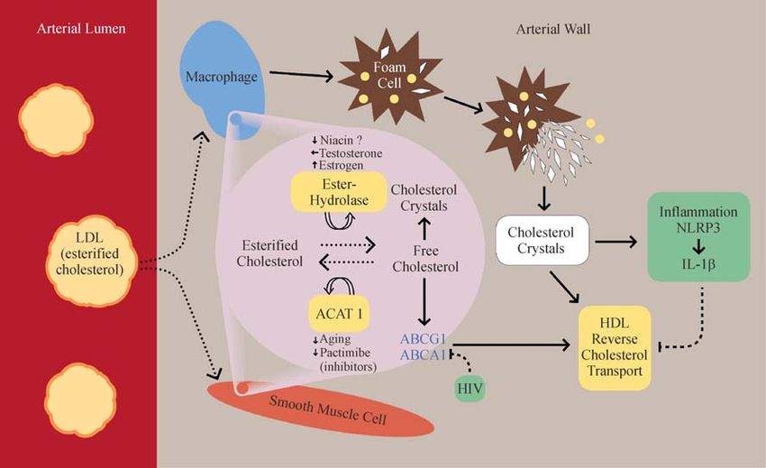
84                                             Ghanem F et al. J Biomed Res, 2017, 31(2)

have not reported CCE and the association between                            and systemically[21-22]. Crystals form early in the
thrombolytic administration and CCE is probably weak                         atherosclerotic process and activate the inflammasome
and the occurrence is difficult to predict[1-2,14] .                          NLRP3 leading to the secretion of IL-1β[22]. This
Although some claim that anticoagulation can worsen                          triggers a systemic response with IL-6. The process is
CCE clinical condition[17], one study using warfarin                         similar to how monosodium urate crystals trigger
compared to no warfarin did not appear to alter the rate                     inflammation in gout[23]. Overall, inflammation leads
of CCE in patients with severe atherosclerotic plaque of                     to arterial wall remodeling and destabilization of
the thoracic aorta visualized by TEE[8]. Use of emerging                     atherosclerotic plaques making them more prone to
risk markers such as elevated C-reactive protein (CRP)                       rupture.
levels and higher resolution imaging techniques may                             The correlation between CCE and aortic athero-
help in detecting the presence of inflamed and unstable                       sclerosis was first suggested by Flory in 1945[4] and was
plaques that are associated with an increased risk for                       readdressed over 30 years later by Kealy who demon-
CCE[12,18].                                                                  strated that in every case of peripheral embolism the
                                                                             aorta had advanced plaques, sometimes with aneurysm
Pathogenesis                                                                 formation[6]. Unstable plaques that are vulnerable to
                                                                             rupture have been defined histologically to have a lipid-
   Cholesterol crystal formation is favored by several                       rich necrotic core, a thin fibrous cap, reduced structural
intra- and extra-cellular factors. Intracellular factors                     support and presence of cellular inflammation that can
include the enhanced activity of cholesterol ester                           weaken the fibrous cap[24]. However, it is cholesterol
hydrolase enzyme or reduction in the Acyl-CoA:                               crystallization within the plaque's necrotic core that
cholesterol acyltransferase (ACAT). Also, reduced                            leads to volume expansion that can rupture the fibrous
HDL and/or dysfunctional HDL contributes to this                             cap. Perforation of the intimal surface by growing sharp
process (Fig. 1)[19]. Local extracellular factors include a                  tipped CCs may also cause erosion and trigger
mild drop in local temperature, increased saturation of                      thrombosis[25,26]. Autopsy findings demonstrate that
cholesterol, hydration of the cholesterol molecule and                       only patients who died of acute myocardial infarction
an alkaline milieu[20]. The formation of CCs is                              had CCs perforating the intima while those who died of
associated with an inflammatory response both locally                         other causes and had significant arterial plaques did not

Fig. 1 Cholesterol transport within cells and extracellular space. Equilibrium between esterified and free cholesterol is noted with
membrane transporters driving free cholesterol into the extracellular space where it is taken up by high-density lipoprotein. Dying foam cells
overloaded with esterified cholesterol and crystals release their content into the extracellular space. Free cholesterol build-up in the extracellular
space leads to crystallization. ABCA1, ABCG1, ATP binding cassette A-1, G-1; ACAT 1, acyl-coenzyme A cholesterol acyltransferase 1; HDL,
high-density lipoprotein; HIV, human immunodeficiency virus; IL-1β, interleukin-1β; LDL, low-density lipoprotein; NLRP3, NLRP3
inflammasome. Reproduced with permission[19].
Cholesterol crystal induced vasculitis                                                       85

have CCs perforating the intima[27]. In an atherosclero-                      The aorta and iliac arteries are the most common
tic rabbit model, those with a greater plaque burden had                   source of particulates that cause embolization followed
more CCs, serum inflammation, macrophage infiltration                        by the femoral arteries, while the popliteal and
and thrombosis[21]. Also, in the rabbit model, light and                   subclavian arteries are rarely involved which may
scanning electron microscopy demonstrated CCs in the                       explain the rarity of upper extremity involvement in
inflamed fibrous cap and perforating the intima at the                       CCE[30]. One of the more critical sources for emboliza-
sites of plaque rupture[21,28]. Moreover, CCs can readily                  tion is the carotid arteries (Fig. 2). These emboli often
puncture the vasa vasorum within the plaque causing                        travel to the cerebral and retinal micro- and macro-
intra-plaque hemorrhage that can contribute to plaque                      circulation causing cerebral inflammation and/or tran-
rupture[25,27]. Also, iatrogenic causes such as catheter                   sient ischemic events as well as strokes. Denudation or
manipulation during intra-arterial procedures or the                       rupture of plaques allows their underlying extracellular
increase in local shear stress with elevated blood                         cholesterol-rich matrix free access to disseminate into
pressure can be other factors contributing to plaque                       the arterial bloodstream[1]. These microemboli of CCs
disruption[12,29].                                                         measure about 50 to 200 µm and can lodge into distal

Fig. 2 Imagings of cholesterol crystals. A: Low power scanning electron micrograph of carotid artery plaque. B: Surface scanning of the
plaque demonstrates extensive cholesterol crystals. C: Fluorescence image of cholesterol crystals on the intimal surface of the artery using
Bodipy stain for cholesterol crystals (Courtesy of Dr. G. Abela). D and E: Fundoscopyof Hollenhorst plaques of embolized cholesterol crystals in
retinal arteries (arrows, Modified from Elizabeth Gauger, MD and Toni Venckus, CRA, University of Iowa; http://webeye.ophth.uiowa.edu/
eyeforum/atlas/pages/Hollenhorst-plaque.htm).
86 Ghanem F et al. J Biomed Res, 2017, 31(2)
capillaries, arterioles and small arteries. This can also perforating the fibrous cap in atherosclerotic plaques
lead to narrowing or obliteration of small arterial lumens and extensive amount of CCs released into the
resulting in ischemia or infarction[16]. circulation. Also, in an early study Eliot et al.
In addition to mechanical injury, CCE can create recognized this problem and attempted to use polarized
inflammatory and endothelial vascular reactions. These light on frozen preparations to detect CCs[31].
changes were reproduced by intravenous injection of
CCs in the ear veins of rabbits and performing Clinical Presentation
histologic examination of the lungs at various inter-
vals[4,31]. In these models, an early panarteritis (24-72 h) Although any site of the body can be affected by
was demonstrated with mononuclear and eosinophilic CCE, the aorta and iliac arteries are the most commonly
cell infiltration in addition to giant cells adjacent to CCs recognized sources of embolization followed the
and adventitial leukocytes. Later (2-7 days), endothelial femoral arteries (the popliteal and subclavian arteries
proliferation with intravascular fibrosis occurs and CCs are rarely involved) which explain the rarity of upper
are engulfed by giant cells[1,31]. The later changes have extremity involvement in CCE[30]. However, the
been found to persist to the last interval examination at carotids are another common source of crystal embo-
160 days[31]. Other pathologic changes may occur and lization to the brain causing neurological symptoms
include formation of intravascular thrombosis (24-72 h), (Fig. 2)[6,30].
and the penetration of the vascular endothelium by
crystals with secondary adventitial fibrosis (days to
Constitutional symptoms
months)[1]. The end product of CC embolism are
inflammation and injury with eventual fibrosis and The presenting symptoms and signs of disseminated
obliteration of the arterial lumen[17,31]. CCE are often nonspecific and range from subtle to
Although it has been known that CCs could be found catastrophic. Disseminated CCE may produce constitu-
in or attached to the surface of the intima creating local tional and systemic symptoms that include fever, weight
inflammation[4], we also demonstrated that CCs can loss, fatigue, myalgia and anemia of chronic disease[1].
damage the endothelium and reduce its vasodilator Skin and the kidneys are the most common organs
response (Fig. 3)[32]. Moreover, this effect was due to affected by CCE and their manifestations are often used
the scraping of the intimal surface of the artery by sharp as clinical diagnostic criteria for CCE. Histology is
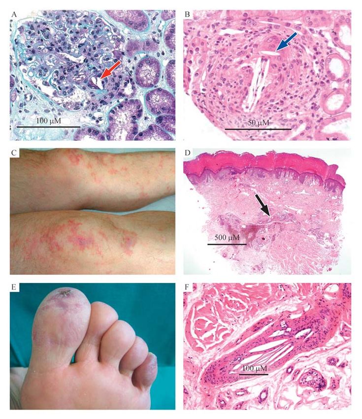
CCs traveling downstream. Intravascular CCs were consistent with a pseudovasculitis with giant cell
thought to be insoluble and resist the scavenger effects reaction (Fig. 4).
of the macrophages[1,31]. The following is a brief review of the most frequent
end-organs involved and their clinical presentations.
Histopathology Skin: Cutaneous manifestations are the most com-
mon finding in CCE patients with frequency ranging
Cholesterol crystal embolization can be defined by between 35% and 96%[16-17]. Livedo reticularis is the
histopathology as the presence of the typical lesions most common skin manifestation followed by gangrene,
found in arterial lumens as first described by Flory in cyanosis, ulceration and less frequently nodules and
1945[4]. The arterial lumen is filled with large CCs purpura[1,16]. The lower extremities are most commonly
spaces (dissolved during the preparation) surrounded by involved, and except for gangrene and ulcerations the
hyperplastic intimal tissue and giant cells. Cholesterol cutaneous lesions associated with CCE are generally
emboli of various ages can be found suggesting that the bilateral[16].
process of embolism is recurrent[2]. The external Livedo reticularis is a red-blue discoloration of the
diameter of the arteries plugged with cholesterol emboli skin distributed in a net-like arrangement that represents
vary from 150 µm to 1100 µm in diameter[13]. Since a cyanotic response pattern commonly associated with
CCs are readily dissolved when using standard methods intravascular obstruction of small vessels[34]. It occurs
with ethanol dehydration for preparing histologic suddenly in cases of CCE and is quite extensive
sections, the amount of CCs has been greatly under- extending from the feet to the buttocks and trunk.When
estimated by light microscopy[15,25,27]. However, by it occurs in the elderly, it implies the presence of
using vacuum dehydration in tissue preparation for CCE[16]. Other causes of livedo reticularis include
scanning electron microscopy or just fresh tissue with coagulopathies like cryoglobulinemia and vasculitic
fluorescence microscopy, Abela et al. were able to syndromes like polyarteritis nodosa[34].
demonstrate the vast extent and effect on tissue Toe involvement is manifested by blue or purple
injury by CCs[15,25–27,33]. This demonstrates CCs discoloration that has traditionally been considered theCholesterol crystal induced vasculitis 87 Fig. 3 Microscopic images with scanning electron microscopy and confocal microscopy of arterial surface injury. A-F: scanning electron microscopy; G-H: confocal microscopy. Circulating saline control (A, C, E, and G) vs. circulating cholesterol crystals (B, D, F, and H). No intimal injury is present with control arteries but extensive injury is present with cholesterol crystals. Cholesterol crystals are seen embedded and disrupting the intimal surface of the artery (D, F, H) with circulating cholesterol crystals. Modified and reproduced with permission[31]. sine qua non for diagnosis. The etiology is probably Eosinophilia is commonly seen with renal CCE (in up to microvascular ischemia which can be seen as well in 80% of patients) but is transient, lasting only a few coagulopathies and vasculitides[16]. days[30]. The urine sediment is usually benign (with few Kidney: Renal involvement is the second most cells and a minimum amount of proteinuria) which common manifestation of CCE due to proximity of helps differentiate renal CCE from systemic vasculi- the kidneys to the abdominal aorta and their large blood tis[5,17]. Less common presentation is a chronic, slowly supply, and could be found in up to 50% histologically progressive renal impairment with absence of extrarenal proven CCE[1]. The presentation can range from signs. The chronic course is commonly attributed to subclinical illness to fulminant disease although it is hypertensive nephrosclerosis or ischemic nephropathy, mostly subacute arising within 1 week of an CCE event which frequently co-exists with cholesterol emboli. This with progressive worsening of renal function[1,17,30]. often clinically silent crystal showering, frequently goes
88                                            Ghanem F et al. J Biomed Res, 2017, 31(2)

Fig. 4 Histology of cholesterol crystals consistent with a pseudovasculitis with giant cell reaction. A: Intraglomerular renal cholesterol
crystals; B: cholesterol crystals in an arcuate artery with encasement of a crystal by a giant cell and pseudovasculitis inflammatory reaction
(arrow); C: purpuric lesion over knees; D: skin biopsy with cholesterol crystals; blue toe (E) embolic crystals (F) in skin arterioles. Modified and
reproduced with permission [17].

underdiagnosed because extrarenal signs are absent and                         Gastrointestinal System: The frequency of gastro-
renal biopsy samples are not taken[17]. Infrequently,                       intestinal (GI) findings among CCE patients ranges
patients may have accelerated hypertension but rarely a                     from 10 to 36%[5,35]. Two patterns of disease can be
renal infarction[1,17]. One cannot exclude the possibility                  identified: acute catastrophic multiorgan presentation
of contrast nephrotoxicity as a contributor to post-                        and chronic and more indolent GI disease. The most
procedural nephropathy. A useful approach is to                             common manifestations are abdominal pain, diarrhea,
consider the creatinine increase within several days to                     and GI blood loss resulting from bowel ischemia,
be due to contrast nephrotoxicity and that at 1-2 weeks                     mucosal ulcerations, hepatocellular liver disorder, and/
to suggest CCE[12].                                                         or pancreatitis[31,35]. Cholecystitis gall bladder involve-
   Renal atheroembolic disease follows a variable                           ment, although rare, tends to be clinically significant
course. Renal replacement therapy is needed in about                        especially in the elderly, with a spectrum ranging from
28%–61% of patients with acute or subacute disease,                         chronic acalculous cholecystitis to acute, necrotizing
with partial recovery expected in 20%–30%. This is                          and ischemic disease[35].
probably due to resolution of inflammation or reversal                          Coronary Arteries: Embolization of CCs within the
of concurrent ischemic acute tubular necrosis[5,17].                        coronary arterial circulation has not been extensively
Cholesterol crystal induced vasculitis                                                         89

investigated[36]. However, a few case reports demon-                        damage to end organs to be detected clinically or by
strate that CC emboli into the coronary arteries have                       imaging and laboratory methods. Laboratory findings
been associated with sudden cardiac death[17]. Debris                       are usually nonspecific and include leukocytosis,
released during plaque rupture within the coronary                          thrombocytopenia, anemia and raised concentrations
arteries includes not only CCs but also platelets, cell                     of inflammatory markers (erythrocyte sedimentation
debris, fibrin and red blood cells. CCs and platelets are                    rate (ESR) or C-reactive protein)[1,17]. Transient eosi-
some of the various ejected materials that can trigger an                   nophilia tends to occur more in patients with renal
inflammatory response. In fact during myocardial                             involvement[1,12,17]. In addition, laboratory abnormal-
infarction, the arterial lumen could be occluded entirely                   ities may reflect specific organ injury such as azotemia,
by CCs released during plaque rupture[26]. However, the                     hyperamylasemia (due to pancreatic or bowel involve-
great amount of CCs present in coronary lumen                               ment), elevated creatine kinase due to muscular
following plaque rupture has been greatly under-                            involvement, and elevated transaminases due to hepatic
estimated due to the standard tissue processing methods                     injury[1]. In cases where hypocomplementemia is
with ethanol that dissolves CCs (Fig. 5)[15,37].                            present, there is a need to rule out embolic disease
   Central Nervous System: Central nervous system                           from bacterial endocarditis[5]. Occasionally, antemor-
manifestations can range from transient ischemic attack                     tem histological examination may be necessary for
and acute stroke to a gradual deterioration of neurolo-                     definite diagnosis; biopsies have mostly come from
gical function and dementia[38-39].                                         skin, muscle or kidney with a few via other tissue types
   Release of CCs into the cerebral circulation breaks                      like the colon[1,5,33,35].
down the blood brain barrier and initiates an inflamma-                         Fundoscopic eye examination may be the easiest and
tory response without evidence of ischemia[40]. This can                    most reliable method to evaluate if there is CCs
result in loss of memory and other integrative cerebral                     showering and is often associated with other sites of
functions.                                                                  embolic events[17]. Other noninvasive imaging mod-
   Brain imaging reveals mostly multiple, small                             alities such as TEE, computed tomography (CT) and
ischemic lesions and border zone infarcts[39]. Retinal                      magnetic resonance imaging (MRI) have gained an
involvement can be occasionally demonstrated by                             increasing role in confirming the diagnosis of CCE.
Hollenhorst plaques which are bright, refractile lesions                    TEE has become the standard test in diagnosing thoracic
indicative of CCE. They can occur in asymptomatic                           aortic disease due to its portability and lack of ionizing
patients but are a poor predictor of future embolic                         radiation or need for contrast agents. TEE can diagnose
events[41].                                                                 the higher risk complex plaques defined by their
   Other vascular beds including coronary, pulmonary,                       protuberance (≥4 mm), ulceration or the presence of
testes, prostate, thyroid and adrenal glands are rarely                     mobile components (Fig. 6)[8,42]. Non-calcified plaques
manifested clinically and their involvement is con-                         may be more lipid-laden and probably associated with
firmed by pathology[1].                                                      higher embolization risk[8]. Transcranial Doppler
                                                                            (TCD) has been shown to detect particulates ejected
Diagnosis                                                                   from ruptured carotid plaques[43]. Moreover, simulta-
                                                                            neous TCD and carotid ultrasound may provide
 Cholesterol crystal embolization becomes evident                           additional insights into potentially unstable plaques[32].
when showers of cholesterol microemboli cause enough                           Both CT and MRI offer a more complete evaluation

Fig. 5 Scanning electron micrograph of coronary artery from patient who died during an acute myocardial infarction. A and B:
Circumflex artery is totally occluded with thrombus loaded with cholesterol crystals (arrows). Crystals are evident when ethanol is not used in the
tissue preparation. Modified and reproduced with permission [19].
90 Ghanem F et al. J Biomed Res, 2017, 31(2)
patients with evidence of atherosclerosis that underwent
a recent cardiovascular procedure.
Management
Prognosis of CCE is generally poor with in-hospital
mortality ranging between 5 and 16%[12,30,48] and up to
81% in autopsy case series[1]. Treatment is mostly
supportive and includes, treating underlying heart
failure and hypertension, nutritional support and if
needed renal replacement therapy with ultrafiltration
and hemodialysis[48]. Despite its weak link to CCE,
anticoagulation has been advocated to be stopped or not
initiated in patients with CCE[1,17,30,48]. Unfortunately,
the highly anticipated ARCH trial that evaluated the
Fig. 6 Transesophageal ultrasound of the ascending aorta from
an asymptomatic patient examined for mitral regurgitation.
efficacy of aspirin plus clopidogrel versus warfarin in
Ulcerated plaque is visibly lining the aorta with overlying thrombus patient with aortic arch atherosclerotic plaques >4 mm
(arrows) (Courtesy Dr. N. Thormeire). did not achieve statistical power to demonstrate a
difference between either intervention. However,
of the aorta and the chance to image other potential aspirin plus clopidogrel had a lesser primary endpoint
arterial source[44]. Nonenhanced CT can assess for both of vascular death compared to the warfarin group[49].
calcium deposits and areas of hypoattenuation adjacent Thus, in the absence of clear correlation of antiplatelet
to aortic wall and the same 4 mm thickness for therapy to inducing CCE, their use should follow
protruding plaque can be used to identify high risk established guidelines.
patients. It is recommended though, that any positive Animal studies have shown that statins (HMG-CoA
nonenhanced CT study be followed with a confirmatory reductase inhibitors) can reduce matrix metalloprotei-
contrast-enhanced CT or TEE [45] . Gadolinium- nase expression in atheromas and may lead to an
enhanced 3D MRA is also a useful modality to visualize increase in fibrous cap thickness in association with
plaques in contiguous ascending aorta, which cannot be collagen accumulation[50]. In humans, established statin
seen with TEE due to tracheal interposition[44]. therapy can decrease the incidence of plaque rupture as
Currently conventional catheter angiography is being detected by intravascular ultrasound[51]. In a retro-
used less frequently in favor of less invasive imaging spective analysis of patients with severe aortic athero-
techniques. However, if therapeutic endovascular pro- sclerosis seen on TEE, the use of statins was associated
cedures are contemplated, a catheter angiogram is with protective effect on atheroembolic recurrence rate,
needed as a "road map". Depending on the concentra- though, no significant benefit of warfarin or antiplatelet
tion of iodine used, plaques can be seen as "filling drugs was detected[8]. In a prospective observational
defects" within the contrast-enhanced lumen or inferred study of patients with spontaneous CCE, statin treat-
from irregularities of the luminal surface[44]. Also ment was associated with protective effects both when
intravascular imaging techniques like optical coherence already in place at the time of diagnosis and when
tomography (OCT) have been shown to identify initiated after diagnosis[5]. In addition, statins have been
macrophage accumulations and large, extracellular shown to reduce CRP[52] and dissolve CC in human
cholesterol crystal plates in the vascular endothe- plaques (Fig. 7) [53]. Anecdotally, simvastatin has been
lium[46]. Recently, a newer advancement called micro reported to improve renal dysfunction caused by CCE,
– OCT, which has 10 times higher resolution than but further prospective studies are needed to investigate
conventional OCT, has been shown to image the usefulness of statins after the onset of symptoms.
endovascular subcellular structures including individual Indirect evidence from acute coronary syndromes
macrophages and cholesterol crystal within those suggests that higher doses of statins provide better
macrophages[47]. Due to lack of specific clinical protection[52,54]. Although older reports have linked the
presentations or diagnostic tests, a high index of use of steroid to poor prognosis[1], the results of more
suspicion should remain in patients presenting with recent studies with steroid therapy have suggested that
livedo reticularis, cyanotic toes and/or subacute renal low dose might be beneficial[48]. A recent retrospective
failure. This becomes especially important in elderly study of steroid therapy in patients with renal CCECholesterol crystal induced vasculitis 91 Fig. 7 Statins treatment dissolves cholesterol crystals in human plaques. A: Carotid plaque with clusters of cholesterol crystals protruding from the intima. B: Similar cholesterol crystals breaking down and dissolving following incubation with simvastain. C: Cholesterol crystals as commonly seen perforating the intimal surface in human carotid plaque from a patient not on statin therapy. D: Cholesterol crystals from patient on atorvastatin treatment prior to carotid endarterectomy demonstrates thinned down crystals with edges that have irregular borders and some are bent as typically seen with dissolving crystals. Modified and reproduced with permission[52]. showed a good renal outcome at 4 weeks; however, this or those with mostly an aortoiliac embolic source treatment did not have a favorable effect on long-term showed significant healing of ischemic ulcerations and renal outcome[55]. decrease of further embolization[58-59]. Finally, mod- It is also helpful to minimize invasive therapies in ifications in endovascular and surgical approaches can patients known to have severe atherosclerosis or to have additional benefits to the use of protection devices. investigate its presence before major cardiovascular These devices include distal filters and occlusion procedures. Preoperative 64-slice multidetector CT scan balloons that have been successfully used in carotid has been used in CABG patients to evaluate the and to a lesser extent in renal artery interventions with a incidence of pathologic lesions of the aorta and its lower incidence of procedural adverse events[60-61]. The major branches. The finding of significant atherosclero- 'no touch' technique can be used as well during renal sis can offer the opportunity to modify risk assessment, artery stenting to minimize rubbing of the guiding CABG procedure itself, perioperative management or catheter against the aorta and reduce the potential for follow-up plan in about half of patients[56]. Surgical cholesterol embolization[29]. Of note, thromboembolic removal or endovascular isolation of the source of complications in off-pump coronary bypass operations emboli could offer a definitive treatment option. are comparable with those in cardiopulmonary bypass Surgeries to correct an embolic source (ligation and operations[62]. bypass, endarterectomy and graft revision) could be achieved with low mortality except when the embolic Summary source is located in the suprarenal aorta[57]. Endovascular interventions with covered stents, Cholesterol crystal embolization results from plaque endoluminal stent grafts, or stent graft components rupture with distal embolization of CCs that originate have been used to exclude the embolic source. Limited mainly in the aorta and iliac arteries. This can occur experience in patients with abdominal aortic aneurysm either spontaneously or from iatrogenic trauma and lead
92                                             Ghanem F et al. J Biomed Res, 2017, 31(2)

to tissue injury. The embolic CCs can trigger tissue                           atheromatousplaques. Am J Pathol, 1945, 21(3): 549–565.
inflammation and a vasculitis-like picture. The most                       [5] Scolari F, Ravani P, Gaggi R, et al. The challenge of diagnosing
common clinical presentation are progressive renal                             atheroembolic renal disease: clinical features and prognostic
dysfunction and livedo reticularis. Laboratory findings                         factors. Circulation, 2007, 116(3): 298–304.
are not specific and non-invasive testing with ultra-                      [6] Kealy WF. Atheroembolism. J Clin Pathol, 1978, 31(10): 984–
sound, CT and MRI are preferred to invasive angio-                             989.
graphy. Occasionally, histological examination may be                     [7] Ramirez G, O'Neill WMJr, Lambert R, et al. Cholesterol
necessary for a definite diagnosis. Use of fundoscopy                           embolization: a complication of angiography. Arch Intern Med,
can be valuable in making a diagnosis of CCE since it                          1978, 138(9): 1430–1432.
can detect CC emboli in the retinal arteries which is                     [8] Tunick PA, Nayar AC, Goodkin GM, et al. , and the NYU
often associated with the systemic condition. Although                         Atheroma Group. Effect of treatment on the incidence of stroke
undetected CCs showering may seem benign, it is                                and other emboli in 519 patients with severe thoracic aortic
important to recognize this entity because it often has a                      plaque. Am J Cardiol, 2002, 90(12): 1320–1325.
diffuse multi-organ involvement and carries a very high                   [9] Sharma PV, Babu SC, Shah PM, et al. Changing patterns of
mortality related to cardiovascular events. Treatment is                       atheroembolism. Cardiovasc Surg, 1996, 4(5): 573–579.
mainly supportive with considerations of using high                       [10] Lin PH, Bush RL, Conklin BS, et al. Late complication of
dose statins and possibly steroids. Surgical removal or                        aortoiliac stent placement- atheroembolization of the lower
endovascular isolation of the source of emboli is an                           extremities. J Surg Res, 2002, 103(2): 153–159.
alternative therapeutic option but that carries a high risk.              [11] Blauth CI, Cosgrove DM, Webb BW, et al. Atheroembolism
Overall, CCE can be a difficult diagnosis to make and is                        from the ascending aorta. An emerging problem in cardiac
often associated with adverse outcomes.                                        surgery. J Thorac Cardiovasc Surg, 1992, 103(6): 1104–1111.,
                                                                               discussion 1111–1112.
Acknowledgements                                                          [12] Fukumoto Y, Tsutsui H, Tsuchihashi M, et al. , and the
  Support was provided in part from Michigan State                             Cholesterol Embolism Study(CHEST) Investigators. The
University, The Jean P. Schultz Biomedical Research                            incidence and risk factors of cholesterol embolization syn-
Endowment; Clinical and Translational ScienceInstitute                         drome, a complication of cardiac catheterization: a prospective
at Michigan State University; Seed Funds from the                              study. J Am Coll Cardiol, 2003, 42(2): 211–216.
Department of Medicine, College of Human Medicine;                        [13] Keeley EC, Grines CL. Scraping of aortic debris by coronary
Graduate Medical Education,Inc. East Lansing, Michi-                           guiding catheters: a prospective evaluation of 1,000 cases. J Am
gan and Edward W. Sparrow Hospital, Lansing,                                   Coll Cardiol, 1998, 32(7): 1861–1865.
Michigan.                                                                 [14] Blankenship JC, Butler M, Garbes A. Prospective assessment
                                                                               of cholesterol embolization in patients with acute myocardial
Conflict of Interest                                                            infarction treated with thrombolytic vs conservative therapy.
                                                                               Chest, 1995, 107(3): 662–668.
   G.A. is a speaker and recipient of grants from Merck.                  [15] Nasiri M, Janoudi A, Vanderberg A, et al. Role of cholesterol
He is a participant at Merck's US Thrombosis Advisory                          crystals in atherosclerosis is unmasked by altering tissue
Board and Atherosclerosis Global Therapeutic Experts                           preparation methods. Microsc Res Tech, 2015, 78(11): 969–
Forum. He is a speaker for Amgen, Daiichi Sankyo and                           974.
consultant for Kowa pharmaceuticals.                                      [16] Falanga V, Fine MJ, Kapoor WN. The cutaneous manifesta-
   F.G., D.V., J.K., S.D. N.T., and S.A. have no conflicts                      tions of cholesterol crystal embolization. Arch Dermatol, 1986,
to declare.                                                                    122(10): 1194–1198.
                                                                          [17] Scolari F, Ravani P. Atheroembolic renal disease. Lancet, 2010,
                                                                               375(9726): 1650–1660.
References                                                                [18] Aziz K, Berger K, Claycombe K, et al. Noninvasive detection
                                                                               and localization of vulnerable plaque and arterial thrombosis
[1]   Fine MJ, Kapoor W, Falanga V. Cholesterol crystal emboliza-              with computed tomography angiography/positron emission
      tion: a review of 221 cases in the English literature. Angiology,        tomography. Circulation, 2008, 117(16): 2061–2070.
      1987, 38(10): 769–784.                                              [19] Janoudi A, Shamoun FE, Kalavakunta JK, et al. Cholesterol
[2]   Cross SS. How common is cholesterol embolism? J Clin                     crystal induced arterial inflammation and destabilization of
      Pathol, 1991, 44(10): 859–861.                                           atherosclerotic plaque. Eur Heart J, 2016, 37(25): 1959–1967.
[3]   Panum PL. Experimentelle Beitrage zur Lehre von der Embolie.        [20] Vedre A, Pathak DR, Crimp M, et al. Physical factors that
      Virchows Arch Pathol Anat Physiol, 1862, 25(3): 308–310.                 trigger cholesterol crystallization leading to plaque rupture.
[4]   Flory CM. Arterial occlusions produced by emboli from eroded             Atherosclerosis, 2009, 203(1): 89–96.
Cholesterol crystal induced vasculitis 93
[21] Patel R, Janoudi A, Vedre A, et al. Plaque rupture and microvascular obstruction in acute coronary thrombosis and
thrombosis are reduced by lowering cholesterol levels and sudden coronary death: relation to epicardial plaque histo-
crystallization with ezetimibe and are correlated with fluor- pathology. J Am Coll Cardiol, 2009, 54(23): 2167–2173.
odeoxyglucose positron emission tomography. Arterioscler [37] Abela GS, Shamoun F, Vedre A, Pathak DR, Shah I, Dhar G,
Thromb Vasc Biol, 2011, 31(9): 2007–2014. Leffler D. Extent of Cholesterol Crystals in Coronary Artery
[22] Duewell P, Kono H, Rayner KJ, et al. NLRP3 inflammasomes Aspirates During Acute Myocardial Infarction. J Am CollCar-
are required for atherogenesis and activated by cholesterol diol, 55;Suppl A,109,2010.
crystals. Nature, 2010, 464(7293): 1357–1362. [38] Laloux P, Brucher JM. Lacunar infarctions due to cholesterol
[23] Martinon F, Pétrilli V, Mayor A, et al. Gout-associated uric acid emboli. Stroke, 1991, 22(11): 1440–1444.
crystals activate the NALP3 inflammasome. Nature, 2006, 440 [39] Ezzeddine MA, Primavera JM, Rosand J, et al. Clinical
(7081): 237–241. characteristics of pathologically proved cholesterol emboli to
[24] Lendon CL, Davies MJ, Born GV, et al. Atherosclerotic plaque the brain. Neurology, 2000, 54(8): 1681–1683.
caps are locally weakened when macrophages density is [40] Rapp JH, Pan XM, Neumann M, et al. Microemboli composed
increased. Atherosclerosis, 1991, 87(1): 87–90. of cholesterol crystals disrupt the blood-brain barrier and reduce
[25] Abela GS. Cholesterol crystals piercing the arterial plaque and cognition. Stroke, 2008, 39(8): 2354–2361.
intima trigger local and systemic inflammation. J Clin Lipidol, [41] Bunt TJ. The clinical significance of the asymptomatic
2010, 4(3): 156–164. Hollenhorst plaque. J Vasc Surg, 1986, 4(6): 559–562.
[26] Abela GS, Aziz K. Cholesterol crystals rupture biological [42] Finkelhor RS, Youssefi ME, Lamont WE, et al. Embolic risk
membranes and human plaques during acute cardiovascular based on aortic atherosclerotic morphologic features and aortic
events—a novel insight into plaque rupture by scanning spontaneous echocardiographic contrast. Am Heart J, 1999,
electron microscopy. Scanning, 2006, 28(1): 1–10. 137(6): 1088–1093.
[27] Abela GS, Aziz K, Vedre A, et al. Effect of cholesterol crystals [43] King A, Markus HS. Doppler embolic signals in cerebrovas-
on plaques and intima in arteries of patients with acute coronary cular disease and prediction of stroke risk: a systematic review
and cerebrovascular syndromes. Am J Cardiol, 2009, 103(7): and meta-analysis. Stroke, 2009, 40(12): 3711–3717.
959–968. [44] Krinsky GA. Diagnostic imaging of aortic atherosclerosis and
[28] Phinikaridou A, Hallock KJ, Qiao Y, et al. A robust rabbit its complications. Neuroimaging Clin N Am, 2002, 12(3): 437–
model of human atherosclerosis and atherothrombosis. J Lipid 443.
Res, 2009, 50(5): 787–797. [45] Tenenbaum A, Garniek A, Shemesh J, et al. Dual-helical CT for
[29] Feldman RL, Wargovich TJ, Bittl JA. No-touch technique for detecting aortic atheromas as a source of stroke: comparison
reducing aortic wall trauma during renal artery stenting. with transesophageal echocardiography. Radiology, 1998, 208
Catheter Cardiovasc Interv, 1999, 46(2): 245–248. (1): 153–158.
[30] Baumann DS, McGraw D, Rubin BG, et al. An institutional [46] Tearney GJ, Regar E, Akasaka T, et al. , and the International
experience with arterial atheroembolism. Ann Vasc Surg, 1994, Working Group for Intravascular Optical Coherence Tomogra-
8(3): 258–265. phy (IWG-IVOCT). Consensus standards for acquisition,
[31] Eliot RS, Kanjuh VI, Edwards JE. Atheromatous embolism. measurement, and reporting of intravascular optical coherence
Circulation, 1964, 30: 611–618. tomography studies: a report from the International Working
[32] Gadeela N, Rubinstein J, Tamhane U, et al. The impact of Group for Intravascular Optical Coherence Tomography
circulating cholesterol crystals on vasomotor function: implica- Standardization and Validation. J Am Coll Cardiol, 2012, 59
tions for no-reflow phenomenon. JACC Cardiovasc Interv, (12): 1058–1072.
2011, 4(5): 521–529. [47] Kashiwagi M, Liu L, Chu KK, et al. Feasibility of the
[33] Abela GS, Shamoun F, Farooq MU, et al. A Novel Method of Assessment of Cholesterol Crystals in Human Macrophages
Identifying Unstable Plaque by Simultaneous Carotid Ultra- Using Micro Optical Coherence Tomography. vanZandvoort M,
sound and Trans-Cranial Doppler. J Am Coll Cardiol, 2010, 55 ed. PLoS ONE, 2014,9(7):e102669.
(Suppl A): 161. [48] Belenfant X, Meyrier A, Jacquot C. Supportive treatment
[34] Donohue KG, Saap L, Falanga V. Cholesterol crystal improves survival in multivisceral cholesterol crystal embo-
embolization: an atherosclerotic disease with frequent and lism. Am J Kidney Dis, 1999, 33(5): 840–850.
varied cutaneous manifestations. J Eur Acad Dermatol [49] Amarenco P, Davis S, Jones EF, et al. , and the Aortic Arch
Venereol, 2003, 17(5): 504–511. Related Cerebral Hazard Trial Investigators. Clopidogrel plus
[35] Ben-Horin S, Bardan E, Barshack I, et al. Cholesterol crystal aspirin versus warfarin in patients with stroke and aortic arch
embolization to the digestive system: characterization of a plaques. Stroke, 2014, 45(5): 1248–1257.
common, yet overlooked presentation of atheroembolism. Am J [50] Fukumoto Y, Libby P, Rabkin E, et al. Statins alter smooth
Gastroenterol, 2003, 98(7): 1471–1479. muscle cell accumulation and collagen content in established
[36] Schwartz RS, Burke A, Farb A, et al. Microemboli and atheroma of watanabe heritable hyperlipidemic rabbits. Circu-94 Ghanem F et al. J Biomed Res, 2017, 31(2)
lation, 2001, 103(7): 993–999. tomographic angiography performed for preoperative evalua-
[51] Otsuka F, Hibi K, Kusama I, et al. Impact of statin pretreatment tion before coronary artery bypass grafting. Eur J Cardiothorac
on the incidence of plaque rupture in ST-elevation acute Surg, 2010, 37(6): 1346–1352.
myocardial infarction. Atherosclerosis, 2010, 213(2): 505–511. [57] Keen RR, McCarthy WJ, Shireman PK, et al. Surgical
[52] Ray KK, Cannon CP, Cairns R, et al. , and the PROVE IT-TIMI management of atheroembolization. J Vasc Surg, 1995, 21(5):
22 Investigators. Relationship between uncontrolled risk factors 773–780., discussion 780–781.
and C-reactive protein levels in patients receiving standard or [58] Carroccio A, Olin JW, Ellozy SH, et al. The role of aortic stent
intensive statin therapy for acute coronary syndromes in the grafting in the treatment of atheromatous embolization
PROVE IT-TIMI 22 trial. J Am Coll Cardiol, 2005, 46(8): syndrome: results after a mean of 15 months follow-up. J
1417–1424. Vasc Surg, 2004, 40(3): 424–429.
[53] Abela GS, Vedre A, Janoudi A, et al. Effect of statins on [59] Shames ML, Rubin BG, Sanchez LA, et al. Treatment of
cholesterol crystallization and atherosclerotic plaque stabiliza- embolizing arterial lesions with endoluminally placed stent
tion. Am J Cardiol, 2011, 107(12): 1710–1717. grafts. Ann Vasc Surg, 2002, 16(5): 608–612.
[54] Ridker PM, Danielson E, Fonseca FA, et al. , and the JUPITER [60] Campbell JE, Stone PA, Bates MC. Efficacy of embolic
Study Group. Rosuvastatin to prevent vascular events in men protection devices in renal artery stenting. J Cardiovasc Surg
and women with elevated C-reactive protein. N Engl J Med, (Torino), 2010, 51(5): 747–754.
2008, 359(21): 2195–2207. [61] Iyer V, de Donato G, Deloose K, et al. The type of embolic
[55] Nakayama M, Izumaru K, Nagata M, et al. The effect of low- protection does not influence the outcome in carotid artery
dose corticosteroids on short- and long-term renal outcome in stenting. J Vasc Surg, 2007, 46(2): 251–256.
patients with cholesterol crystal embolism. Ren Fail, 2011, 33 [62] Cartier R, Robitaille D. Thrombotic complications in beating
(3): 298–306. heart operations. J Thorac Cardiovasc Surg, 2001, 121(5):
[56] Park KH, Lee HY, Lim C, et al. Clinical impact of computerised 920–922.
RECEIVE IMMEDIATE NOTIFICATION FOR EARLY
RELEASE ARTICLES PUBLISHED ONLINE
To be notified by e-mail when Journal early release articles are
published online, sign up at JBR-pub.org.You can also read