Chronic Nasal Disease in Dogs: Nothing to Sneeze at! Veterinary Specialty & Emergency Center - VSEC
←
→
Page content transcription
If your browser does not render page correctly, please read the page content below
Chronic Nasal Disease in Dogs:
               Nothing to Sneeze at!
    Alan R. Klag, DVM, DACVIM, Chief of Internal Medicine
Peter Chapman, BVetMed (Hons), DECVIM-CA, DACVIM, MRCVS

         Veterinary Specialty & Emergency Center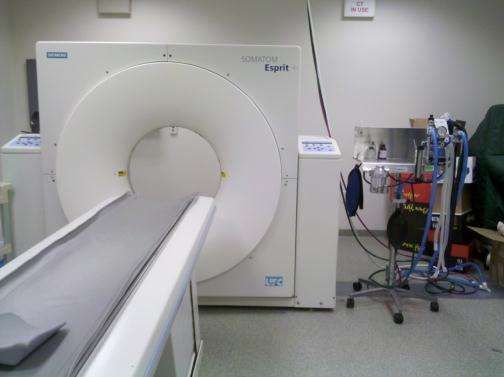
Overview
•   Introduction
•   Anatomy
•   History and Presentation
•   Equipment
•   Diagnostic Workup
•   Differential Diagnoses
•   Treatment and Prognosis by Etiology
•   Clinical Cases
Introduction
• Challenges in obtaining diagnosis

• Incidence

• Need for advanced imaging
  studies
Anatomy
• Nasal passage extends caudally from external
  nares to cribiforme plate and nasopharynx
• Divided in half by septum-cartilage cranially
  and bone caudally
• Contains thin scrolls of cartilage and bone
  called conchae or turbinates
  – Serve to warm, humidify and filter air as it passes
    through the nasal cavity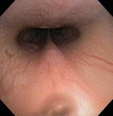
Anatomy Cont.
• Ventral, middle and dorsal nasal meatus
  – ventral meatus communicates with nasopharynx
    via choanae or internal nostrils
• Nasopharynx is delineated ventrally by soft
  palate and dorsally by vomer bone.
• Cribiforme plate separates caudal nasal cavity
  from rostral olfactory bulbs of brain hence a
  major problem if breached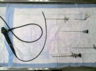
Anatomy Cont.
• Frontal sinuses, lateral maxillary recesses and
  sphenoidal sinuses all communicate with nasal
  cavity
  – not able to be visualized routinely during
    endoscopy
  – importance of combining CT scanning with
    endoscopic procedure.
History/Presentation

• Sneezing, nasal discharge, epistaxis, nasal
  congestion/stertor, gagging, reverse sneezing,
  coughing

• Unilateral vs bilateral

• Chronic vs acute
History/Presentation Cont.

• Signalment-tumors in older dogs, fungal
  rhinitis in German Shepherds and other
  dolichocephalic larger breeds
• History/presentation can be very helpful in
  determining location (nasal cavity vs
  nasopharyx) as well as most likely diagnoses
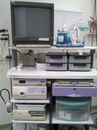
Equipment
• Camera adaptor, Processor, Xenon light source
  – Olympus
Equipment Cont.

• Rigid endoscopes
  – Stortz (rhinoscopy)
• Flexible endoscope
  – Olympus (nasopharyngeal endoscopy)
Equipment Cont.
Equipment Cont.
Equipment Cont.
• CT scanner
  – Siemens Somatom Esprit+
Diagnostic Workup
• Baseline
– CBC/chem-rule out thrombocytopenia, severe metabolic
issues (complicating factors), anemia (not common even
with epistaxis), helps assess as candidate for anesthesia.
• PT/PTT (if epistaxis)
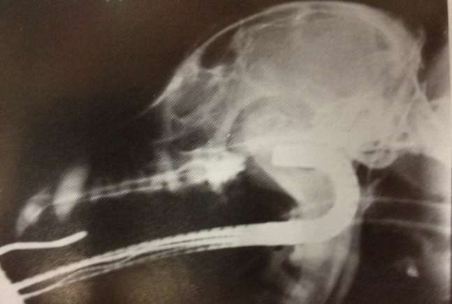
• Nasal rads
– limited value,
– must be anesthetized in most cases to obtain good films
– Much less informative than CT
– If CT declined, nasal radiographs at time of endoscopy.Diagnostic Workup Cont.
• Rhinoscopy/NP endoscopy + CT of skull and sinuses +/-
chest cavity=gold standard
– CT can image areas where rhinoscopy cannot( i.e. frontal and
accessory sinuses, bone, caudal nasal cavity)
• Helps localize problem and determine extent of invasiveness
– CT of chest much more sensitive in detecting metastatic disease
compared to chest rads
• additional cost to image chest no more expensive than 3 view chest
rads
– Pulmonary metastasis of nasal tumors is uncommon (10% or
less) but implications are important, especially if aggressive
therapy such as radiation therapy will be considered
• Therefore, CT of chest typically done where suspicion for nasal tumor
is high or if nasal tumor found on nasal CTDiagnostic Workup Cont.
• Why not CT alone (without rhinoscopy)?
– CT identifies tumor but will not allow for histopath diagnosis
• important for treatment planning and prognosis
• In most cases does not help establish cause of non neoplastic disease
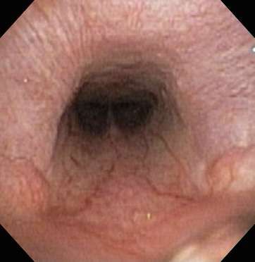
• Rhinoscopy/NP endoscopy
– allows for detailed evaluation of nasal cavity and nasopharynx
for tumor, foreign body
– allows for careful assessment of mucosal changes/allows for
tissue biopsy
• is critical in establishing specific tumor diagnoses as well as nature of
non neoplastic disease
• Oral/dental exam if oronasal fistula on differential listDifferential Diagnoses
• Neoplasia
– nasal cavity, frontal sinus, nasopharyngeal,
extension of bone/maxillary
• Nasal polyps
– extremely rare in dogs
• Nasal foreign body
• Fungal rhinitis
• Lymphoplasmacytic rhinitisDifferential Diagnoses Cont.
• Allergic rhinitis
• Bacterial rhinitis/sinusitis
– usually secondary/opportunistic
• Tooth root abscess
• Nasal mites
• Diseases of the rhinarium
– can be confused with nasal cavity disease if
bleeding or crustingDifferential Diagnoses Cont.
Unilateral Bilateral Age Breed/size Discharge Epistaxis
Neoplasia + rare older* variable 0-4+ common
Polyps + rare older variable 0-4+ common
FB + - younger variable 3-4+ rare
Fungal + + younger larger 4+ common
LP + + variable variable 1-2+ occasional
Allergic - + younger variable 3-4+ no
Bacterial + + variable variable 3-4+ rare
Tooth abscess + - older variable 3-4+ rare
Mites + + variable variable 0-1+ no
Rhinarium + + variable variable 0-1+ variable
*exception-lymphoma, fibrosarcomaTreatment and Prognosis
• Neoplasia-Non lymphoma: Radiation therapy
– Most common=carcinoma
– Radiation therapy for carcinoma-median survival
12-16 months with full course radiation.
– If no radiation-NSAIDS?
– With no treatment euthanasia typically necessary
within 3-4 months of diagnosis due to
uncontrollable epistaxisTreatment and Prognosis Cont.
• Neoplasia-Lymphoma
– Uncommon
– typically focal and not part of systemic condition
– Chemotherapy-full course sequential vs more
conservative
– benefit to radiation therapy + chemotherapy vs
chemotherapy alone?
– 6-12 months with chemotherapy is typical for those
that show response (80+%)
– 6-12 weeks with palliative prednisoneTreatment and Prognosis Cont.
• Nasal Polyps
– Very rare in dogs
– Can be extensive and cause remodeling/lysis in
nasal cavity so caution in ruling out without
biopsy!
– Treatment is surgical (rhinotomy)
– Low grade fibrosarcomas?Treatment and Prognosis Cont.
• Nasal foreign body
– Relatively uncommon
– Usually plant material or food
– Can trigger secondary bacterial rhinitis
– Treatment consists of removal (usually able to
remove endoscopically) along with appropriate
antibiotic therapy (ideally guided by c/s results)
– Prognosis is excellent until the next one.Treatment and Prognosis Cont.
• Fungal rhinitis
– Treatment consists of nasal infusion of clotrimazole
under anesthesia
– Can be combined with sinus trephanation for
instillation of clotrimazole solution and cream if
needed
– Sinus trephanation alone if CT shows infection to be
limited to frontal sinuses
– Approximate 85% cure rate with 1-3 infusions
– Oral antifungals are expensive
• must be given long term and have lower cure rate.Treatment and Prognosis Cont.
• Lymphoplasmacytic rhinitis
– Difficult to treat/no uniformly effective treatment.
– Make sure to rule out underlying disease with
rhinoscopy and CT
– Different cause in different individuals?
– Typically try intranasal steroids (Rhinocort)
• If not effective, can try oral steroids, or chlorambucil,
antifungals?
– Typically controlling condition, not curing it
– Prognosis is excellent for survival but very guarded for
resolution of signsTreatment and Prognosis Cont.
• Allergic rhinitis
• Relatively uncommon
• Very responsive to high dose steroids
– 0.75mg/kg Bid initially with slow taper
– Will often relapse if weaned off steroids so usually
maintain on low EOD dosing
– Intranasal steroids?
– Prognosis is excellentTreatment and Prognosis Cont.
• Bacterial rhinosinusitis
– Treat underlying condition!!
– Rare cases of primary bacterial infection require 6-
12 weeks of antibiotics selected based on c/s
– Prognosis usually depends on underlying
conditionTreatment and Prognosis Cont.
• Tooth root abscess
– Extraction of affected teeth/ appropriate
antibiotic therapy ideally based on c/s results
– Warn client of possible ongoing concurrent nasal
condition if signs do not resolve with dental
treatment!
– Prognosis is excellent if no concurrent primary
nasal diseaseTreatment and Prognosis Cont.
• Nasal mites
– Uncommon
– Pneumonyssoides caninum
– 1mm length
– Transmission probably through direct transmission
– Diagnosis by direct visualization of yellow white mites at
external nares or in nasal passage/nasal discharge
– Rarely can be only in sinuses
– Can be treated with Revolution q2 weeks x 3, Interceptor
0.5-1.0mg/kg q1-2 weeks x3, Ivermectin 400mcg/kg SQ or
PO q2 weeks x3
– Treat all animals in household!Treatment and Prognosis Cont.
• Diseases of rhinarium
– Lupus, pemphigus, mucocutaneous pyoderma,
vasculitis, trauma (chemical, thermal, physical)
and other conditions of rhinarium that cause
bleeding or crusting can be confused with
intranasal disease.
• Dr Spiegel!You can also read