Complete Avulsion of the Hoof Capsule and Subsequent Testicular Degeneration in a Criollo Stallion
←
→
Page content transcription
If your browser does not render page correctly, please read the page content below
Acta Scientiae Veterinariae, 2021. 49(Suppl 1): 677.

                                                                  CASE REPORT                                                   ISSN 1679-9216
                                                                   Pub. 677

                         Complete Avulsion of the Hoof Capsule and Subsequent
                              Testicular Degeneration in a Criollo Stallion

              Mariana Andrade Mousquer ¹, Bruna da Rosa Curcio ¹, Vitória Müller ², Eliza Piemolini¹,
                          Camila Gervini Wendt ¹ & Carlos Eduardo Wayne Nogueira ¹

                                                                ABSTRACT

Background: Complete avulsion of the hoof in horses, also known as exungulation, is not a commonly reported injury and
usually leads to euthanasia due to the great amount of tissue loss, intense pain, secondary complications, expensive and
lengthy treatment. It can involve deep structures and cause different complications leading to chronic lameness. In stallions
affected by such injury, the reproductive tract and performance may also be affected. The aim of this study was to report
a case of complete avulsion of the right front hoof in a Criollo stallion and subsequent bilateral testicular degeneration.
Case: A 10-year-old Criollo stallion was referred to the Veterinary Clinical Hospital of the Federal University of Pelotas
(HCV- UFPel) with a complete avulsion of the left front hoof. At admission, the stallion had clinical parameters compat-
ible with intense pain and blood loss. Evaluation of the wound demonstrated that the distal end of the third phalanx (P3)
was exposed but no fracture was detected on radiological evaluation. No other structure was apparently affected. Initially,
anti-inflammatory (phenylbutazone) and opioid (morphine) was given for pain control and supportive fluid therapy was
started to restore hydration. Antibiotic (Sulfamethoxazole with trimethoprim) was administered for 10 days. Continued
therapy with phenylbutazone, pentoxifylline, omeprazole and supplementation with methionine, biotin and zinc was also
given. Local treatment was carried out by cleaning the wound, applying an antimicrobial ointment and dressing it with
a bandage. Wound management was adapted according to the evolution and healing process. The stallion was kept in
stall rest during its hospitalization time. In the second month after the injury, accumulation of liquid in the scrotum was
observed. Clinical and ultrasound evaluation lead to a presumptive diagnosis of testicular degeneration. The stallion was
discharged after three months when the wound was almost healed and the hoof had started to grow. Six month later, a
follow up by the referring vet showed that the hoof was almost completely grown and the x-ray assessment demonstrated
a dorsal rotation and resorption of the distal end of the third phalanx.
Discussion: The stallion of this report had a complete avulsion of the hoof capsule caused by trauma. Conservative treat-
ment was established including wound cleaning and dressing to avoid contamination, control of pain and inflammation,
antimicrobial care and supplementation to support hoof growth. Time period for wound healing and hoof growth was in
agreement with other cases described previously. Bone sequestrum of the distal end of the third phalanx, and detachment
of a fragment were observed in this case, followed by bone resorption. The stallion was closely monitored to prevent
laminitis in the contralateral limb and no alterations were detected during the treatment period. Testicular degeneration was
observed, probably caused as a consequence of hoof avulsion and due to a long period of stall rest. Degenerative alterations
in testicles interfere with thermoregulation and spermatogenesis, affecting semen quality and reproductive performance.
Rotation of the third phalanx was also observed six months later caused by the hoof loss. In conclusion, the patient of
this report had a complete regrowth of the hoof capsule although a long intensive treatment was necessary to achieve this
result. As a consequence, testicles degeneration may happen impairing its function as a stallion.
Keywords: exungulation, hoof trauma, degenerative changes.

                                                        DOI: 10.22456/1679-9216.112847
Received: 8 April 2021                                       Accepted: 13 July 2021                                 Published: 4 September 2021
1
 Departamento de Clínicas Veterinária, Faculdade de Medicina Veterinária & Faculdade de Biotecnologia, CDTec, Universidade Federal de Pelotas
                                                                           2

(UFPel), Capão do Leão, RS, Brazil. CORRESPONDENCE: M.A. Mousquer [mmousquer.vet@gmail.com] & C.E.W. Nogueira [cewn@terra.com.br].
Departamento de Clínicas Veterinária - Hospital de Clínicas Veterinárias - UFPel. CEP 96010-900 Capão do Leão, RS, Brazil.

                                                                      1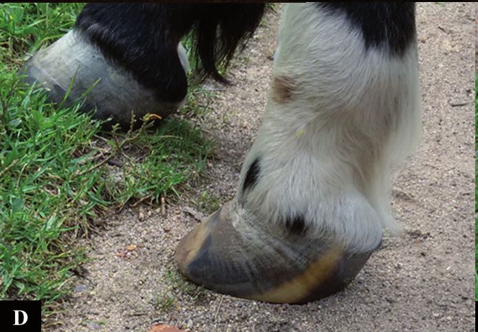
M.A. Mousquer, B.R. Curcio, V. Müller, et al. 2021. Complete Avulsion of the Hoof Capsule and Subsequent Testicular Degeneration in
a Criollo Stallion.                                                                     Acta Scientiae Veterinariae. 49(Suppl 1): 677.

                       INTRODUCTION                                   was estimated in 10%. Hematologic evaluation reve-
         Complete avulsion of the hoof in horses may                  aled anemia and neutrophilic leukocytosis. Closely
limit function, interfere with performance and euthana-               evaluation of the wound revealed that the distal end
sia may be advised in some cases [20]. It is a relatively             of the third phalanx (P3) was exposed. Radiographic
atypical injury with just few reports on literature [9-               evaluation was performed and, besides exposure of P3,
11,18]. The origin can be traumatic or can be caused                  no fracture was identified and no other structure was
by traumatic, septic or inflammatory alterations in the               apparently affected.
coronary band. Even when significant loss of germinal                          Phenylbutazone1 [Fenilbutazona OF - 4.4 mg/
tissue occurs, the foot has the capacity to heal with                 kg, IV] and morphine2 [Dimorf - 0.2 mg/kg, IV] were
establishment of proper treatment, although slower                    administered initially to manage pain. Support fluid
than other tissues [20].                                              therapy was given to restore hydration. Sulfamethoxa-
         This injury can involve different and deep                   zole with trimethoprim3 [Borgal - 15 mg/kg, IV, q 12 h]
structures in the hoof capsule. Implication of deep                   was given for 10 days, phenylbutazone4 [Equipalazone
structures should be evaluated by clinical and imaging                pó - 2.2 mg/kg, PO] was administered as needed and
exams [19]. Osteomyelitis, distal phalanx fractures,                  omeprazole5 [Omeprazol - 5 mg/kg, PO, q 24 h] were
septic arthritis and degenerative disease are common                  continued for 30 days. Pentoxifylline5 [Pentoxilifina -
complications [20]. Treatment is lengthy and expensive                7,5 mg/kg, PO, q 12 h] was associated on the seventh
and the prognosis is guarded to poor [18,20].                         day and was given for 30 days. Wound treatment was
         In addition, when such injury occurs in stallions,           also performed daily by cleansing the wound with
it is a concern to preserve reproductive performance.                 water, drying it and applying nitrofurazone ointment5
Musculoskeletal disorders and long periods of stall rest              (nitrofurazona). A bandage made with cotton and gauze
can lead to development of testicular degeneration [3,6].             or a diaper was put to prevent dirt from entering. In the
Testicular degeneration is the main cause of subferti-                first month of treatment, there was an improvement in
lity and infertility [3,23]. It has multifactorial causes,            the healing process and the patient was already able to
however, alteration in testicular thermoregulation is                 support the limb on the ground (Figure 2A). Detach-
known as the main triggering factor [1,3]. Depending                  ment of the distal end of the third phalanx was also
on the cause and the exposure time, the degeneration                  observed and the fragment was removed.
can also vary as temporary or permanent [6].                                   After 2 months, local treatment was adapted
         Therefore, the aim of this study was to report               due to the improvement in the healing process (Figure
a case of complete avulsion of the right front hoof in a              2B). The foot started to be soaked in a diluted povidone
Criollo stallion and subsequent bilateral testicular dege-            iodine solution6 (Riodeine 0,2%) and petroleum jelly5
neration. To the best of our knowledge this is the first              (Vaselina) was applied around the wound, the same
report of a front hoof exungulation in a Criollo stallion.            dressing was continued. Supplementation with biotin,
                                                                      methionine and zinc7 [Bio Hoof - 20 g/day, PO] was
                             CASE
                                                                      given during all hospitalization time. During all the
         A 10-year-old Criollo stallion was referred to               treatment, the stallion was maintained in stall rest, with
the Veterinary Clinical Hospital of the Federal Uni-                  a high rice husk bed. The horse was able to alternate its
versity of Pelotas (HCV- UFPel) during the breeding                   position from recumbent to standing by itself.
season. The horse presented a complete avulsion of                             In the second month of hospitalization, accu-
the left front hoof with third phalanx exposure due to                mulation of liquid in the scrotum was observed. The
a wire fence accident (Figure 1).                                     testicles were palpated and a softening in the consis-
         At admission, the stallion was alert, demons-                tency of both testicles was identified. No alterations on
trating intense pain and non-weight-bearing lameness.                 size and position were observed. Ultrasound revealed
At the initial clinical exam, the heart rate (HR) was 100             a hydrocele and no other abnormalities were found.
bpm, respiratory rate (RR) was 60 bpm, body tempe-                    Two attempts to collect semen were made using an
rature was 39ºC, mucous membranes were pale with                      artificial vagina and two different mares in heat, but
a capillary refill time (CRT) of 4 s, intestinal sounds               failed because the horse did not have libido. Phar-
were diminished in all quadrants and dehydration                      macological induction of ejaculation was also tried

                                                                  2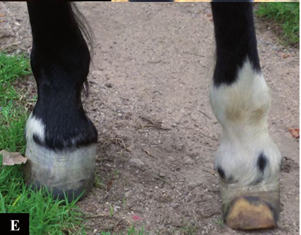
M.A. Mousquer, B.R. Curcio, V. Müller, et al. 2021. Complete Avulsion of the Hoof Capsule and Subsequent Testicular Degeneration in
a Criollo Stallion. Acta Scientiae Veterinariae. 49(Suppl 1): 677.
but it was not successful. A presumptive diagnosis the stratum germinativum [15]. The hoof wall growth
of testicular degeneration was made based on clinical occurs from the stratum germinativum of the coronary
evaluation. Testicular biopsy was not performed to corium and reached approximately 6-10 mm per month
confirm the diagnosis. [8,21]. In our patient, the hoof reached 1.3 cm during
Three months later, the stallion was discharged the 3-month period in which the horse was hospitalized.
since the wound was almost healed and the hoof had In the case reported here, the time for wound
grown 1.3 cm (Figure 2C). The owner was advised to healing (3 months) and hoof growth (6 months) was
continue wound dressing and veterinary monitoring in agreement with the time reported in the literature
at home. Six months later, the referring veterinarian [8,20]. According to Fessler [8] and Stashak [20], it
reported that the hoof was almost completely grown takes generally 3 to 5 months to heal and 9 to 12 months
and radiograph evaluation showed resorption of the
to the complete growth of a new hoof. Other reports
distal end of the third phalanx and rotation.
describe a complete growth of a new hoof capsule in
DISCUSSION a period of 4 [11] and 6 [10] months.
The ground proximity of this injury also incre-
The patient of this report had a complete avul-
ases the risks of contamination and delay healing. The-
sion of the hoof capsule caused by trauma. Although
intensive and lengthy therapy had to be established and refore, protection of the wound with either a bandage or
the patient developed testicular degeneration, it had a cast is recommended [17,20]. Conservative treatment
a successful recovery with complete hoof regrowth. instituted in this case was similar to those described
The hoof wall is responsible for supporting before [8,22], including soaking the foot in antiseptic
the horse’s weight and absorption of shock when solutions, wound bandaging changed regularly to mo-
contacting the ground [7]. The epithelium of the hoof nitor possible local infections and maintenance of the
is composed of three recognized layers: the stratum horse in high bed stalls to improve its comfort. The use
basale, stratum spinosum and stratum corneum in whi- of a rigid cast is also recommended in order to acce-
ch the stratum basale and stratum spinosum constitute lerate the healing process and weight bearing [10,20].
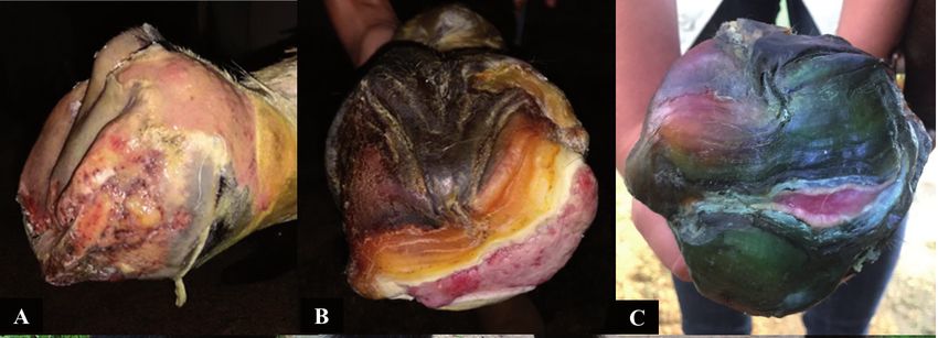
Figure 1. A & B- Day of admission at the Veterinary Clinical Hospital - UFPel. Complete avulsion of the hoof capsule with exposure of the third phalanx.
3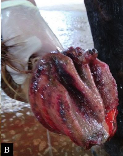
M.A. Mousquer, B.R. Curcio, V. Müller, et al. 2021. Complete Avulsion of the Hoof Capsule and Subsequent Testicular Degeneration in
a Criollo Stallion.                                                                     Acta Scientiae Veterinariae. 49(Suppl 1): 677.

Figure 2. Healing evolution of the wound. A- One month of treatment. B- Two months of treatment. C- Three months of treatment. D & E- One year
after the injury.

         Antimicrobial therapy was administered to con-                   of the structural arrangement that connects the hoof
trol possible infection and long-term anti-inflammatory                   wall to the coffin bone, which involves insensitive
therapy was used to improve horse’s well-being through                    and sensitive laminae that also maintain the phalanx
pain control, reduce inflammation and laminar damage                      lined up. Exungulation brakes up this connection and
[13]. Pentoxifylline was also included in the treatment                   rotation of the coffin bone may occur [7]. Even when
although its actions are still controversial. It is suggested             no complications happen, the horse can remain lame
that this drug inhibits the release of metalloproteinases                 because of the amount of tissue loss [7,19,20].
and decreases blood viscosity by acting on platelets                               Support limb laminitis (SLL) is another
and red blood cells [13,14]. Oral supplementation with                    concern in conditions requiring excessive weight
biotin and methionine, as used in our patient, was also                   bearing in the contralateral limb [2]. Because of that,
reported to be helpful in hoof injuries [24].                             the stallion was closely monitored for digital pulses,
         Several conditions can interfere in the recovery                 warmth in the hoof, bulging in the sole and depression
of a horse with complete hoof avulsion, as the develop-                   at the coronary band during its hospitalization time,
ment of osteomyelitis, the risk of articular involvement                  however, no alterations were observed. The authors
leading to septic arthritis, third phalanx fractures and                  also believe that the horse’s behavior of self-protection,
degenerative diseases [20]. In our patient, exposure of                   which was observed since the beginning of treatment,
the third phalanx and the detachment of a fragment was                    helped to minimize its chances of developing SLL.
observed, probably caused by a bone sequestrum. Six                       Horses that are able to relieve weight from the con-
months after the injury, radiograph evaluation showed                     tralateral limb have a lower risk of developing SLL
resorption of the distal end of the third phalanx and                     [2,16]. Jackson [11] reported a mare with complete
a dorsal rotation. Rotation probably happens because                      hoof avulsion that was maintained in a sling during

                                                                      4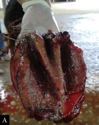
M.A. Mousquer, B.R. Curcio, V. Müller, et al. 2021. Complete Avulsion of the Hoof Capsule and Subsequent Testicular Degeneration in
a Criollo Stallion.                                                                     Acta Scientiae Veterinariae. 49(Suppl 1): 677.

10 h/day to relieve weight from the contralateral limb                lation and spermatogenesis, which depending on the
with no development of SLL.                                           duration and severity, may affect semen quality [1,3].
         Alterations in the reproductive tract of the                          In conclusion, the outcome for exungulation
stallion were noticed in the second month of hospita-                 in horses is difficult to predict and several disorders
lization, including fluid accumulation in the scrotum,                can occur as a consequence. The patient of this report
softened consistency of both testicles and lack of libido.            had a complete regrowth of the hoof capsule although
Hydrocele and testicular degeneration diagnosis were                  a long intensive treatment was necessary to achieve
made presumptively based on the horse’s clinical pre-                 this result. The commitment of the veterinary team and
sentation and ultrasound exam. Although semen evalu-                  owners was also a key point for a successful outcome.
ation is important for diagnosis, attempts for collection             As a consequence, testicles degeneration may happen
were not successful due to lack of libido and response                impairing its function as a stallion.
to chemical stimulation. Similarly, biopsy was not
                                                                      MANUFACTURERS
performed to avoid postoperative complications [3].
         According to Blachard et al. [4] and Blachard                  Ouro Fino Saúde Animal Ltda. Cravinhos, SP, Brazil.
                                                                      1

                                                                      2
                                                                        Cristália Produtos Químicos Farmacêuticos Ltda. Cotia, SP, Brazil.
et al. [5], softening in the consistency with no neces-               3
                                                                        Merck Sharp & Dohme Saúde Animal Ltda. São Paulo, SP, Brazil.
sary alteration in size of testicles may be an indicative             4
                                                                        Vansil Indústria, Comércio e Representações Ltda. Dascalvado, SP,
of a degenerative process. Low libido in experienced                    Brazil.
                                                                      5
                                                                        Vetpharma Medicamentos Veterinários Ltda. Pelotas, RS, Brazil
breeding stallions can also be identified in animals with             6
                                                                        Rioquímica S.A. São José do Rio Preto, SP, Brazil.
testicular degeneration, musculoskeletal discomfort or                7
                                                                        Vetnil Indústria e Comércio de Produtos Veterinários Ltda.
intensive pain [6,12]. Hydrocele can be a consequence                   Louveira, SP, Brazil.
of musculoskeletal injuries and lack of exercise. In addi-            Declaration of interest. The authors report no conflicts of
tion, a hot climate may also contribute to its formation              interest. The authors alone are responsible for the content and
[3]. This disorder interferes in testicular thermoregu-               writing of paper.

                                                           REFERENCES

 1 Alvarenga M.A. & Papa F.O. 2009. Principais distúrbios reprodutivos observados em garanhões no Brasil. Revista
   Brasileira de Reprodução Animal. 6: 204-209.
 2 Baxter G.M. 2017. Supporting limb laminitis. In: Belknap J.K. & Geor R. (Eds). Equine laminitis. Oxford: Wiley-
   Blackwell, pp.210-213.
 3 Beard W. 2011. Abnormalities of the Testicles. In: McKinnon A.O., Squires E.L. Vaala W.E. & Varner D.D. (Eds).
   Equine reproduction. 2nd edn. Ames: Wiley-Blackwell, pp.1161-1665.
 4 Blanchard T.L., Johnson L. & Roser A.J. 2000. Creased germ cell loss rates and poor semen quality in stallions with
   idiopathic testicular degeneration. Journal of Equine Veterinary of Equine Science. 20(4): 263-265.
 5 Blanchard T.L., Johnson L., Varner D.D., Rigby S.L., Brinsko S.P., Love C.C. & Miller C. 2001. Low daily sperm
   output per ml of testis as diagnostic criteria for testicular degeneration in stallions. Journal of Equine Veterinary of
   Equine Science. 21(1): 33-35.
 6 Brinsko S.P., Blanchard T.L., Varner D.D., Schumacher J., Love C.C., Hinrichs K. & Hartman D. L. 2011.
   Examination of the Stallion for Breeding Soundness. In: Brinsko S.P., Blanchard T.L., Varner D.D., Schumacher J.,
   Love C.C., Hinrichs K. & Hartman D.L. (Eds). Manual of Equine Reproduction. 3rd edn. St. Louis: Mosby Elsevier,
   pp.176-206.
 7 Brooks J. & Pusey A. 2010. Horse anatomy for osteopaths. In: Pusey A., Brooks J. & Jenks A. (Eds). Osteopathy and
   the Treatment of Horses. Oxford: Wiley-Blackwell, pp.4-39.
 8 Fessler J.F. 1989. Hoof Injuries. Veterinary Clinics of North America: Equine Practice. 5(3): 643-664.
 9 Fonteque J.H., Souza A.F., Shade J., Muller T.R. & Gehrcke M.I. 2017. Exungulation followed by fracture and
   avulsion of the phalanx in mare. Veterinária e Zootecnia. 24(3): 509-514.
10 Gresti A., Zani D.D., D’arpe L. & Scandella M.A. 2008. Singular case of traumatic total hoof capsule avulsion.
   Equine veterinary Education. 20(8): 406-410.

                                                                  5
M.A. Mousquer, B.R. Curcio, V. Müller, et al. 2021. Complete Avulsion of the Hoof Capsule and Subsequent Testicular Degeneration in
a Criollo Stallion. Acta Scientiae Veterinariae. 49(Suppl 1): 677.
11 Jackson L.L. 1969. Regrowth of an Equine Hoof Following Traumatic Removal. Iowa State University Veterinarian.
31(2): 44-47.
12 McDonnel S.M. 1999. Libido, Erection, and Ejaculatory Dysfunction in Stallions. Compendium on Continuing Educa-
tion for the Practicing Veterinarian - North American Edition. 21(3): 263-266.
13 Mitchell C.F., Fugler L.A. & Eades S.C. 2015. The management of equine acute laminitis. Veterinary Medicine:
Research and Reports. 6: 39-47.
14 Moyer W., Schumacher J., Schumacher J. & Carter G.K. 2008. Are Drugs Effective Treatment for Horses with
Acute Laminitis? Proceedings of the Annual Convention of the AAEP. 54: 337-340.
15 Parks A.H. 1999. Equine Foot Wounds: General Principles of Healing and Treatment. Proceedings of the Annual
Convention of the AAEP. 45: 180-187.
16 Redden R.F. 2004. Preventing Laminitis in the Contralateral Limb of Horses with Nonweight-Bearing Lameness.
Clinical Techniques in Equine Practice. 3: 57-63.
17 Riggs C.M., Proudman C.J., Hughes I. & Caldwe M. 1995. Management of traumatic, partial hoof avulsion in 2
horses. Equine Veterinary Education. 7(3): 140-144.
18 Ruzickova P., Trencart P. & Laverty S. 2016. Spontaneous hoof capsule loss following lacerations of the equine
distal limb. Equine Veterinary Education. 29(9): 472-477.
19 Schumacher J. & Stashak T.S. 2008. Management of Wounds of the Distal Extremities. In: Stashak T.S. & Theoret
C.L. (Eds). Equine wound management. 2nd edn. Ames: Wiley-Blackwell, pp.375-425.
20 Stashak T.S. 1989. Management of lacerations and avulsion injuries of the foot and pastern region and hoof wall
cracks. Veterinary clinics of North America: Equine practice. 5(1): 195-220.
21 Theoret C.L. 2008. Physiology of Wound Healing. In: Stashak T.S. & Theoret C.L. (Eds). Equine Wound Manage-
ment. 2nd edn. Ames: Wiley-Blackwell, pp.5-28.
22 Thomassian A. 2005. Enfermidades dos Cavalos. 4.ed. São Paulo: Livraria Varela, pp.178-179.
23 Turner R.M.O. 2007. Pathogenesis, Diagnosis, and Management of Testicular Degeneration in Stallions. Clinical Tech-
niques in Equine Practice. 6: 278-284.
24 Young J.H. 1988. Hoof wall avulsion: Three case reports. Equine Veterinary Science. 8(6): 420-423.
CR677
http://seer.ufrgs.br/ActaScientiaeVeterinariae
6You can also read