Complications, prognostic factors, and long-term outcomes for dogs with brachycephalic obstructive airway syndrome that underwent H-pharyngoplasty ...
←
→
Page content transcription
If your browser does not render page correctly, please read the page content below
Complications, prognostic factors, and long-term
outcomes for dogs with brachycephalic obstructive
airway syndrome that underwent H-pharyngoplasty
and ala-vestibuloplasty: 423 cases (2011–2017)
Julien P.R. Carabalona, DVM*; Kevin Le Boedec, DVM, MS; Cyrill M. Poncet, DVM

Centre Hospitalier Vétérinaire Frégis, Arcueil, France

*Corresponding author: Dr. Carabalona (juliencarabalona@hotmail.com)

OBJECTIVE
To describe the H-pharyngoplasty procedure, report the outcomes of dogs with brachycephalic obstructive airway syndrome
(BOAS) treated with ala-vestibuloplasty and H-pharyngoplasty with a CO2 laser, and identify prognostic factors.
ANIMALS
423 dogs.
PROCEDURES
Medical records of dogs admitted for BOAS from 2011 to 2017 were reviewed. Dogs were included if they were treated with
ala-vestibuloplasty and H-pharyngoplasty with a CO2 laser. Signalment, physical examination findings, grades at admission of
clinical signs associated with respiratory and digestive systems, diagnostic test results, postoperative treatments, and short-term
follow-up results were extracted from medical records. Long-term follow-up of > 12 months was evaluated via questionnaire.
Generalized ordered logistic regression was used for bivariable and multivariable analyses.
RESULTS
Overall mortality rate was 2.6%. Median duration of follow-up was 36 months (12 to 91 months), and 341 (80.6%) dog owners
completed the questionnaire. Major complications included respiratory distress (2.1%), heatstroke (0.5%), and bronchopneu-
monia (0.5%). No dogs required revision surgery. Improvement in signs associated with the respiratory and digestive systems
was reported in 72% and 34% of the dogs, respectively, and owners’ satisfaction was high (97.1%). Risk of death increased by
29.8% (95% CI, 11.8% to 50.7%) for every 1-year increase in age.
CONCLUSION AND CLINICAL RELEVANCE
H-pharyngoplasty was possible in all dogs with BOAS, including those previously treated with conventional surgery and was
associated with low morbidity and improved respiratory and digestive signs. H-pharyngoplasty combined with ala-vestibuloplasty
may be an alternative treatment for even the most severely affected dogs.

S   urgery to alleviate upper airway obstruction is con-
    sidered the standard of care for dogs with brachy-
cephalic obstructive airway syndrome (BOAS).1,2
                                                                       excised remains unknown. Furthermore, the best
                                                                       way to manage other anomalies such as aberrant tur-
                                                                       binates, inflamed tonsils, everted laryngeal saccules,
Nevertheless, despite an improved understanding of                     macroglossia, laryngeal collapse, and redundant pha-
the underlying pathophysiology of BOAS and despite                     ryngeal mucosa that may affect recovery also remains
surgical intervention, some dogs have abnormalities                    unknown.13–17
that cannot be corrected.1,3–5 Various surgical tech-                       In 1982, Harvey10 described the first palatoplasty
niques have been used2,3,6–12 to address BOAS, and                     procedure that alleviated laryngeal obstruction by
various materials have been used for resecting exces-                  shortening the soft palate. In 2008, Findji and Dupré2
sive tissue, including Metzenbaum scissors, electro-                   described folded-flap palatoplasty, which in addi-
surgery, vessel-sealing devices, CO2 and diode lasers,                 tion to addressing the length of the soft palate also
and harmonic scalpels.                                                 addressed problems related to its thickness (hyper-
     The many anomalies that comprise BOAS sug-                        plasia). Brdecka et al6 in 2008 and Dunié-Mérigot
gest its complexity. Although staphylectomy and ala-                   et al3 in 2010 proposed more extensive staphylecto-
plasty are recommended for the treatment of airway                     mies, but pharyngeal obstruction caused by redun-
obstruction, some dogs do not improve following                        dant pharyngeal mucosa was still not addressed.
these procedures.5 The appropriate amount of the                       Recently, multilevel surgeries that involve alaplasty
soft palate and the wing of the nostril that must be                   and palatoplasty associated with ≥ 1 of the following

                                               JAVMA | JAN 15, 2022 | VOL 260 | NO. S1                                          S65

                                                                                           Unauthenticated | Downloaded 02/03/22 07:06 PM UTC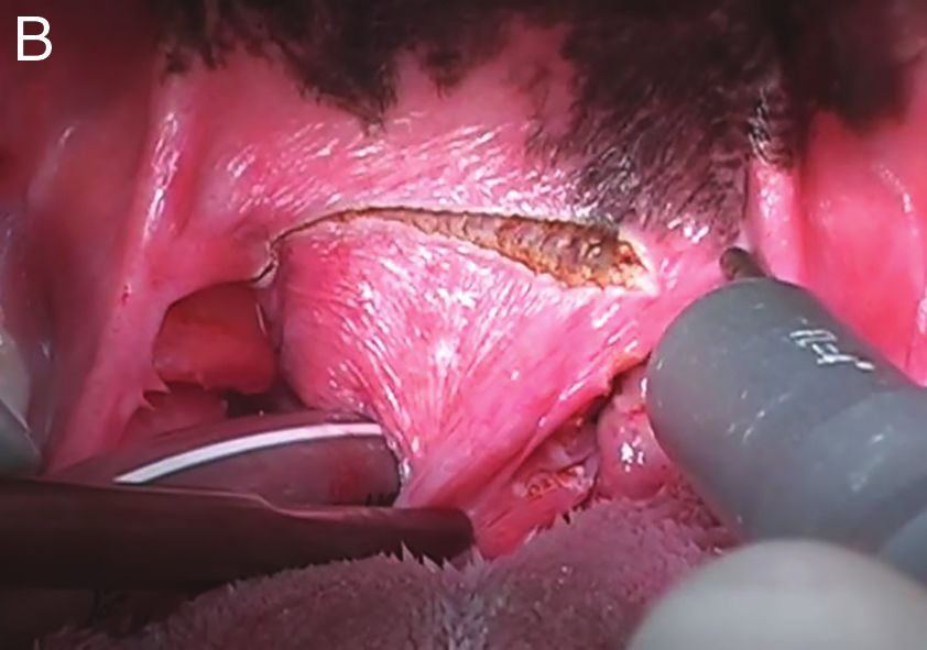
procedures—laser-assisted turbinectomy, vestibulo-         serum biochemical analysis, and thoracic radiography
plasty, removal of laryngeal saccules, partial tonsil-     were performed, with the latter to assess for tracheal
lectomy (ie, removal of the everted portion of the         hypoplasia and pulmonary lesions. Each dog was pre-
tonsil), and partial cuneiformectomy (laryngoplasty        medicated on arrival to the hospital with IM adminis-
[trimming of deformed or collapsed cuneiform pro-          tration of 0.05 mg of acepromazine/kg (0.02 mg/lb),
cesses])—have been proposed to improve nasal and           0.2 mg of dexamethasone/kg (0.09 mg/lb), 0.01 mg
pharyngeal clearance. These procedures are associ-         of glycopyrrolate/kg (0.005 mg/lb), 0.5 mg of raniti-
ated with improved outcomes, compared with tra-            dine/kg (0.23 mg/lb), and 0.5 mg of metoclopramide/
ditional surgeries such as alaplasty and cut-and-sew       kg (0.23 mg/lb). After preoxygenation (with anes-
staphylectomy.1,18 Laser-assisted turbinectomy and         thetic mask over the nose and mouth), anesthesia
vestibuloplasty, in combination with a multilevel sur-     was induced with 4 to 8 mg of propofol/kg (1.81 to
gery or after an unsatisfactory response to conven-        3.63 mg/lb) and each dog was orotracheally intu-
tional surgery, have yielded good outcomes for the         bated. Anesthesia was maintained with isoflurane in
relief of nasal obstruction.1,18,19                        100% oxygen.
     With the aim of attenuating the redundant pha-             The second step consisted of esophagogastro-
ryngeal mucosa and optimizing relief from pharyngeal       duodenoscopy, pharyngolaryngoscopy, and surgery.
obstruction, a new surgical technique, H-pharyngo-         Esophagogastroduodenoscopy plus tissue biopsy
plasty (which included tonsillectomy plus pharyngo-        acquisition was performed by a board-certified inter-
plasty) with ala-vestibuloplasty, had been designed        nal medicine specialist (KLB) for dogs with preop-
as part of a standardized multilevel approach (SMA).       erative grade 3 clinical signs associated with the
Therefore, the goals of the study reported here were       digestive system or preoperative grade 2 clinical signs
to describe this surgical technique for dogs with          that progressively worsened. Pharyngolaryngoscopy
BOAS, report patient outcome following this proce-         was performed by a board-certified surgeon (CMP)
dure as part of an SMA, and identify prognostic fac-       for all dogs. Each dog was briefly extubated for pha-
tors. An additional goal was to determine whether          ryngoesophageal examination. Length and thickness
H-pharyngoplasty could be used in dogs that had            of the soft palate were subjectively evaluated through
previously undergone other surgeries for BOAS.             laryngoscopy and direct palpation. Tonsils were visu-
The hypothesis was that H-pharyngoplasty with ala-         ally inspected for signs of inflammation and eversion,
vestibuloplasty as part of an SMA would improve            and laryngeal saccules were assessed for eversion and
signs attributable to BOAS, minimize postoperative         the presence of granulomas on their surface. Laryn-
morbidity, and result in good long-term outcomes.          geal collapse was graded as previously described.20
                                                                Each dog was placed in sternal recumbency with
Materials and Methods                                      its mouth wide open and its tongue taped rostrally
                                                           to expose the oropharynx. Wet gauze was placed
     Medical records of all dogs diagnosed with BOAS       behind the soft palate to prevent collateral damage
from June 2011 to July 2017 were reviewed. Signal-         from the laser beam. The surgeon and accompanying
ment, history, presenting complaints, thoracic radio-      staff wore safety glasses when the laser was in use,
graphic findings, procedural information as part of        and a smoke evacuator was used to prevent inhala-
the SMA, and follow-up information were recorded.          tion of the laser plume.
Study inclusion required that all dogs were treated by          Tonsillectomy and staphylectomy, part of the
the same board-certified surgeon (CMP). Dogs that          H-palatoplasty procedure, were performed with a
underwent surgery performed by another surgeon or          6- to 12-W noncontact superpulsed, scanned-mode
did not undergo an SMA or had missing data in their        small-size CO2 laser (Space Vet, Deka, Manchester,
medical records were excluded from the study.              NH). A deep circumferential incision was made in
     Clinical signs associated with the respiratory and    the tonsillar fossa to completely remove both tonsils.
digestive systems were graded during the preopera-         The caudal tip of the soft palate was grasped with for-
tive visit according to a grading scale that categorized   ceps and pulled ventrally, which put tension on it and
snoring, exercise intolerance, syncope, regurgitation,     facilitated the cut. The incision line on the soft palate
vomiting, and ptyalism by their frequency as follows:      formed an arch between the ventrolateral-most aspect
never, < 1X/mo, 1X/wk, 1X/d, > 1/d, or constantly          of each tonsillar crypt (Figure 1; Supplementary
(Supplementary Appendix S1). Every dog owner               Video S1). The top of the arch was at the level of the
was provided an explanation about and asked to indi-       soft palate along a virtual line that joined the most
cate the frequency of each clinical sign and then to       rostral aspect of each tonsillar fossa. The entire length
assign a grade. Overall preoperative grade for each        of the oral mucosa of the soft palate was incised up
dog for each body system was determined on the             to the nasopharyngeal mucosa. When the incision on
basis of the highest grade assigned to one of the clini-   the soft palate reached the lateral aspect of the tonsil-
cal signs associated with that body system.                lar crypts, the incision was extended caudally on the
     The SMA consisted of a 3-step protocol. For the       pharyngeal mucosa on both sides to free the soft palate
first step, each dog had a complete physical examina-      from its lateral attachments in the pharyngeal region.
tion before hospitalization, and before surgery, CBC,      The final incision roughly had an inverted U shape.

S66                                JAVMA | JAN 15, 2022 | VOL 260 | NO. S1

                                                                                Unauthenticated | Downloaded 02/03/22 07:06 PM UTC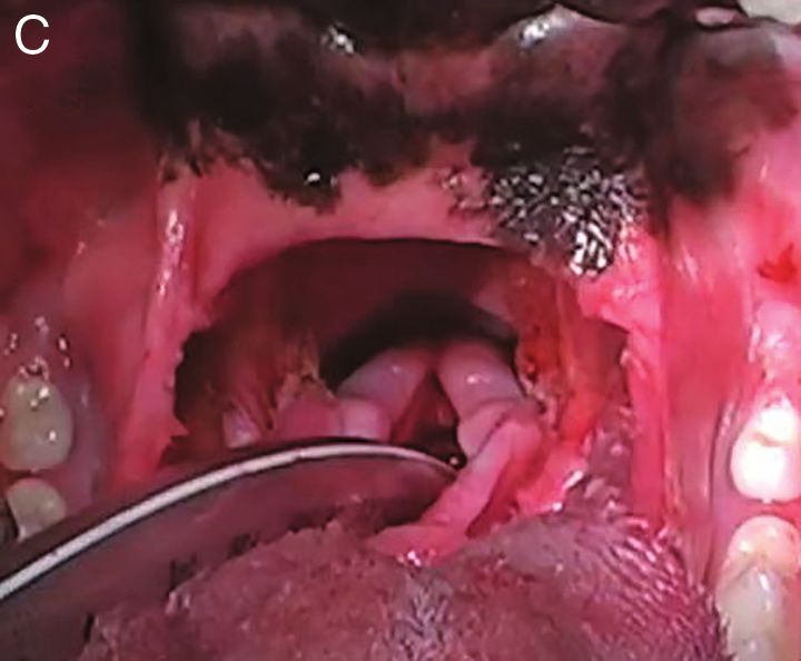
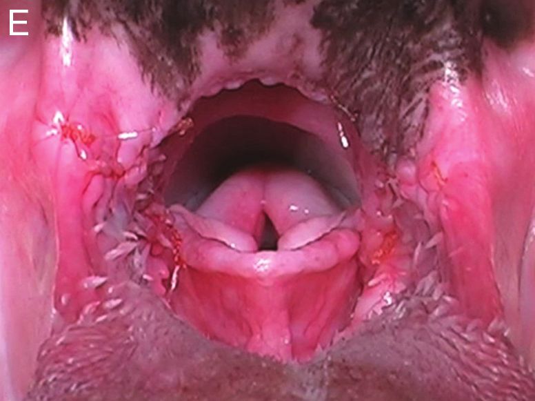
Figure 1—Schematic drawings (A and D)
                                                                          and intraoperative photographs (B, C, and E)
                                                                          of the H-pharyngoplasty technique for a dog
                                                                          with brachycephalic obstructive airway syn-
                                                                          drome. A—Dotted line indicates the arched
                                                                          incision line between the ventrolateral-most
                                                                          aspect of each tonsillar crypt. B—Note the
                                                                          incision of the soft palate made with a CO2
                                                                          laser. C—Photograph obtained following
                                                                          bilateral tonsillectomy and staphylectomy.
                                                                          D—Schematic drawing of suture placement
                                                                          following bilateral tonsillectomy and staphy-
                                                                          lectomy. E—Photograph obtained immedi-
                                                                          ately following surgery. In all panels, the top
                                                                          of the dog’s head is to the top and the dog’s
                                                                          right is to the left. Drawings by Dr. Julien
                                                                          P. R. Carabalona.

Closure was achieved with 3 simple continuous                   The third step was postoperative recovery in the
suture patterns with absorbable suture material 4-0        intensive care unit. After surgery, each dog remained
(Caprosyn [Polyglytone 6211], Medtronics, Dublin,          intubated as long as possible. Each dog was closely
Ireland) in an H shape (H-pharyngoplasty). The verti-      monitored during hospitalization, especially during
cal lines of the H were created by apposition of the       the first 24 hours after surgery, and water and food
free borders of the tonsillar crypts for the most ros-     were incrementally reintroduced over a 12-hour
tral half of the suture and by apposition of the free      period after surgery. Each dog was discharged from
borders of the oropharyngeal mucosa for the most           the hospital 24 hours after surgery unless complica-
caudal half of the suture. The pharyngeal mucosa was       tions occurred. The duration of hospitalization and
pulled cranially before being sutured in the continu-      the presence of any postoperative complications were
ity of the tonsillar crypt. These sutures smoothed the     recorded. Respiratory distress, temporary tracheos-
redundant pharyngeal mucosa. The horizontal line of        tomy, signs of heatstroke, and signs and radiographic
the H consisted of apposition of the nasopharyngeal        findings consistent with aspiration pneumonia were
mucosa with the oropharyngeal mucosa of the free           considered major complications, and vomiting, regur-
border of the remaining soft palate.                       gitation, and nasal discharge were considered minor
     Vertical wedge ala-vestibuloplasty was performed      complications.
as previously described.18,21 The nares were sprayed            Treatment with 0.7 to 1.0 mg of omeprazole/
with xylocaine immediately before beginning the            kg (0.32 to 0.45 mg/lb), PO, every 12 hours; 0.5 mg
procedure. Three to 4 simple interrupted sutures           of prednisolone/kg, PO, every 12 hours; and 0.3 to
with absorbable monofilament suture (Caprosyn              0.5 mg of metoclopramide/kg (0.14 to 0.23 mg/lb),
[Polyglytone 6211], Medtronics, Dublin, Ireland)           PO, every 8 hours, was recommended for 6 days. If
were used to close the incisions. Everted laryngeal        inflammatory gastrointestinal (GI) disease was diag-
saccules were not removed unless a granuloma was           nosed on the basis of histiologic examination of GI
observed on their surface.                                 tissue samples obtained via endoscopy, affected
     At the end of surgery on the basis of the surgeon’s   dogs received 1 mg of prednisolone/kg (rather than
clinical impression, a nasotracheal tube was placed        0.5 mg/kg), PO, every 12 hours, and then at decreas-
for postoperative oxygen supplementation, especially       ing dosages every 2 weeks, such that prednisolone
when the surgeon expected a complicated recovery           was administered for a total of 6 to 8 weeks; received
because of laryngeal collapse, laryngeal edema, obe-       15 mg of metronidazole/kg (6.8 mg/lb), PO, every
sity, or other pharyngeal or laryngeal abnormalities       12 hours, for 2 weeks; and were fed a hydrolyzed pro-
observed during pharyngolaryngoscopy.                      tein diet.

                                      JAVMA | JAN 15, 2022 | VOL 260 | NO. S1                                        S67

                                                                                Unauthenticated | Downloaded 02/03/22 07:06 PM UTC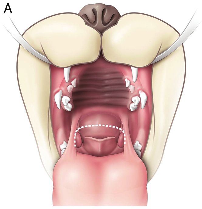
Follow-up examination at the hospital was per-       was only considered. Hence, postoperative improve-
formed 2 weeks after surgery or earlier if complica-      ment was indicated by a score of 1. The evaluated
tions occurred. Correspondences with dog owners           factors were age, sex, body weight within a breed,
and their dog’s primary care veterinarian through         breed, preoperative clinical signs associated with
email and summaries of telephone conversations            the respiratory and digestive systems, and grade of
were also recorded when an in-hospital follow-up          laryngeal collapse. French Bulldog was selected as
examination was not possible. Postoperative grades        the referent breed because it was the most common
for clinical signs associated with the respiratory and    brachycephalic breed presented to the authors’ hos-
digestive systems were obtained through a question-       pital. Other brachycephalic breeds were specified in
naire administered by one of the authors (JPRC) with      the statistical analysis on the basis of previous stud-
an online survey tool (Survey Monkey, SVMK Inc,           ies and the authors’ experience that postsurgical
San Mateo, CA; Supplementary Appendix S2) at              outcome varies for various breeds. Results were pre-
least 12 months after surgery. Owners graded the          sented as ORs and 95% CI, with the OR a reflection
clinical signs in relation to those assigned preopera-    in the change in odds of postoperative improvement
tively, either as unchanged, worse, or improved. In an    of signs associated with the respiratory or digestive
attempt to obtain accurate postoperative grades and       systems observed with each incremental change in
avoid recall bias, owners were asked to evaluate their    the evaluated prognostic variable. If the OR was > 1
dog at the time they were completing the question-        or < 1, an improvement or worsening, respectively,
naire, not how their dog was 12 months after surgery.     in signs associated with the respiratory or digestive
Additionally, dog owners were asked whether they          systems was expected postoperatively with each
were satisfied with the surgery and whether they          incremental change of the prognostic variable. All
would recommend it to a dog owner whose dog was           variables with a value of P < 0.1 in the bivariable anal-
similarly affected.                                       yses were selected for inclusion in the multivariable
                                                          analysis.21 Only those variables that remained signifi-
Statistical analyses                                      cant in the multivariable analysis were considered as
     Categorical data (eg, sex and breed) were reported   independent prognostic factors. Furthermore, the
as frequencies and percentages. Continuous data (eg,      associations between possible prognostic factors and
age at presentation and duration of hospitalization)      survival rate were evaluated by use of a log-rank test
were reported as median, minimum, and maximum             for categorical variables or a Cox proportional haz-
values because of deviation from the Gaussian distri-     ards regression model for continuous variables. All
bution (ie, data not normally distributed).               dogs that died regardless of the time between surgery
     To explore possible prognostic factors, the          and death (ie, perioperative death and death before
grades that were based on the dog owner’s answers         and after the first reexamination) were included in
to the questionnaire during the preoperative visit        the analysis. Results were presented as change in
and the grades that were based on the dog owner’s         the risk of death observed with each incremental
answers to the questionnaire > 12 months after sur-       change in the evaluated prognostic variables. Only
gery were used. Questionnaires from dog owners            bivariable analysis was performed for survival rate
whose dogs had died at the time of final follow-up        because the number of deaths was small to perform
were not included in the analyses to avoid recall bias.   a multivariable analysis. Statistical analyses were
A clinical improvement score for the 3 clinical signs     performed with commercially available software
associated with the respiratory system (snoring, exer-    (STATA, version 14.0; StataCorp LP, College Station,
cise intolerance, and syncope) that were included         TX). Values of P < 0.05 were considered significant.
in the questionnaire was determined by subtract-
ing the preoperative grade from the postoperative
grade (obtained at the time of the final follow-up
                                                          Results
examination). If the calculation yielded a negative            Of 532 dogs that underwent surgery for BOAS dur-
value, a grade of –1 was recorded. If the calculation     ing the study period, 423 met the inclusion criteria.
yielded a positive value, a grade of 1 was recorded. If   Breeds represented by at least 2 dogs included French
the calculation yielded a value of 0, a grade of 0 was    Bulldog (n = 216 [51.1%]), Pug (87 [20.6%]); English
recorded. A clinical improvement grade was assigned       Bulldog (80 [18.9%]), Boston Terrier (13 [3.1%]),
analogously for the 3 clinical signs associated with      Cavalier King Charles Spaniel (11 [2.6%]), Pekingese
the digestive system (vomiting, regurgitation, and        (4 [0.9%]), Shih Tzu (3 [0.7%]), and Boxer (3 [0.7%]).
ptyalism) that were included in the questionnaire.        In total, 315 (74.5%) dogs were male and 108 (25.5%)
Generalized ordered logistic regression was used for      were female. Median body weight was 12 kg (range,
bivariable and multivariable analyses of factors that     2.2 to 56.0 kg), and median age at the time of surgery
were significantly associated with improved scores        was 29 months (range, 4 to 145 months).
for signs associated with the respiratory and diges-           Thirty-two (7.6%) dogs had already undergone
tive systems. Because improvement scores of –1 and        surgery for BOAS elsewhere, and their owners
0 indicated that treatment was ineffective, worsen-       brought their dogs to the hospital independent of or
ing or unchanged outcome versus improved outcome          referred by their primary care veterinarian because

S68                               JAVMA | JAN 15, 2022 | VOL 260 | NO. S1

                                                                               Unauthenticated | Downloaded 02/03/22 07:06 PM UTC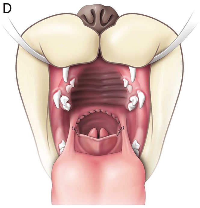
of a poor response to the previous surgical interven-                 Short- and long-term outcomes
tion or reoccurrence of clinical signs. No clinical                        Complications were observed in 51 (12.1%) dogs.
grades were available for these dogs before and after                 Major complications included respiratory distress
the first surgical intervention. Six dogs had alaplasty               (n = 9 [2.1%]), aspiration pneumonia (2 [0.5%]), signs
alone that was performed 3 to 12 months earlier with                  of heatstroke (2 [0.5%]), and the need for a temporary
no or only mild improvement and subsequent reoc-                      tracheostomy (1 [0.2%]). One dog was euthanized
currence of clinical signs soon after surgery. Seven                  because of severe aspiration pneumonia. Two other
dogs had a palatoplasty alone that was performed                      dogs had 1 episode each of aspiration pneumonia. Of
9 to 24 months earlier, and only 1 of these 7 dogs                    these, one dog had an episode 2 days after surgery,
                                                                      recovered quickly with oxygen supplementation
improved during the year following surgery before
                                                                      through a nasotracheal tube and medical support,
reoccurrence of clinical signs. One dog had had pala-                 and was discharged from the hospital 2 days later. The
toplasty performed 24 months before alaplasty had                     second dog died because of aspiration pneumonia
been performed with subsequent mild improvement                       6 months after surgery. Minor complications included
in clinical signs. This dog was admitted to the hos-                  vomiting (26 [6.1%]), regurgitation (16 [3.8%]), and
pital 12 months after alaplasty with grade 3 clinical                 nasal discharge (8 [1.9%]). Of the dogs with postop-
signs associated with the digestive and respiratory                   erative vomiting and regurgitation, 9 (2.1%) required
systems, extensive oral ulcers, and grade 2 laryngeal                 rehospitalization so medications could be admin-
collapse. The other 18 dogs had both palatoplasty                     istered IV rather than PO. These 9 dogs underwent
and alaplasty performed 5 to 48 months earlier; 11 of                 esophagogastroduodenoscopy before or after surgery
these dogs specifically had folded flap palatoplasty.                 to investigate the cause of the clinical signs associated
     Esophagogastroduodenoscopic and pharyngolar-                     with the digestive system and were discharged from
yngoscopic anomalies were summarized (Table 1).                       the hospital when vomiting and regurgitation had
All dogs had grade 2 to grade 3 clinical signs associ-                stopped for at least 24 hours. Considering the entire
                                                                      cohort of 423 dogs, median duration of hospitaliza-
ated with the respiratory system. H-pharyngoplasty
                                                                      tion was 1 day (range, 12 hours to 9 days), with most
was successfully performed in all dogs, including
                                                                      dogs (365 [86.3%]) discharged 1 day after surgery.
those that had previously undergone surgery else-                          A total of 341 (80.6%) dog owners completed the
where, and no dog required revision surgery during                    questionnaire at the time of the final follow-up exami-
the long-term follow-up period. One Cavalier King                     nation (median, 36 months; range, 12 to 91 months).
Charles Spaniel with grade 2 respiratory clinical signs               No dog required revision surgery during the long-
only had an elongated soft palate with unremarkable                   term follow-up period. Most dog owners (331 [97.1%])
tonsils. The other 3 dogs that had unremarkable ton-                  were satisfied with the outcome of the surgery, and
sils had grade 2 to grade 3 respiratory signs. Eight                  most (336 [98.5%]) would recommend the surgery to
dogs underwent unilateral laryngeal sacculectomy.                     the owners of similarly affected dogs. After surgery,
                                                                      72.6% (244/336 dogs) showed improvement in respi-
                                                                      ratory signs (after vs before surgery: grade 1 or 2 vs
Table 1—Anomalies observed preoperatively during pha-                 grade 3, or grade 1 vs grade 2) and 34.1% (115/337
ryngolaryngoscopy (n = 423) and esophagogastroduodenos-               dogs) showed improvement in GI signs (after vs
copy (94) in dogs that had brachycephalic obstructive airway          before surgery: grade 1 or 2 vs grade 3, or grade 1 vs
syndrome.
                                                                      grade 2; Table 2).
Anomaly                                         No. (%) of dogs            Oxygen was supplemented through nasotracheal
Everted laryngealsaccules†                         397 (93.9)         tubes for 48 dogs before recovery from anesthesia.
Tonsillar eversion                                 419 (99.1)         Three of these dogs died postoperatively before
Elongated and hyperplastic soft palate‡            422 (99.8)
Grade of laryngeal collapse
                                                                      hospital discharge because of severe respiratory dis-
  1                                                125 (29.6)         tress, with 2 that had acute cardiorespiratory col-
  2                                                202 (47.8)         lapse before a tracheostomy could be performed and
  3                                                 70 (16.5)         unsuccessful resuscitation. A phase of overexcite-
  None                                              26 (6.1)
Esophagogastroduodenal lesion                       94 (22.2)
                                                                      ment followed by a heatstroke-like episode and car-
Lymphoplasmacytic esophagitis                       24 (5.7)          diorespiratory collapse were suspected. Both were
Lymphoplasmacytic gastritis                         38 (8.9)          French Bulldogs with preoperative grade 3 respira-
Severe chronic lymphoplasmacytic gastritis          47 (11.1)         tory signs and grade 2 digestive signs. The third dog
Lymphoplasmacytic duodenitis                        40 (9.5)
Severe chronic lymphoplasmacytic duodenitis         25 (5.9)
                                                                      was an obese Pug with preoperative grade 3 respi-
Helicobacter bacteria (stomach)                     22 (5.2)          ratory signs and grade 2 laryngeal collapse. Despite
Hiatal hernia                                        5 (1.2)          oxygen supplementation through the nasotracheal
                                                                      tube, this dog experienced severe respiratory distress
    The denominator for all percentage calculations is 423. †Eight
(1.9%) dogs also had granulomas on the surface of their laryngeal
                                                                      24 hours after surgery, and a tracheostomy was per-
saccules. ‡The 1 dog excluded from this group had only an elongated   formed. This dog died 48 hours after surgery because
soft palate.                                                          of intractable respiratory compromise. The other

                                              JAVMA | JAN 15, 2022 | VOL 260 | NO. S1                                           S69

                                                                                           Unauthenticated | Downloaded 02/03/22 07:06 PM UTC
Table 2—Grades for clinical signs associated with the respiratory and gastrointestinal (GI) systems.
Respiratory signs GI signs
No. (%) with
No. (%) with exercise No. (%) with No. (%) with No. (%) with No. (%) with
Variable snoring intolerance syncope regurgitation vomiting ptyalism
Preoperative grade*
1 15 (4) 63 (14.9) 331 (78.3) 333 (79) 315 (74.5) 325 (76.8)
2 107 (25) 161 (38.1) 0 (0) 46 (11) 66 (15.6) 58 (13.7)
3 301 (71) 199 (47) 92 (21.7) 44 (10) 42 (9.9) 40 (9.5)
Postoperative grade†
1 223 (66.4) 154 (45.8) 336 (100) 281 (83.4) 276 (81.9) 242 (84.3)
2 88 (26.2) 122 (36.3) 0 (0) 31 (9.2) 54 (16) 36 (10.7)
3 25 (7.4) 60 (17.9) 0 (0) 25 (7.4) 7 (2.1) 17 (5)
Difference‡
2 162 (48.2) 61 (18.2) 70 (20.8) 16 (4.7) 17 (5) 21 (6.2)
1 114 (33.9) 128 (38.1) 0 (0) 40 (11.9) 50 (14.8) 42 (12.5)
0 51 (15.2) 101 (30.1) 266 (79.2) 245 (72.7) 239 (70.9) 234 (69.4)
–1 9 (2.7) 35 (10.4) 0 (0) 23 (6.8) 29 (8.6) 28 (8.3)
–2 0 (0) 11 (3.3) 0 (0) 13 (3.9) 2 (0.6) 12 (3.6)
Adjusted difference grades
1 276 (82.1) 189 (56.3) 70 (20.8) 56 (16.6) 60 (19.8) 63 (18.7)
–1 or 0 60 (17.9) 147 (43.7) 266 (79.2) 281 (83.4) 270 (80.2) 274 (81.3)
*Data represent 423 dogs. †Data represent 336 dogs for respiratory signs and 337 dogs for gastrointestinal signs. ‡Difference between pre-
and postoperative respiratory signs determined for 336 dogs and between pre- and postoperative gastrointestinal signs for 337 dogs.
45 dogs were discharged from the hospital after 1 to 8.63; 95% CI, 4.52 to 16.47; P < 0.001). Furthermore,
4 days of oxygen supplementation. the higher the preoperative snoring grade, syncope
No dogs died intraoperatively. Death occurred was more likely to improve postoperatively (adjusted
before hospital discharge in 5 of 423 (1.2%) dogs; OR, 1.99; 95% CI, 1.08 to 3.67; P = 0.03).
besides the 3 dogs that received supplemental oxygen Dogs that had higher preoperative grades for
through a nasotracheal tube, a fourth dog died follow- regurgitation (adjusted OR, 22.86; 95% CI, 8.96 to
ing an episode of respiratory distress, and 1 died for 58.32; P < 0.001; Table 2), vomiting (adjusted OR,
unknown reasons. An additional 6 (1.4%) dogs died 172.64; 95% CI, 35.89 to 830.49; P < 0.001), and ptya-
postoperatively between the time of hospital dis- lism (adjusted OR, 117.52; 95% CI, 29.68 to 465.34;
charge and the first follow-up examination because of P < 0.001) were more likely to show improvement in
respiratory distress (n = 2), an unknown cause (2), these signs postoperatively. Regurgitation was more
aspiration pneumonia (1), or vasculitis at the base of likely to improve postoperatively in dogs with higher
the tongue (1). The overall mortality rate was 2.6% preoperative ptyalism grades (adjusted OR, 2.51; 95%
(11/423). In the long-term follow-up period, 22 dogs CI, 1.07 to 5.85; P = 0.03) but less likely to improve
died, with only 3 that died for reasons related to BOAS. postoperatively in dogs with higher laryngeal col-
lapse grades (adjusted OR, 0.12; 95% CI, 0.02 to 0.69;
Prognostic factors P = 0.02). No other variables were associated with
Results from the bivariable and multivariable improvement in respiratory and digestive clinical
analyses are summarized (Supplementary Tables signs in the other multivariable analyses.
S1 and S2). From the multivariable analyses, snoring Results from the bivariable analyses for postop-
was less likely to improve in Pugs after surgery than erative survival were summarized (Supplementary
in French Bulldogs (adjusted OR, 0.32; 95% CI, 0.14 to Table S3). Perioperative death and death before
0.72; P = 0.006). Dogs that had higher preoperative and after the first reexamination were considered
snoring grades were more likely to show improve- together. Only age was significantly (P < 0.001) asso-
ment in snoring postoperatively (adjusted OR, 10.15; ciated with an increased risk of death, such that the
95% CI, 5.06 to 20.37; P < 0.001). Exercise toler- risk of death increased by 29.8% (95% CI, 11.8% to
ance was less likely to improve after surgery in Pugs 50.7%) as a dog’s age increased by 1 year. Sex, body
(adjusted OR, 0.37; 95% CI, 0.19 to 0.71; P = 0.003) weight, breed, preoperative respiratory and digestive
and English Bulldogs (adjusted OR, 0.06; 95% CI, 0.01 system signs, and presence and grade of laryngeal
to 0.29; P = 0.001) than in French Bulldogs, but as collapse did not significantly impact the risk of death.
body weight increased for English Bulldogs, exercise
tolerance was more likely to improve after surgery
(adjusted OR, 1.18; 95% CI, 1.06 to 1.31; P = 0.003).
Discussion
Dogs that had higher preoperative exercise intoler- The SMA was found to be applicable to all dog
ance grades were more likely to show improvement breeds, regardless of their grades, for management
in exercise tolerance postoperatively (adjusted OR, of their clinical signs associated with the respiratory
S70 JAVMA | JAN 15, 2022 | VOL 260 | NO. S1
Unauthenticated | Downloaded 02/03/22 07:06 PM UTCand digestive systems, and the SMA resulted in cut-and-sew staphylectomy and vertical wedge rhino-
improvement in respiratory signs in most dogs. plasty, but the results of that study are questionable
Although 65.5% (220/336 dogs) still had postopera- because rhinoplasty and cut-and-sew staphylectomy
tive respiratory grades of 2 or 3, most dogs (72.6% alone may not have been sufficient for some dogs
[244/336]) had improved respiratory signs, complica- with BOAS, as discussed previously, and persistent
tion rates were low (12.1%, including 2.6% mortality airway obstruction may lead to persistent laryngeal
rate), and owner satisfaction was high (97.1%). The edema and everted laryngeal saccules. Moreover,
design of the surgical technique, H-pharyngoplasty, the everted laryngeal saccules were edematous in all
which included bilateral tonsillectomy and extensive 10 dogs (moderate, n = 4; severe, 3) and mildly
staphylectomy, allowed the soft palate to be short- fibrotic in only 4 dogs (mild, 3; moderate, 1). Thus,
ened and may have optimized relief from oro- and everted laryngeal saccules may not be irreversible
nasopharyngeal obstruction. The incision boundaries and better relief from airflow resistance and medi-
provided a large pharyngeal clearance area, and the cal support may improve or eliminate this condition.
plasty allowed the redundant pharyngeal mucosa to Another recent study26 shows that morbidity and
be flattened (ie, following H-pharyngoplasty, a defect duration of hospitalization increased for dogs fol-
is created in the pharyngeal mucosa, and suture puts lowing laryngeal sacculectomy (vs those that did not
the mucosa under slight tension such that the redun- undergo laryngeal sacculectomy). Yet, not all dogs
dant mucosal fold flattens). in that study also had staphylectomies and therefore
Preoperative findings, postoperative improve- increased morbidity and duration of hospitalization
ment, and complication rates in the present study may not be specifically attributable to the sacculecto-
were similar to those previously reported.1,3,5,7,18,22,23 mies. Furthermore, the benefits of laryngeal sacculec-
Tonsillectomy has been shown to be a safe proce- tomy alone are unknown; therefore, on the basis of
dure with low complication rates.1,7,23 Previous stud- the available data and authors’ experiences, everted
ies5,15,17,24,25 also reveal that redundant pharyngeal laryngeal saccules were not systematically removed.
mucosa contributes to airway obstruction. In a recent Dogs with mild anomalies (eg, soft palate either
study,1 conventional multilevel surgery has an 8-fold only elongated or only thickened, or normal tonsils)
higher risk of a poor prognosis versus modified multi- had moderate to severe preoperative clinical signs.
level surgery. The modified multilevel surgery involves Conversely, some dogs with severe anomalies had
vestibuloplasty, partial cuneiformectomy, and folded mild preoperative clinical signs. These dogs may
flap palatoplasty. The benefit of partial cuneiformec- have had favorable results with conventional treat-
tomy was questioned by the authors of that study,1 ment but considering the severity of their clinical
and they could not determine whether the procedure signs or anomalies, respectively, they underwent an
was effective in dogs affected by BOAS. Conversely, SMA and enrolled in the present study. However, dogs
vestibuloplasty and folded flap palatoplasty provide with mild anomalies and mild clinical signs may still
better relief from airway obstruction than alaplasty benefit from less extensive surgery. Visual inspection
alone and standard staphylectomy.1,2 Therefore, less and imaging of the lesions may be altered by anes-
aggressive surgeries, such as conventional multilevel thesia depth, head positioning, and the degree of
surgery, may lead to persistent airway obstruction or mouth opening and may have impacted grading of
airflow turbulence in the pharyngeal and laryngeal the lesions.
regions, resulting in poorer outcomes or postopera- Laryngeal collapse had no impact on respiratory
tive relapse of clinical signs. The SMA is also a multi- grade. This finding was consistent with the finding of
level surgery that involved extensive staphylectomy a previous study5 in which global improvement was
and bilateral tonsillectomy plus an original pharyn- noted following palatoplasty and alaplasty in dogs that
goplasty and vertical wedge alaplasty with vestibu- suffered from laryngeal collapse. H-pharyngoplasty
loplasty. Subjectively, SMA may optimize relief from seemed to improve the condition of dogs with severe
nasopharyngeal and oropharyngeal obstruction by laryngeal collapse.
thinning and shortening of the soft palate and by Each dog that was diagnosed with an inflamma-
resection of the tonsils and the action on the redun- tory GI disease on the basis of histologic examination
dant pharyngeal mucosa by the associated plasty, of GI tissue samples received prednisolone, metro-
respectively. This configuration may have explained nidazole, omeprazole, and a hydrolyzed diet in an
the favorable results in the dogs in the present study attempt to resolve and prevent recurrence of vomit-
but further studies, especially those that include diag- ing, regurgitation, and gastroesophageal reflux. The
nostic imaging, are warranted. acidic gastric content may have caused inflammation
Everted laryngeal saccules were not system- in the laryngeal and pharyngeal regions, which may
atically excised from the dogs in the present study. have contributed to airway obstruction. Some dogs
Only 8 laryngeal saccules (1 saccule/dog; 8 dogs) with severe GI signs, including extensive oral ulcers,
were removed because they had granulomas on their had important laryngeal and pharyngeal edema and
surface.26 This strategy is controversial, and sup- severe respiratory signs. Managing GI disease with
porting evidence is lacking. A 2012 study27 reveals appropriate medical treatment may improve both GI
that everted laryngeal saccules did not resolve after and respiratory signs and may prevent postoperative
JAVMA | JAN 15, 2022 | VOL 260 | NO. S1 S71
Unauthenticated | Downloaded 02/03/22 07:06 PM UTCcomplications.28,29 However, determining whether obtain objective results.30 This is demonstrated in
improvement in GI signs was because of medical the present study by the discrepancy between owner
treatment or upper airway surgical interventions was satisfaction (97.1%) and dogs with global improve-
difficult. Surgical interventions likely had a positive ment in their respiratory signs (72.6%). Yet, because
effect on GI signs; because medical treatment was each owner assessed their dog both pre- and post-
only for a relatively short-term period, GI signs would operatively with explanations for each clinical sign,
have likely recurred prior to the long-term follow-up the results obtained were directly comparable and
examination if surgical treatment did not have a posi- relatively reliable. Comparative studies coupled with
tive effect on GI signs. whole-body plethysmography or other diagnostic
When comparing pre- and postoperative grades tests are warranted to better understand BOAS and
for each respiratory and GI sign, grades tended to the optimal treatment.31,32 To avoid recall bias, ques-
decrease from grades 2 and 3 to grade 1. The number tionnaires completed by the owners whose dogs
of dogs with grade 2 snoring or with grade 2 vomit- had died at the time of the final follow-up examina-
ing was proportionally higher postoperatively, likely tion were not considered. This may have created an
because snoring and vomiting grades for a larger overestimation of the percentage of postoperative
number of dogs changed from grade 3 to grade 2, improvement, but this bias should be limited consid-
versus that changed grade 2 to grade 1. Some dogs ering that only 5 dogs died before hospital discharge
had worsening grades (3.3% for respiratory system and only 3 dogs died from their BOAS after hospital
signs and 6.5% for digestive system signs). These discharge. The retrospective nature of the study was
results may be explained by a limitation of the surgi- another limitation. Postoperative follow-up was not
cal procedure, in that the total amount of mucosa standardized as it would have been in a prospective
that can be safely excised from the pharyngeal area study. Lesions were only assessed through pharyngo-
is bounded. The lack of the sensitivity of the grading laryngoscopy and thoracic radiography; CT was not
system proposed by Poncet et al4 may be another performed. Dogs with no to mild improvement after
explanation. Approximately 27.4% and 65.8% of the SMA may have benefited from additional diagnostic
dogs did not have improved or had worse respira- imaging or surgery.
tory and digestive grades, respectively. Yet, most In conclusion, SMA that included the new tech-
had improved clinical signs despite unchanged
nique H-pharyngoplasty was appropriate for most
grades. For example, a dog that vomited < 1X/d pre-
dogs with BOAS, even those that had undergone pre-
operatively and vomited < 1X/mo postoperatively
vious surgeries, regardless of breed, age, or severity
was still classified as having grade 2 GI signs. Like-
of their respiratory and GI signs. The SMA was associ-
wise, dogs that presented with snoring several times
ated with an improvement in respiratory and GI signs
per day and constantly were classified grade 2 and
grade 3, respectively, despite only a slight difference and low morbidity and mortality rates. Prognostic fac-
between the grades. These facts may have caused tors were identified, and the provision of appropriate
bias in the statistical analysis. supportive care may help to decrease the complica-
Snoring and exercise intolerance were less tion rates.
likely to improve in Pugs, compared with French
Bulldogs, after surgery. This finding may be owing Acknowledgments
to the difference in conformation of the nasal cavi- No external funding was used for this study. The authors
ties between these breeds. Nasal cavity obstruction declare that there were no conflicts of interest.
was not addressed with SMA, and those dogs might Presented in part as an abstract at the 27th Annual Meeting of
have benefited from laser-assisted turbinectomy. Pugs the European College of Veterinary Surgeons, Athens, July 2018.
also had higher body condition scores than French The authors thank Loïc Desquilbet, Head of the Biologic and
Pharmaceutic Sciences Department of the National Veterinary
Bulldogs, and the higher body condition score may School of Alfort, for his assistance with the statistical analysis.
have affected improvement in snoring and exercise
tolerance. Regurgitation was less likely to improve
postoperatively in dogs with higher laryngeal col- References
lapse grades. Constant laryngeal obstruction caused 1. Liu NC, Oechtering GU, Adams VJ, et al. Outcomes and
by laryngeal collapse may lead to persistent increased prognostic factors of surgical treatments for brachyce-
intrathoracic negative pressure, which may facili- phalic obstructive airway syndrome in 3 breeds. Vet Surg.
2017;46:271–280.
tate regurgitation. Risk of death increased by 29.8%
2. Findji L, Dupré GP. Folded flap palatoplasty for treatment
as a dog’s age increased by 1 year, which may be of elongated soft palates in 55 dogs. Wien Tierarztl Mschr.
explained by more severe clinical signs at the time of 2008;95:56–63.
surgery with advanced age and less effective compen- 3. Dunié-Mérigot A, Bouvy B, Poncet C. Comparative use of CO2
satory mechanisms in older dogs. laser, diode laser and monopolar electrocautery for resec-
The main limitation of the present study was tion of the soft palate in dogs with brachycephalic airway
obstructive syndrome. Vet Rec. 2010;167:700–704.
that the clinical evaluation of the dogs was subjec- 4. Poncet CM, Dupre GP, Freiche VG, et al. Prevalence of gastro-
tive, which is inherent to most studies on BOAS, and intestinal tract lesions in 73 brachycephalic dogs with upper
use of questionnaires is not a reliable technique to respiratory syndrome. J Small Anim Pract. 2005;46:273–279.
S72 JAVMA | JAN 15, 2022 | VOL 260 | NO. S1
Unauthenticated | Downloaded 02/03/22 07:06 PM UTC5. Torrez CV, Hunt GB. Results of surgical correction of abnor- brachycephalic obstructive airway syndrome. Vet Surg.
malities associated with brachycephalic airway obstruc- 2019;48:79–87.
tion syndrome in dogs in Australia. J Small Anim Pract. 20. Leonard HC. Collapse of the larynx and adjacent structures
2006;47:150–154. in the dog. J Am Vet Med Assoc. 1960;137:360–363.
6. Brdecka DJ, Rawlings CA, Perry AC, et al. Use of an elec- 21. Bursac Z, Gauss CH, Williams DK, et al. Purposeful selection
trothermal, feedback-controlled, bipolar sealing device for of variables in logistic regression. Source Code Biol Med.
resection of the elongated portion of the soft palate in dogs 2008;3:17.
with obstructive upper airway disease. J Am Vet Med Assoc. 22. Dupré G, Heidenreich D. Brachycephalic syndrome. Vet Clin
2008;233:1265–1269. North Am Small Anim Pract. 2016;46:691–707.
7. Cook DA, Moses PA, Mackie JT. Clinical effects of the use of a 23. Belch A, Matiasovic M, Rasotto R, et al. Comparison of the
bipolar vessel sealing device for soft palate resection and ton- use of LigaSure versus a standard technique for tonsillectomy
sillectomy in dogs, with histological assessment of resected in dogs. Vet Rec. 2017;180:196.
tonsillar tissue. Aust Vet J. 2015;93:445–451. 24. Monnet E. Brachycephalic airway syndrome. In: Slatter D,
8. Michelsen J. Use of the harmonic scalpel for soft palate resec- ed. Textbook of Small Animal Surgery. 3rd ed. Saunders;
tion in dogs: a series of three cases. Aust Vet J. 2011;89:511–514. 2003:808–813.
9. Ellison GW. Alapexy: an alternative technique for repair of ste- 25. Pink JJ, Doyle RS, Hughes JML, et al. Laryngeal collapse
notic nares in dogs. J Am Anim Hosp Assoc. 2004;40:484–489. in seven brachycephalic puppies. J Small Anim Pract.
10. Harvey C. Upper airway obstruction surgery part II: soft 2006;47:131–135.
palate resection in brachycephalic dogs. J Am Anim Hosp 26. Hughes JR, Kaye BM, Beswick AR, et al. Complications fol-
Assoc. 1982;18:538–544. lowing laryngeal sacculectomy in brachycephalic dogs.
11. Brdecka D, Rawlings C, Howerth E, et al. A histopatho- J Small Anim Pract. 2018;59:16–21.
logical comparison of two techniques for soft palate resec- 27. Cantatore M, Gobbetti M, Romussi S, et al. Medium-term
tion in normal dogs. J Am Anim Hosp Assoc. 2007;43: endoscopic assessment of the surgical outcome following
39–44. laryngeal saccule resection in brachycephalic dogs. Vet Rec.
12. Davidson EB, Davis MS, Campbell GA, et al. Evaluation of car- 2012;170:518.
bon dioxide laser and conventional incisional techniques for 28. Poncet CM, Dupre GP, Freiche VG, et al. Long-term results
resection of soft palates in brachycephalic dogs. J Am Vet of upper respiratory syndrome surgery and gastrointestinal
Med Assoc. 2001;219:776–781. tract medical treatment in 51 brachycephalic dogs. J Small
13. Vilaplana Grosso F, Ter Haar G, Boroffka SA. Gender, weight, Anim Pract. 2006;47:137–142.
and age effects on prevalence of caudal aberrant nasal tur- 29. Fenner JVH, Quinn RJ, Demetriou JL. Postoperative regurgi-
binates in clinically healthy English bulldogs: a computed tation in dogs after upper airway surgery to treat brachyce-
tomographic study and classification. Vet Radiol Ultra- phalic obstructive airway syndrome: 258 cases (2013–2017).
sound. 2015;56:486–493. Vet Surg. 2020;49:53–60.
14. Bernaerts F, Talavera J, Leemans J, et al. Description of origi- 30. Packer R, Hendricks A, Burn C. Do dog owners perceive the
nal endoscopic findings and respiratory functional assess- clinical signs related to conformational inherited disorders
ment using barometric whole-body plethysmography in as “normal” for the breed: a potential constraint to improving
dogs suffering from brachycephalic airway obstruction syn- canine welfare. Anim Welf. 2012;21:81–93.
drome. Vet J. 2010;183:95–102. 31. Villedieu E, Rutherford L, Ter Haar G. Brachycephalic
15. Penfold MJ, Van Der Zee J, Hartman MJ. Laryngopharyngeal obstructive airway surgery outcome assessment using
mucosal fold causing upper airway obstruction in a dog. Vet the 6-minute walk test: a pilot study. J Small Anim Pract.
Rec Case Rep. 2019;7:1–4. 2019;60:132–135.
16. Jones BA, Stanley BJ, Nelson NC. The impact of tongue 32. Riggs J, Liu N, Sutton DR, et al. Validation of exercise test-
dimension on air volume in brachycephalic dogs. Vet Surg. ing and laryngeal auscultation for grading brachycephalic
2020;49:512–520. obstructive airway syndrome in pugs, French Bulldogs, and
17. Poncet C, Freiche V. Brachycephalic airway obstruction syn- English Bulldogs by using whole-body barometric plethys-
drome. In: Bonagura JD, Twedt DC, eds. Kirk’s Current Vet- mography. Vet Surg. 2019;48:488–496.
erinary Therapy XV. Elsevier; 2014:649–653.
18. Oechtering GU, Pohl S, Schlueter C, et al. A novel approach
to brachycephalic syndrome. 2. laser-assisted turbinectomy
(LATE). Vet Surg. 2016;45:173–181.
Supplementary Materials
19. Liu NC, Genain MA, Kalmar L, et al. Objective effective- Supplementary materials are posted online at the journal
ness of and indications for laser-assisted turbinectomy in website: avmajournals.avma.org
JAVMA | JAN 15, 2022 | VOL 260 | NO. S1 S73
Unauthenticated | Downloaded 02/03/22 07:06 PM UTCYou can also read