Age-related hearing loss in dogs and treatment with Vibrant Soundbridge middle ear implant
←
→
Page content transcription
If your browser does not render page correctly, please read the page content below
© Veterinary Sciences Tomorrow November 2010

Age-related hearing loss in dogs and treatment
with Vibrant Soundbridge middle ear implant
   Gert ter Haar

Hearing and hearing loss
Hearing is a fascinating phenomenon and a very important sensory function in humans and
animals. Purves described the auditory system as one of the engineering masterpieces of the
body, with an array of miniature acoustical detectors packed into a space no larger than a
pea. Although humans are highly visual creatures, much of human communication,
socializing, learning, and listening is mediated by the auditory system and indeed, under
some circumstances, deafness can be even more debilitating than blindness [25]. It is much
more difficult to establish the impact of partial hearing loss or complete deafness in dogs than
in humans.
Hearing loss is a common disorder in many breeds of dogs and auditory dysfunction and its
clinical consequences can vary from mild to severe. Dogs with unilateral hearing loss can
have difficulty in localizing the source of a sound [33]. They are not suited as working dogs for
blind and deaf people, or rescue and police work, but are not themselves severely
handicapped. However, dogs with bilateral hearing loss are unable to anticipate dangers such
as motor vehicles and they may consequently fall victim to serious or fatal injury [33]. In
addition, they seem to be easily startled and have an increased tendency to bite.
Furthermore, deaf puppies require specialized training and are therefore usually euthanized.
Acquired hearing loss in humans is receiving growing attention because of its detrimental
effects on the affected individual’s psychosocial situation, including social isolation,
depression, and loss of self-esteem. Hearing impairment has also been implicated as a
cofactor in senile dementia [8]. The psychosocial effects of hearing impairment in dogs are
not known, but undoubtedly hearing loss contributes to the lethargy, depression, and lack of
interest in interaction with the environment that is commonly observed in old dogs [12].
Another serious behavioral side effect of acquired deafness in dogs is exaggerated barking
[12]. Similar to acquired hearing loss in humans, which affects not only the quality of life of
those with hearing loss but also that of their loved ones, it is to be expected that hearing
disabilities in dogs strongly affect the intimate relationship with the owner, due to the
interactive nature of the habitual vocal communication [10]. Timely recognition and correct
diagnosis of hearing impairment are therefore mandatory for both audiological rehabilitation of
the patient and counseling of the owner.
Much less is known about hearing impairment, hearing capacity, frequency range of hearing,
and threshold audiograms in dogs than in humans and laboratory animals. This is due to the
unpopularity of dogs as a model in hearing research, problems associated with behavioral
testing, and the fact that objective hearing tests were only introduced in veterinary medicine
rather recently. As in humans, hearing loss in dogs can have central or peripheral causes.
Central deafness can theoretically result from a variety of retrocochlear lesions but is very
rare in veterinary practice [33]. Bilateral central deafness requires bilateral lesions of the
auditory cortex or lesions of such a significant portion of the brainstem or midbrain that
significant clinical signs beyond deafness are to be expected [33].
Peripheral hearing loss in dogs is much more common and has been classified as inherited or
acquired, conductive or sensorineural, and congenital or late onset [33]. The most frequently
observed forms are acquired conductive hearing loss as a result of chronic otitis externa and
media, congenital (inherited) sensorineural hearing loss (SNHL) and acquired SNHL including
age-related hearing loss (ARHL) or presbycusis, noise-induced hearing loss (NIHL), and
ototoxicity [33]. With a thorough physical examination including otoscopy (video 1), the
differentiation between conductive and sensorineural hearing loss can usually be determined
[36]. Advanced imaging with CT or MRI is necessary, however, to definitely rule out
conduction deafness and to devise a plan for treatment. Though essential for the diagnostic
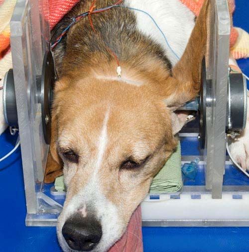
work-up of all patients with hearing disorders, these two techniques can only identify
morphological abnormalities of the petrous bone, middle ear, and inner ear. Diagnosis of
functional abnormalities requires hearing tests.

Video 1. Demonstration of an ear examination in a healthy dog with inspection and palpation of the auricle and ear
canal including otoscopy, showing a normal ear canal with intact tympanic membrane.

Hearing assessment
Hearing evaluation in humans can be relatively straightforward, using behavioral tests in
which a variety of tones with different levels of loudness are presented. Albeit prone to some
subjectivity, in addition to hearing a specific tone the person being tested can describe the
sensation of hearing or the lack of it using this technique [25]. Behavioral studies have been
performed on a very small scale in dogs [9] and although the sensation of hearing cannot be
determined in this species, studies have shown that dogs can hear frequencies up to 45 kHz,
which is considerably higher than heard by humans [9]. Training dogs to respond reliably and
repetitively to different auditory stimuli is time-consuming, even for research purposes, and is
therefore not applicable to veterinary practice, where client-owned patients with hearing
disabilities have to be examined (video 2, 3).

Video 2. Behavioural assessment of a dog using a whistle demonstrating a lack of response after auditory
stimulation, but a clear response on tactile stimulation indicating that the dog is deaf.

Video 3. Behavioural assessment of a dog using a whistle demonstrating an immediate response after a soft auditory
stimulation and the poor reproducibility of the test because of lack of interest of the dog.
Hearing can be assessed with greater objectivity by several methods, including impedance
audiometry, evoked response audiometry, and electrocochleography. Brainstem evoked
response audiometry (BERA), initially developed for hearing assessment in very young
children when behavioral tests could not be performed, is the technique most commonly used
in veterinary medicine. With this technique, the consistent changes in electrical activity in the
brainstem following auditory stimulation can be recorded from scalp electrodes [13] (figure 1).
Although clinically relevant information can be obtained with BERA, this technique only
evaluates the functional integrity of the peripheral auditory system but not sound awareness.
It must be borne in mind that hearing is a much more complex phenomenon than can be
appreciated by hearing assessment with BERA.
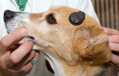
Figure 1. Recording of BERA with the dog in sternal recumbency under a light plane of anesthesia. Three recording
electrodes are inserted subcutaneously. Stimulations are delivered to the ear via a flexible transmission tube into the
vertical ear canal.

Causes of hearing loss
Conductive hearing loss in dogs is usually the result of chronic otitis externa and media and is
therefore amenable to treatment. The diagnostic work-up and treatment of these patients and
the use of BERA for diagnosis of the associated conductive hearing loss and for evaluation of
the effects of medical treatment and surgery on hearing have been reviewed in the veterinary
literature [20] [43]. Congenital sensorineural deafness is the most extensively studied form of
deafness in dogs (especially Dalmatians) and many studies have been published in the past
20 years on the etiopathogenesis of this disease and its diagnosis with BERA [11] [34]. The
acoustic signal used to diagnose congenital deafness usually consists of a click, which is a
short sound containing many frequency components, thus stimulating a large part of the
cochlea. Brainstem evoked response audiometry using click stimulation is useful for
differentiating sensorineural from conduction deafness and for demonstrating complete
deafness as is the case in the congenital inherited form in dogs. However, frequency-specific
information is needed to assess the extent of sensorineural deafness and its possible origin,
such as NIHL, ototoxicity, and ARHL, each of which can be partial and frequency specific.
Acquired SNHL in dogs has received little attention in the veterinary literature, with the
exception of that due to ototoxicity. There have been several reports on ototoxicity in dogs,
demonstrating the effect on hearing of commonly used ototoxic agents and stressing the
importance of early detection using BERA [7] [33]. There have been no reports on NIHL in the
veterinary literature and but few on ARHL [14] [15] [27], nor have there been any reports of
frequency-specific thresholds for these forms of acquired hearing loss in dogs.
Acquired sensorineural hearing loss (SNHL) is a very common cause of hearing impairment
in dogs, second only to congenital SNHL [33], yet it has received little attention in the
veterinary literature. The diagnosis of acquired, frequency-specific hearing impairment
requires objective electrophysiological tests that evaluate the entire audible frequency range
in dogs. The technique most commonly used in veterinary medicine for hearing assessment
has been brainstem-evoked response audiometry (BERA) using click stimuli (CS) [35].
However, clicks stimulate a large portion of the cochlea and are therefore not frequency
specific, which renders this technique unsuitable for the diagnosis of acquired SNHL. A
method was developed in our laboratory for frequency-specific assessment of the cochlea in
dogs using BERA with tone burst stimulation (TS). Tone bursts ranging from 1 to 32 kHz were
created and used in this study [37]. Our results demonstrate that the greatest sensitivity of the
dog’s ear is at 12 and 16 kHz, which is in very close agreement with results of behavioral
tests [9] (figure 2). Various methods could have been used to test frequency-specific areas of
the cochlea, such as the use of CS with high-pass noise or notch-noise masking, or the use
of direct stimulation with tone bursts. The latter was selected for this study because it had
been shown to be easy to perform and to yield reliable information about pure tone thresholds
[21]. A concern in using tone bursts is spectral splatter [28]. This means that the BERA
threshold obtained using tone bursts is not solely determined by the response of the neurons
at the nominal frequency but also that of neurons at the side lobe frequencies. In patients with
hearing loss, this could lead to an underestimation of the loss at the nominal frequency of the
tone burst [28]. Based on our results, however, and their close agreement with results of
behavioral tests, we concluded that reproducible information on frequency specificity of the
canine cochlea can be obtained by TS and that this report provides a normative database
with stimulus variables needed to evaluate frequency-specific hearing loss in dogs.

Figure 2. Threshold audiogram showing mean threshold values of 10 healthy dogs with a mean age of 6 years. X =
left ear, O = right ear. The standard deviation of mean threshold values ranges between 6.7 and 24.6 dB.
Another technique was reported recently for measuring frequency-specific thresholds in dogs,
using auditory steady-state evoked potentials [18]. This appears to be a valid method but
frequencies above 8 kHz were not tested. It may be found to be applicable when data at
higher carrier frequencies are available. In addition to BERA, the use of otoacoustic
emissions (OAEs) has been advocated to assess cochlear function in humans [31]. OAEs
can not only be obtained quickly but can also be applied very objectively to evaluate outer
hair cell integrity. They can, for example, identify ototoxicity earlier than BERA [31]. In dogs,
valid spontaneous and evoked OAEs have been recorded, but the reports are scarce and the
clinical applicability and usefulness in frequency-specific hearing assessment in dogs has yet
to be demonstrated [29] [31].
Age related hearing loss in dogs
It is generally assumed that the hearing of dogs becomes impaired with advancing age, yet
there are few reports to support this. ARHL is claimed to be the most frequent cause of
acquired SNHL in dogs [33] and reports of decreased hearing in aged dogs have been
documented by auditory brainstem-evoked responses, but using CS or one stimulus level
only [14] [15] [27]. No thresholds have been reported in aged dogs with impaired hearing by
use of either CS or TS. We hypothesized that hearing in dogs, as in humans, becomes
impaired with ageing across the entire frequency range, but primarily in the high-frequency
area, and can be assessed quantitatively by BERA using TS. The results verifying this
hypothesis are presented in the form of a cross-sectional and a longitudinal study of age-
related changes in audiograms in dogs obtained by the technique are described [37]. The
most important conclusion of our cross-sectional study is that auditory thresholds at all
frequencies tested were significantly higher in geriatric than in young and middle-aged dogs
and were likely to be the result of presbycusis (figure 3). Although differences were significant
at all frequencies, the most dramatic increase in thresholds was seen at middle to high
frequencies (8-32 kHz) [38], which is similar to that in human presbycusis [5] [16].

Figure 3. Tone audiogram indicating mean threshold values in 3 groups of 10 dogs: group I (solid lines) mean age
1.9 years, group II (dashed lines) mean age 5.7 years, group III (dotted lines) mean age 12.7 years. The standard
deviation of the data sets (mean thresholds for 1–32 kHz) ranged between 5.2–12.2 for group I, between 6.7–24.6 for
group II, and between 11.0–15.6 for group III (X = left ear, O = right ear).
The audiograms of the dogs included in the longitudinal study show a progressive increase in
thresholds associated with ageing, starting around 8-10 years of age and being most
pronounced in the middle- to high-frequency region (8-32 kHz) (figure 4). Thresholds were
significantly higher at a mean age of 12 years than at a mean age of 6 years for 8, 12, 16, 24,
and 32 kHz (P < 0.05). The average increase between 10 and 12 years of age was around 10
dB at 1, 2, and 4 kHz, and ranged from 15 dB at 8 kHz to over 22 dB at 12, 16, 24, and 32
kHz [38]. This difference between low and high frequencies is also observed in longitudinal
studies in presbycusis in humans [5] [16] From these studies, it appears that a significant
reduction in hearing capacity occurs from the age of 60 years onward and begins at the high
frequencies (6-16 kHz), but gradually encompasses the entire frequency range. Furthermore,
between the ages of 70 and 80 years the reduction in hearing amounts to 1-2 dB per year,
depending on the specific frequency tested [5] [16]. The threshold increases which we
observed in dogs are 10 times greater than those reported in humans. This may in part be
explained by technical differences between studies (for practical purposes we decreased
stimulus intensity in steps of 10 dB), and in part by differences between species. Dogs having
a much shorter lifespan, any age-related deterioration might occur at a more rapid rate than in
man.
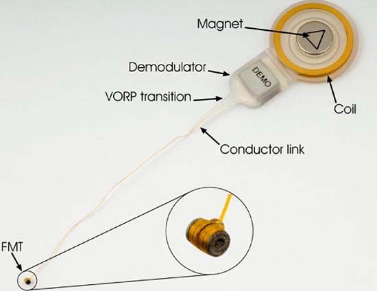
Figure 4. Longitudinal tone audiograms of the left ear of 1 dogs from the longitudinal group at octave frequencies
from 1 to 32 kHz. This dog entered the study at 7 years of age.
The cross-sectional study also revealed a significantly higher threshold in the middle-aged
dogs than in the young dogs at 4 kHz, while the thresholds at other frequencies did not differ
[38]. Kennel noise exposure might be responsible for this finding. The middle-aged dogs had
been housed in our facilities during their entire lives and were thus exposed to the noise of
their own barking. NIHL in humans typically occurs in the 2-6 kHz range, with initial changes
at 6 kHz and the greatest increase in threshold at 4 kHz [16] [23]. This suggests that the
increased threshold in the middle-aged dogs at 4 kHz could have been the result of noise
damage. These dogs had been used in the first study [35] to establish normative data. In
retrospect, they cannot be considered to be healthy, normal-hearing dogs, since at 4 kHz they
had a significantly higher threshold than a group of very young dogs. However, since there
was a significant difference in auditory thresholds between the geriatric and young dogs, and
over time within the group of middle-aged dogs, we conclude that presbycusis does exist in
dogs, as in humans. The impairment can be demonstrated and its progression can be
followed using BERA with frequency-specific, tone burst stimulation of the cochlea [38].
Our observations and conclusions are derived from a small group of medium-sized dogs and
therefore not necessarily applicable to all dogs. Future studies should be carried out on a
larger scale in client-owned dogs, not housed in kennels and not exposed to loud recreational
and environmental noise, to determine the actual prevalence of this disorder. To demonstrate
or exclude differences in effects of ageing on hearing between dogs of different sizes, with
different lifespans, several more groups would have to be included in these studies. In future
studies it would also be advisable to combine BERA with measurements of otoacoustic
emissions, as discussed above.

Histology of the inner ear of dogs with hearing loss
Schuknecht proposed that the histological form of ARHL can be determined from the shape of
the audiogram [26]. Age-related loss of hair cells and spiral ganglion cells (SGCs) has been
reported in dogs [15] [27]. Knowles et al. found loss of SGCs in a group of deaf dogs in which
auditory thresholds were completely absent [15] and Shimada et al. reported varying degrees
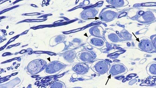
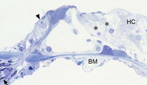
of loss of SGCs, and atrophy of the organ of Corti and the stria vascularis, in all dogs over 12
years of age, predominantly in the basal turn of the cochlea [27]. Although the latter findings
would seem to imply a high-frequency hearing loss of the mixed type in dogs, comparable to
that found in most cases of human ARHL, frequency-specific hearing thresholds were not
determined. Having found audiometric proof of frequency-specific age-related changes in
hearing in dogs, we further hypothesized that cochlear lesions in dogs with ARHL would be
similar to those in humans as well, and that the severity of the histological changes would be
reflected in tone audiograms.
The results are published of a study on age-related changes in the cochlea in relation to
BERA-derived thresholds [39]. Cochlear lesions were found in all 10 geriatric dogs studied.
Not only was significant loss of SGCs found in this study, but also of outer hair cells (OHCs)
and a reduction in stria vascularis cross-sectional area (SVCA) (figure 5a, b). Histological
abnormalities were found primarily in the basal turn, consistent with the occurrence of the
largest threshold shifts and highest absolute BERA-derived thresholds in the middle- to high-
frequency regions (8-32 kHz). It was concluded that the degeneration of the OHCs and SGCs
observed in the basal turn was primarily responsible for the elevated hearing thresholds,
similar to findings in humans [8]. The SVCA was smaller in all cochlear turns in the geriatric
dogs, similar to gerbils, where degeneration of the stria vascularis is an early event in ARHL,
usually beginning at both the base and apex and extending to midcochlear regions with
advancing age [33]. While the reduction in SVCA in all turns could explain the loss of hearing
sensitivity over the entire frequency range in the geriatric dogs, it could not explain the
difference between low- and middle-to-high-frequency loss. A mixture of lesions—OHC loss,
SGC loss, and reduction in SVCA—thus seemed to be the best explanation for the
audiometric results. Our hypothesis that cochlear lesions would be similar to those found in
humans with ARHL, which is most often the mixed type [22], was therefore accepted [39].

Figure 5a. Midmodiolar section through the upper basal turn (B2) of the cochlea of a geriatric dog. In the organ of
Corti the OHCs have been replaced by supporting cells (asterisks). The IHC (arrowhead) is still present. Note that the
number of nerve fibers in the osseous spiral lamina is reduced (arrow). (BM) basilar membrane, (HC) Hensen’s cells.
Figure 5b. In the spiral ganglion there is obvious loss of SGCs and nerve fibers. In the remaining SGCs there is
shrinkage of the perikarya (arrowheads), intracellular vacuolation, and lipofuscin inclusions (arrows).
Despite Schuknecht’s statements, several studies have failed to show a close
correspondence between audiometric data and cochlear pathology and this study was no
exception [40]. The auditory thresholds found in these dogs did not indicate whether the
histological abnormalities were primarily sensory, neural, or strial in nature. This is most likely
due to the fact that there were mixed lesions in all cases, with variable degrees of OHC loss,
IHC loss, SGC loss, and reduction in SVCA. Individual audiograms did, however, reflect the
severity and location of cochlear lesions in all cases. In general, cochlear lesions were more
extensive in dogs with more advanced hearing loss. It was concluded that tone audiograms
could therefore be used to diagnose and characterize ARHL in dogs, for they not only
indicated the severity of hearing loss but also the extent and location of the mixed cochlear
lesions [39].
Electron microscopic examination of the stria vascularis and the SGCs will be needed to
determine the exact nature and importance of the observed microscopic abnormalities in the
cochleas of the geriatric dogs, such as the abundant lipofuscin inclusions and intracellular
vacuoles in the remaining SGCs. Furthermore, although there is consensus that the cochlea
is the site of ARHL, the brain is the ultimate organ of hearing and perception, and the central
auditory system is also known to be affected by ageing [42]. Neurophysiological studies of
cochlear nucleus neurons have shown lower glycine-mediated inhibition, reflected in
increased firing rates in cochlear nucleus neurons from old animals relative to young adults.
In addition, anatomical reductions in neurons of the cochlear nucleus and their output
pathways can occur due to ageing changes in the brain, as well as due to age-dependent
plasticity of the cochlear nucleus in response to the age-related loss of inputs from the
cochlea, particularly from the basal, high-frequency regions [42]. Nerve cell loss, astrogliosis,
and ubiquitin deposition were found in cochlear nuclei of dogs over 10 years of age [27]. Age-
related alterations in GABA synthesis and release are found in the auditory midbrain (superior
olivary complex and inferior colliculus) and primary auditory cortex of humans [3]. It is not
known whether they are primarily the result of ageing of the central pathways or secondary to
peripheral pathology, nor whether they also occur in dogs.

Hearing aid efficacy in dogs
Effective treatment of presbycusis is important for quality of life in geriatric medicine, and
modern hearing aids, middle ear implants, and cochlear implants are valuable aids to
communication for elderly people with hearing loss [8]. Most people with ARHL are fitted with
a conventional hearing aid, but the use of these in dogs has been mentioned only anecdotally
[19], and has not met with much clinical success. People with mild to severe SNHL who do
not benefit from conventional external amplification benefit greatly from implantable auditory
prostheses [6]. There have been no reports on the use of implants in hearing-impaired dogs.
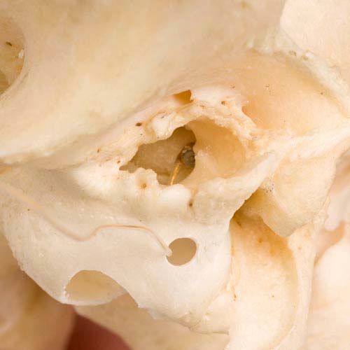
Of the available options, fully implantable systems are of particular interest because there is
less likelihood of loss of the device, occlusion of the ear canal, or induction of otitis externa
with these implants than with nonimplanted hearing aids [1] [6]. However, fully implantable
devices were not commercially available at the start of this study and semi-implantable
systems had to be used. Nevertheless it is expected that continuing technological innovations
will enable conversion to fully implantable systems in the foreseeable future.

Middle ear hearing device
The Vibrant Soundbridge (VSB) is an active, semi-implantable, middle ear hearing device that
has been available in Europe for several years and there have been many reports of its
effectiveness [6] [30]. The VSB consists of an external audio processor (AP) and an
implantable part, the vibrating ossicular prosthesis (VORP). The active vibratory element of
the VSB is a small electromagnetic element, called the floating mass transducer (FMT) (figure
6). Several surgical approaches for implantation have been described, usually with coupling
of the transducer of the VSB to the long process of the incus, to provide a “direct drive” of the
ossicular chain [6]. This could not be duplicated in dogs because of the restricted size of the
epitympanic recess and the size and shape of the incus. In humans, placement of the
transducer of the VSB in the round window niche has also been reported, as an alternative to
clipping the FMT to the incus [4]. We hypothesized that it was technically possible to implant
the Vibrant Soundbridge (VSB) middle ear implant successfully in dogs using a lateral
approach to the tympanic bulla (TB) (figure 7). The results of this feasibility study are
described [40]. The experimental technique was safely executed unilaterally in three dogs
without intraoperative complications, after which computer tomography of the temporal bone
was used to ascertain correct placement of the FMT and the dogs were then euthanized
(figure 8). The hypothesis that the implant surgery was technically feasible without
intraoperative complications using a lateral approach to the TB was accepted. A lateral
approach to the middle ear cavity was used rather than the ventral approach which is
commonly used in dogs because it would enable the FMT to be introduced into the tympanic
bulla and maneuvered into the round window niche, as well as fixation of the VORP, in one
session without repositioning of the patient.

Figure 6. The implanted part of the Vibrant Soundbridge middle ear implant (vibrating ossicular prosthesis or VORP)
consists of a magnet, a coil, and a demodulator. It is connected to the floating mass transducer (FMT) via a
conductor link. The inset shows the FMT in greater detail.

Figure 7. A skull showing where the hole was drilled in the left tympanic bulla for introduction of the FMT and the
position of the FMT in the round window niche viewed through the bony acoustic meatus.

Figure 8. Postoperative CT scan of a dog, 3 months after VSB implant surgery. The scan shows the FMT in the left
round window niche and part of the conducting wire.
It was further hypothesized that the technique would also be feasible in dogs with ARHL, that
the incidence of postoperative complications such as delayed wound healing, wound
infection, or breakage of conductor link wire would be low, that residual hearing would be
unaffected, and that it would be possible to demonstrate improved hearing in the implanted
ear. To verify this hypothesis, three geriatric dogs with ARHL were implanted unilaterally with
a VSB implant and were allowed to recover. The early clinical results of this study with 3
months follow-up are described [41]. No intraoperative or postoperative complications
occurred and recovery was uneventful in all three dogs, except for transient facial nerve
paralysis in 2 of them. As in people, differences in air conducted pre- and postoperative
BERA-derived thresholds were not significant, demonstrating that residual hearing was
unaffected by the surgery. The functionality of the implants was assessed using auditory
steady state evoked potentials. This technique is used to measure the gain of the implant in
humans [24]. Because auditory steady-state responses (ASSRs) can be evoked by amplitude-modulated tones, which are frequency specific and time effective and, compared with transient stimuli, are much less likely to be distorted by amplification in a hearing aid [24]. The feasibility of using ASSRs in dogs, with carrier frequencies up to 8 kHz for experimental assessment of hearing, was reported recently [18] and was therefore used in this study to determine aided thresholds. Since the VSB middle ear implant, developed for human use, has a frequency response between 250 Hz and 7 kHz, there was no need to test frequencies of 8 kHz and above. The VSB implant amplifies all frequencies necessary for normal speech understanding in humans, and this implant could therefore not be used to substitute for the middle- to high-frequency losses found in dogs with ARHL. However, augmentation of the less, yet also significantly, affected low frequencies was considered to be more important, since our goal was to improve communication between dog and owner. ASSR thresholds determined at 1, 2, and 4 kHz were found to decrease progressively with increasing gain of the AP in all three dogs, demonstrating correct functioning of the implants (figure 9). However, the thresholds determined using the maximal gain settings of the AP were lower than the postoperative BERA-derived thresholds. Depending on the frequency and dog tested, the maximum decrease in threshold was 20 dB. The decrease in threshold reported for the VSB in humans with coupling of the FMT to the incus can be as high as 35 dB [30], but our results are similar to those in humans in whom the FMT is also implanted in the round window niche [2] [17]. We therefore accepted our hypothesis and concluded that in this small group of dogs with ARHL, implantation of the VSB with placement of the FMT in the round window niche was a safe surgical procedure, without significant side effects or degradation of residual hearing [41]. The implants functioned satisfactorily and produced a clear improvement in hearing using the maximal gain setting of the AP. Figure 9. The left figure demonstrates the tone audiograms of dog A. Within the audiogram the auditory thresholds at octave frequencies from 1 to 32 kHz determined by BERA are depicted. X = preoperative, O = postoperative, ● = mean threshold values in 10 young dogs with a mean age of 1.9 years (Ter Haar et al., 2008). The right figure demonstrates the postoperative aided ASSR thresholds at octave frequencies from 1 to 4 kHz at AP gain settings of 20 (), 45 (O), 60 (X), and 70 (Δ). The three Beagle dogs used for this study appeared to fully accept the externally worn AP and made no attempts to remove it by shaking the head or pawing at it (figure 10). However, since these dogs had been housed in our kennel facilities during their entire lives and had not been trained to respond to vocal commands, it was not possible to determine on the basis of their behavior whether their hearing was improved after surgery. How well client-owned dogs with ARHL will accept wearing the AP remains to be seen. Since it is held on the dog’s head only by magnetic attraction to the implanted VORP, there may be problems in wearing it in a household environment that were not encountered in the kennel and, if so, a remedy will have to be found. Preventing complete loss of the AP by means of a collar containing a magnet sufficient to catch an AP dislodged from the head is one of the options that have been considered. The current cost of the implant, the surgery, and postoperative follow-up including hearing tests is approximately 10,000 Euros. A definite increase in audibility leading to a substantial improvement in patient-owner communication will have to be demonstrated in future studies to justify a financial investment of this magnitude. Finally, as in humans, an AP with a greater gain than currently available is required for dogs with SNHL exceeding 20 dB.
Figure 10. One of the dogs that received a VSB implant showing the external audio processor held in position by the
attraction of the magnet in the VORP implanted in the temporal muscle.

Conclusions
ARHL is the most common form of acquired SNHL in dogs, yet had received little attention in
the veterinary literature. This thesis describes the diagnosis and audiometric characteristics of
ARHL using BERA with frequency-specific tone burst stimulations. Furthermore, the age-
related histopathological characteristics of the inner ear are described and related to the
BERA-derived auditory thresholds. The feasibility of treatment of ARHL with the VSB middle
ear implant was explored and an experimental trial using this implant was conducted.

References
1. Backous, D.D., Duke, W., 2006. Implantable middle ear hearing devices: current state of technology
and market challenges. Curr. Opin. Otolaryngol. Head Neck Surg. 14, 314-318.
2. Beltrame, M., Martini, A., Prosser, S., Giarbini, N., Streitberger, C., 2009. Coupling the vibrant
soundbridge to cochlea round window: Auditory results in patients with mixed hearing loss. Otol.
Neurotol. 30, 194-201.
3. Caspary, D.M., Ling, L., Turner, J.G., Hughes, L.F., 2008. Inhibitory neurotransmission, plasticity and
aging in the mammalian central auditory system. J. Exp.Biol. 211, 1781-1791.
4. Colletti, V., Soli, S.D., Carner, M., Colletti, L., 2006. Treatment of mixed hearing losses via
implantation of a vibratory transducer on the round window. Internat. J. Audiol. 45, 600-608.
5. Enrietto, J.A., Jacobson, K.M., Baloh, R.W., 1999. Aging effects on auditory and vestibular
responses: A longitudinal study. Am. J. Otolaryngol. 20, 371-378.
6. Fraysse, B., Lavieille, J.P., Schmerber, S., Enée, V., Truy, E., Vincent, C., Vaneecloo, F.M., Sterkers,
O., 2001. A multicenter study of the Vibrant Soundbridge middle ear implant: Early clinical results and
experience. Otol. Neurotol. 22, 952-961.
7. Gallé, H.G., Venker-van Haagen, A.J., 1986. Ototoxicity of the antiseptic combination
chlorhexidine/cetrimide (Savlon): effects on equilibrium and hearing. Vet. Q. 1, 56-60.
8. Gates, G.A., Mills, J.H., 2005. Presbycusis. Lancet 366, 1111-1120.
9. Heffner, H.E., 1983. Hearing in large and small dogs: Absolute thresholds and size of the tympanic
membrane. Behav. Neurosci. 97, 310-318.
10. Hétu, R., Jones, L., Getty, L., 1993. The impact of acquired hearing impairment on intimate
relationships: implications for rehabilitation. Audiology 32, 363-381.
11. Holliday, T.A., Nelson, H.J., Williams, D.C., Willits, N., 1992. Unilateral and bilateral brainstem
auditory-evoked response abnormalities in 900 Dalmatian dogs. J. Vet. Intern. Med. 3, 166-74.
12. Houpt, K.A., Beaver, B., 1981. Behavioral problems of geriatric dogs and cats. Vet. Clin. North Am.
Sm. Anim. Pract. 11, 643-652.
13. Jewett, D.L., Williston, J.S., 1971. Auditory-evoked far field averaged from the scalp of humans.
Brain 94, 681-96.
14. Knowles, K.E., Cash, W.C., Blauch, B.S., 1988. Auditory-evoked responses of dogs with different
hearing abilities. Canad. J. Vet. Res. 52, 394–397.
15. Knowles, K., Blauch, B., Leipold, H., Cash, W., Hewett, J., 1989. Reduction of spiral ganglion
neurons in the aging canine with hearing loss. J. Vet. Med. A 36, 188-199.
16. Lee, F.S., Matthews, L.J., Dubno, J.R., Mills, J.H., 2005. Longitudinal study of pure-tone thresholds
in older persons. Ear Hearing 26, 1-11.
17. Linder, T., Schlegel, C., DeMin, N., van der Westhuizen, S., 2009. Active middle ear implants in
patients undergoing subtotal petrosectomy: new application for the Vibrant Soundbridge device and its
implication for lateral cranium base surgery. Otol. Neurotol. 30, 41-47.
18. Markessis, E., Poncelet, L., Colin, C., Coppens, A., Hoonhorst, I., Deggouj, N., Deltenre, P., 2006.
Auditory steady-state evoked postentials (ASSEPs): A study of optimal stimulation parameters for
frequency-specific threshold measurement in dogs. Clin. Neurophysiol. 117, 1760-1771.
19. Marshall, A.E., 1990. Invited commentary on Knowles, K., 1990. Reduction of spiral ganglion
neurons in the aging canine with hearing loss. Adv. Sm. Anim. Med. Surg. 12, 3-4.
20. McAnulty, J.F., Hattel, A., Harvey, C.E., 1995a. Wound healing and brain stem evoked potentials
after experimental total ear canal ablation with lateral tympanic bulla osteotomy in dogs. Vet. Surg. 24,
1-8.
21. Oates, P., Stapells, D.R., 1997. Frequency specificity of the human auditory brainstem and middle
latency responses to brief tones. I. Highpass noise masking. J. Acoust. Soc. Am. 102, 3597–3608.
22. Ohlemiller, K.K., 2004. Age-related hearing loss: The status of Schuknecht’s typology. Curr. Opin.
Otolaryngol. Head Neck Surg. 12, 439-443.
23. Pedersen, K.E., Rosenhall, U., Möller, M.B., 1989. Changes in pure-tone thresholds in individuals
aged 70-81: Results from a longitudinal study. Audiology 28, 194-204.
24. Picton, T.W., John, M.S., Dimitrijevic, A., Purcell, D., 2003. Human auditory steady-state responses.
Int. J. Audiol. 42, 177-219.
25. Purves, D., Augustine,G.J., Fitzpatrick, D., Katz, L.C., LaMantia, A.-S., McNamara J.O., Williams,
S.M., 2001. The auditory system. In: Purves, D., Augustine, G.J., Fitzpatrick, D. Katz, L.C., LaMantia,
A.-S., McNamara J.O., Williams S.M., (Eds.), Neuroscience, 2nd Edition, Sinauer Associates,
Sunderland, MA, pp. 275-296.
26. Schuknecht, H.F., Gacek, M.R., 1993. Cochlear pathology in presbycusis. Ann. Otol. Rhinol.
Laryngol. 102, 1-16.
27. Shimada, A., Ebisu, M., Morita, T., Takeuchi, T., Umemura, T., 1998. Age-related changes in the
cochlea and cochlear nuclei of dogs. J. Vet. Med. Sci. 60, 41-48.
28. Silman, S., Silverman, C.A., 1991. Brainstem auditory-evoked potentials. In: Silman, S., Silverman,
C.A. (Eds.), Auditory Diagnosis, Principles and Applications. Academic Press Inc, San Diego, California,
pp. 249-297.
29. Sims, M.H., Rogers, R.K., Thelin, J.W., 1994. Transiently evoked otoacoustic emissions in dogs.
Progr. Vet. Neurol. 5, 49-56.
30. Snik, A.F., Mylanus, E.A., Cremers, C.W., Dillier, N., Fisch, U., Gnadeberg, D., Lenarz, T., Mazolli,
M., Babighian, G., Uziel, A.S., Cooper, H.R., O’Conner, H.R., Fraysse, B., Charachon, R., Shehata-
Dieler, W.E., 2001b. Multicenter audiometric results with the Vibrant Soundbridge, a semi-implantable
hearing device for sensorineural hearing impairment. Otol. Clin. North Am. 34, 373-388.
31. Sockalingam, R., Filippich, L., Charles, B., Murdoch, B., 2002. Cisplatin-induced ototoxicity and
pharmacokinetics: preliminary findings in a dog model. Ann. Otol. Rhinol. Laryngol. 8, 745-50.
32. Spicer, S.S., Schulte, B.A., 2002. Spiral ligament pathology in quiet-aged gerbils. Hear. Res. 172,
172-185.
33. Strain, G.M., 1996. Aetiology, prevalence and diagnosis of deafness in dogs and cats. Br. Vet. J.
152, 17-36.
34. Strain, G.M., 2004. Deafness prevalence and pigmentation and gender associations in dog breeds
at risk. The Veterinary Journal 1, 23-32.
35. Ter Haar, G., Venker-van Haagen, A.J., de Groot, H.N.M., van den Brom, W.E., 2002. Click and
low-, middle, and high-frequency toneburst stimulation of the canine cochlea. J. Vet. Intern. Med. 16,
274-280.
36. Ter Haar, G., 2006. Inner ear dysfunction in dogs and cats: Conductive and sensorineural hearing
loss and peripheral vestibular ataxia. Eur. J. Comp. Anim. Pract. 17, 127-135.
37. Ter Haar, G., 2009. Age-related hearing loss in dogs. Diagnosis with Brainstem-Evoked Response
Audiometry and treatment with Vibrant Soundbridge Middle Ear Implant. PhD Thesis, Utrecht University,
The Netherlands.
38. Ter Haar, G., Venker-van Haagen, A.J., van den Brom, W.E., van Sluijs, F.J., Smoorenburg, G.F.,
2008. Effects of aging on brainstem responses to toneburst auditory stimuli: A cross-sectional and
longitudinal study in dogs. J. Vet. Intern. Med. 22, 937-945.
39. Ter Haar, G., de Groot, J.C.M.J., Venker-van Haagen, A.J., van Sluijs, F.J., Smoorenburg, G.F.,
2009a. Effects of aging on inner ear morphology in dogs in relation to brainstem responses to toneburst
auditory stimuli. J. Vet. Intern. Med. 23, 536-543.
40. Ter Haar, G., Mulder, J.J., Venker-van Haagen, A.J., van Sluijs, F.J., Smoorenburg, G.F., 2009b.
Vibrant Soundbridge middle ear implant: Feasibility study in dogs using a lateral approach to the
tympanic bulla. Vet. Surg. (accepted).
41. Ter Haar, G., Mulder J.J., Venker-van Haagen, A.J., Van Sluijs, F.J., Snik, A.F., Smoorenburg, G.F.,
2010. Treatment of age-related hearing loss in dogs with the Vibrant Soundbridge Middle Ear Implant:
Short-term results in 3 dogs. J. Vet. Intern. Med. 24, 557-564.
42. Willott, J.F., 1991d. Aging and the anatomy and physiology of the central auditory system. In: Willot,
J.F. (Ed.), Aging and the Auditory System: Anatomy, Physiology, and Psychophysics. Singular
Publishing Group, San Diego, pp. 99-131.
43. Wolschrijn, C.F., Venker-van Haagen, A.J., Van den Brom, W.E., 1997. Comparison of air- and
bone-conducted brain stem auditory evoked responses in young dogs and dogs with bilateral ear canal
obstruction. Vet. Q. 19, 158-162.
You can also read