Current trends in the biokinetic analysis of the foot and ankle
←
→
Page content transcription
If your browser does not render page correctly, please read the page content below
DOI: https://doi.org/10.30795/jfootankle.2020.v14.1189
Special Article
Current trends in the biokinetic analysis of the
foot and ankle
Leonardo Metsavaht1,2 , Gustavo Leporace1,2
1. Instituto Brasil de Tecnologias da Saúde, Rio de Janeiro, RJ, Brazil.
2. Universidade Federal de São Paulo, Escola Paulista de Medicina, Departamento de Diagnóstico por Imagem, São Paulo, SP, Brazil.
Abstract
Although the importance of studying the anatomy of structures of the ankle and foot joints is fundamental, evidence points to a low
correlation between static and dynamic measurements; this could represent a problem in the study of the functioning of the ankle and
foot during daily activities. The aim of the present study is to review the classic knowledge on ankle and foot biomechanics and present
new concepts of functional biomechanics (3-dimensional biokinetic analysis) in order to clarify their clinical applications in assisting
diagnostic and/or treatment decisions. For this, we performed a literature review and divided the article into 6 sections: (1) functional
biomechanics of the ankle and foot; (2) dynamic joint stability; (3) functional stability mechanisms of the foot; (4) functional stability
mechanisms of the ankle; (5) gait and running biokinetics; (6) the role of proximal joints in ankle and foot movement. At the end of this
article, the reader should be able to understand how the 3-dimensional biokinetic analysis of the ankle and foot can contribute along
with imaging examinations to the clinical setting, thus allowing the construction of a more complete profile of the patient. Such infor-
mation could enable the identification of weaknesses and the implementation of objective interventions for each patient.
Level of Evidence V; Prognostic Studies; Expert Opinion.
Keywords: Activities of daily living; Ankle injury/pathophysiology; Joint instability/pathophysiology; Biomechanical phenomena.
Introduction deformities (performed using radiographs) explained only a
small variation in foot movements during gait, especially in
The foot supports all the weight exerted by the human
children with flexible flat feet. This was possibly related to
body, which can eventually reach 4 to 6 times a person’s nor-
an overload of the ankle and foot both statically and during
mal weight(1). It is the first body segment to absorb the reac
different dynamic activities. These findings suggest that the
tion forces caused the contact against the ground in daily
function of the foot cannot be precisely assessed exclusively
activities and sports movements, and it performs the transfer
of forces through the proximal joints for power generation(2). from manual clinical examinations, provocative tests, and static
The foot is an anatomically complex structure, consisting of radiographic observations, although this is commonly perfor-
various bones and joints, as well as intrinsic and extrinsic med in clinical practice.
muscles. These aspects provide the necessary mobility to absorb Moreover, individuals with similar anatomopathological
forces, along with a high capacity to change rigidity and be a diagnoses have been reported to present different biokine-
robust lever arm for transferring forces to the ground(3). tic findings, demonstrating that the functional assessment
In spite of the importance of studying the anatomy of the of specific movements should be seen as a complementary
ankle and foot joints, evidence indicates a low correlation examination that is essential to the conventional practice of
between static and dynamic foot measurements(4,5). Böhm et ankle and foot specialists(6). The purpose of this article is to
al.(5) (2019) demonstrated that static measurements of foot review the biomechanics of the foot and ankle combining
Study performed at the Instituto Brasil de Tecnologias da Saude, Rio de Janeiro,
RJ, Brazil.
How to cite this article: Metsavaht L, Leporace G.
Correspondence: Gustavo Leporace. 407 Visconde de Pirajá St., Rio de Janeiro, Current trends for the biokinetic analysis of the
RJ, Brazil, Zip Code: 22410-001. E-mail: gustavo@brasilsaude.org.br. Conflicts of
interest: none. Source of funding: none. Date received: August 05, 2020. Date foot and ankle. J Foot Ankle. 2020;14(2):191-6.
accepted: August 05, 2020. Online: August 30, 2020.
Copyright © 2020 - Journal of the Foot&Ankle J Foot Ankle. 2020;14(2):191-6 191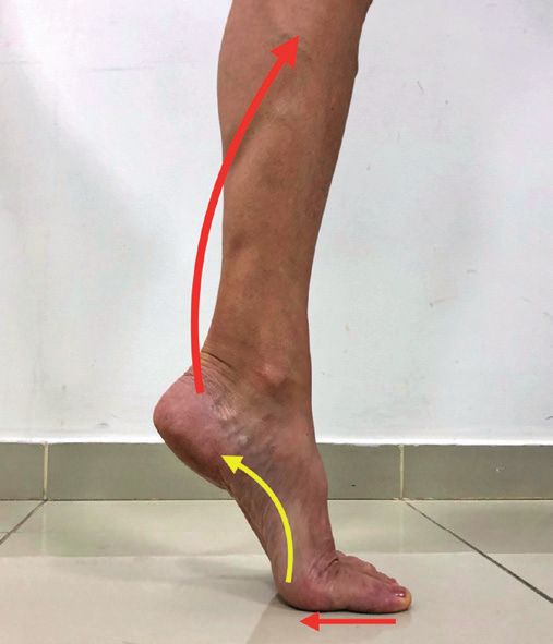
Metsavaht et al. Current trends in the biokinetic analysis of the foot and ankle
classic knowledge with new concepts of functional biome- Functional stability mechanisms of the foot
chanics in order to lay the foundation for the clinical appli-
The main structure that generates stability in the human
cation of 3-dimensional (3D) biokinetic analysis in diagnostic
gait is the foot. Functionally, the foot has 3 main roles:
and/or treatment decisions.
1) To be a stable base of support for movements of the pro-
ximal segments;
Functional biomechanics of the foot and ankle 2) To assist in the absorption of ground reaction forces;
Conventionally, the movements of the ankle and its muscu-
3) To be a powerful lever arm for the ankle muscles during
lar actions are studied either with the tibia as a fixed point for
the propulsion of gait and sport movements.
the free movement of the ankle and foot (usually named an
open kinetic chain [OKC] movement) or with a fixed foot, for The intrinsic and complex role of the plantar arch of the foot
instance against the ground, where the movement would in maintaining stability and mobility has been the subject of
happen in the proximal segments and be named a closed ki- studies in several areas from the Renaissance era, with Leo
netic chain (CKC) movement(7). However, these approaches nardo Da Vinci(14), to anatomists of the last century(15) and pre-
do not fully represent what happens during daily activities. sent day(4). The medial longitudinal arch (MLA) has been the
The concept of functional biomechanics advocates that al- most studied structure because its load sharing system (arch
though one segment will always be the base for the other to load-sharing system) is believed to be essential for the proper
move, both segments can be simultaneously mobile in any functioning of the foot(16). It works as a spring system, chan-
trivial activity. The main difference between conventional and ging foot stiffness and allowing deformation for absorbing
functional biomechanics is that the latter considers that the loads while creating a robust segment for transferring forces
function of joints and segments cannot be separately ob- to the ground. For a more detailed understanding of the role
served. The central nervous system (CNS) works as the ge- of the medial longitudinal arch, please refer to Kirby(16) (2017).
nerator of complex movement patterns based on muscular Despite widespread research on this reductionist 2-dimen-
synergisms, aiming to accomplish a motor task instead of sional (2D) view of the MLA, some of the evidence indicates
accounting for individual muscular actions(8). Following this that it functions as a 3D structure. Some authors suggest that
line of reasoning, 3D biokinetic analyses are meant to identi- the plantar arch should be named “plantar dome,” due to the
fy the role of each anatomical, joint, and muscular structure importance of other passive, active, and neuromuscular struc-
in the functional capacity of an individual throughout his or tures in maintaining the plantar arch(2). This theory has been
her daily activities. To facilitate the understanding of this re- confirmed by cadaveric experiments showing that the resec-
latively new area of study, it is necessary to establish how tion of the plantar fascia reduced foot stiffness by less than
the main pillars of functional biomechanics are applied to the 25%(17). On the other hand, engineering principles demonstra-
study of foot and ankle function. te that even thin structures, when folded in the transverse di-
rection, increase their longitudinal stiffness; this concept that
can be easily demonstrated by a slice of pizza curved across
Dynamic joint stability in our hands. Recently, Venkadesan et al.(18) (2020) applied
Rienmann and Lephart(9) (2002) define dynamic joint stabi- these concepts of transverse arch stiffness and observed that
lity as the ability of a joint to remain or readily resume to its the resection of the transverse arch reduced foot stiffness by
proper alignment through an equalization of forces. Evidence more than 50%, highlighting its important role in the main-
suggests that the control of active joint stability is orchestra- tenance of the plantar dome. For an illustration of the effect
ted by the neuromuscular system and not by isolated muscle of transverse stiffness on longitudinal stiffness, the authors
strength or range of motion(10), highlighting the importance of suggest the following video: https://youtu.be/adt3sH9O_vE.
the CNS as a functional maestro. Another aspect associated with the functioning of the
The ability to generate safe movement and to improve per- plantar arch is the windlass mechanism, which is widely
formance depends on the movement of joints in segments observed in orthopedic clinical practice through the Ja-
with stable bases. Literature on the importance of functional ck’s Test(19). During this test, the hallux extension produces
ankle stability for injury prevention and rehabilitation(2,6,11) is a tension in the plantar aponeurosis, which brings the cal-
extensive and relates chronic ankle instability to a lower ca- caneus closer to the metatarsophalangeal joints(20) (Figure
pacity of generating functional strength by the triceps surae 1). In association with passive structures, the posterior tibial
muscle(11), lower power production during jump propulsion(12), muscle begins to act concentrically and blocks the midtarsal
and a higher risk of ligament and cartilage injuries(13). The- joints to increase foot stiffness(20). Functionally, the windlass
refore, reducing this instability through specific training or mechanism occurs with the hallux as a fixed point and with
surgery is crucial and should be done before the adoption the movement of the metatarsophalangeal joint (Figure 2).
of an overloading activity such as an increase in sports per- This mechanism is initiated by tibiotalar dorsiflexion as the
formance. In order to understand some of the strategies for tibia advances over the talus in the midstance (MS) pha-
reversing non-surgical instabilities, it is necessary to address se of the gait(20). In the terminal stance (TS) phase, load on
the structures that participate in the joint stability of the foot the forefoot region increases, activating the fibularis longus
and ankle. muscle and inducing the windlass mechanism and the elevation
192 J Foot Ankle. 2020;14(2):191-6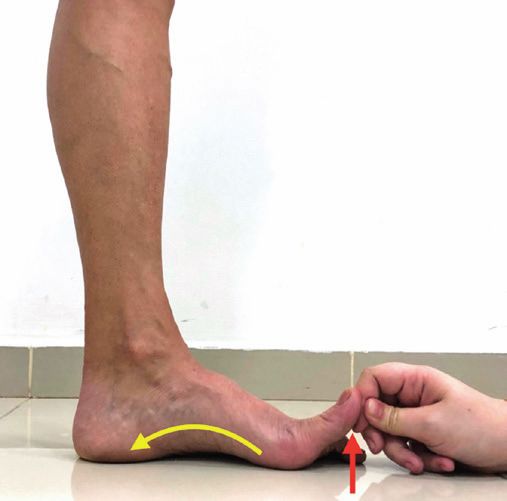
Metsavaht et al. Current trends in the biokinetic analysis of the foot and ankle
of the calcaneus on a rigid forefoot base, thus creating an
effective lever arm to generate propulsion for the second
half of the support phase(20).
Increased mobility of the midfoot and reduced mobility of
tibiotalar dorsiflexion and hallux may impair the windlass
mechanism and contribute to increased foot stiffness in this
phase(21); individuals with flexible flatfeet may not be able to
create a rigid base, causing the axis of movement to move
towards the midtarsal joints so the lever arm is reduced(21). As
a form of compensation, the triceps surae is more intensely
activated and produces more strength; this overload may
lead to painful conditions such as Achilles tendinopathies or
muscle injuries. Moreover, inadequate triceps activation and/or
strength also increases ankle instability(21).
Functional stability mechanisms of the ankle
Several studies have demonstrated changes in the move-
ment patterns of hips, knees, and ankles in individuals with
chronic ankle instability(2,6,22), demonstrating that the same
condition can lead to different motor adaptations and each
case requires individual evaluation. The motor variability
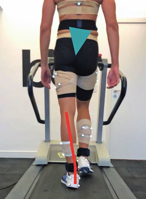
Figure 1. The windlass mechanism demonstrated passively: A
among these individuals may reflect either an attempt to
hallux extension produces a tension in the plantar aponeurosis, which
explore alternative stabilizing strategies or an inadequate
brings the calcaneus closer to the metatarsophalangeal joints.
sensory-motor control(23). In addition, the arthrogenic inhi-
bition of the fibularis longus has been related to continued
instability even after the restoration of triceps surae muscle
strength(24).
According to Hertel et al.(25) (2002), individuals with ankle
instability can be classified into 2 major groups: those with
mechanical ankle instability (MAI) and those with functional
ankle instability (FAI). MAI is defined as a pathological laxi-
ty after ligament injury, while FAI is a subjective symptom
or sensation of instability due to proprioceptive deficits and
changes in neuromuscular functions.
Several clinical tests are commonly used to measure ankle
stability, and subjective measurements of the eversion/in-
version of the heel can be performed with activities such as
unipodal support and walking on a treadmill. However, in ad-
dition to the measurement errors intrinsic to subjective tests,
clinically assessing dynamic joint stability does not guaran-
tee the functional competence of the ankle and foot in daily
tasks and sports. Despite being more affordable and easier
to perform, 2D assessments can present important measure-
ment errors even in individuals with small rotational changes
in the ankle and foot(26). It should also be taken into account
that ankle stability is direction- and task-dependent(24), and
the ability of an individual to maintain joint stability in one di-
rection does not mean he or she will be able to do so in other
directions. It is necessary to evaluate all 3D components of
foot and ankle stability to ensure a safe return to daily acti-
Figure 2. The windlass mechanism actively provoked on termi- vities and sports. Therefore, the functioning of the foot and
nal support phase: The activation of fibularis longus and tibialis ankle should be tested and analyzed in different activities. In
posterior muscles collaborates in maintaining the plantar arch for this review, we will summarize relevant information currently
adequate triceps surae function and ankle stabilization. published on gait and running.
J Foot Ankle. 2020;14(2):191-6 193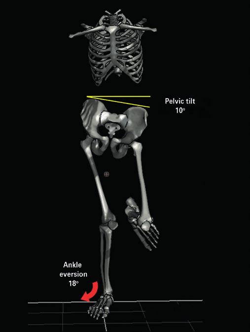
Metsavaht et al. Current trends in the biokinetic analysis of the foot and ankle
Gait and running biokinetics foot or forefoot (midfoot/forefoot strikers), the sural triceps
assumes a primary eccentric role, which may increase the risk
Human gait and running can be divided into stance and ba-
of Achilles tendinopathies and injuries in the tibialis posterior(27)
lance phases. In gait, the support phase is subdivided into
when no proper training is employed.
4 phases: load response (LR), MS, TS, and pre-swing (PS)(1)
(Figure 3). In running, the support phase is subdivided into 2 The ankle rocker phase during MS is characterized by a ro-
phases: LR and propulsion response (Figure 4). During LR in tation of the tibia over the foot, which is fixed on the ground
a non-pathological gait, in the sagittal plane, the foot drops on unipodal support, while the plantar arch is maintained by
(heel rocker) with a plantar flexion movement eccentrically activating the posterior tibialis and intrinsic muscles of the
controlled by the tibialis anterior muscle. In the coronal plane, foot. During TL and PS, the forefoot rocker phase happens
there is an eversion movement of the ankle, eccentrically con- when the heel rises from the ground and begins the pro-
trolled by the tibialis posterior muscle. At this moment, mo- pulsion phase. At this moment, the sural triceps activation,
tion control is achieved by reducing the stiffness of the foot mostly through the soleus muscle, has the important role
and turning it into a structure that is able to absorb mecha- of limiting the anteriorization of the tibia and inducing knee
nical loads(1). In running, the role of eccentric eversion control extension. The fibularis longus depresses the first metatar-
increases due to increased ground reaction forces. In runners sal head and contributes to the formation of the plantar arch
whose initial contact happens with the heel (rearfoot strikers), and stabilization of the ankle joint. In running, the soleus has
the sural triceps has a secondary effect in load absorption by an additional propulsion role since it is responsible for more
preventing excessive advancement of the tibia(1). On the other than 50% of the horizontal acceleration of the runner’s center
hand, in runners whose initial contact occurs with the mid- of mass(1).
1 2 3 4 5 6
Figure 3. The gait cycle. Using the light limb as reference: (1) initial contact; (2) load response (LR); (3)
midstance (MS); (4) terminal stance (TS); (5) pre-swing (PS); (6) swing (Image by Biocinetica Laboratório
do Movimento Ltda, Rio de Janeiro, Brazil).
A B C D
Figure 4. The running cycle. Using the right limb as reference: (A) initial contact; (B) LR; (C) propulsion;
(D) swing (Image by Biocinetica Laboratório do Movimento Ltda).
194 J Foot Ankle. 2020;14(2):191-6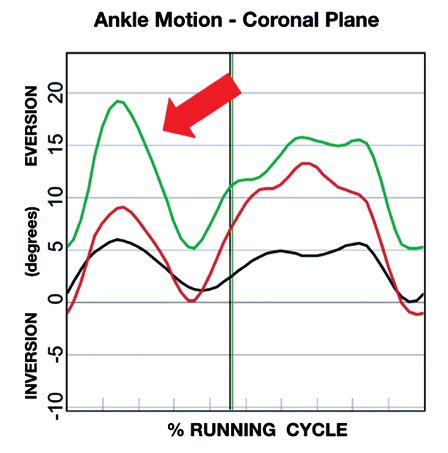
Metsavaht et al. Current trends in the biokinetic analysis of the foot and ankle

The role of proximal joints in ankle and foot movement
  Although it is easy to suppose the influence of neighbor
and distant joints in the control of the foot and ankle, the
identification and measurement of such influences only re-
cently has been deeply studied. Cavalin et al. (28) (2018) found
a strong association between hip adduction and ankle
eversion in healthy runners, where 50% showed a descen-
ding relationship (hip influencing the ankle), 25% showed an
ascending relationship (ankle influencing the hip), and 25%
presented a synchronic relationship over time (Figure 5).
Other authors have also demonstrated the influence of the
ankle dorsiflexion range of motion in femoral medialization,
a movement dysfunction often referred as “dynamic valgus”.
The proximal chain can influence and be influenced by distal
changes and interfere on the loading of joints and segments
as a whole(29) (Figure 6).

Conclusion
  Human motion happens as a system where many variables
may individually or collectively influence the loading, mobi-
lity, and stability of any one joint or segment. Similar to an

                                                                       A

                                                                       B
                                                                     Figure 6. Three-dimensional biokinetic analysis image (A) and graph
                                                                     in degrees (B), in the coronal plane, of a 35-year-old male recreational
                                                                     long-distance runner diagnosed with plantar fasciitis. Peak right ankle
                                                                     eversion (18°) influenced by a peak contralateral pelvic drop (10°) in
                                                                     the stance phase of running. Red arrows: excessive ankle eversion;
Figure 5. Individual walking on a treadmill at 4.3km/h showing, in   yellow lines: excessive contralateral pelvic drop; green line: right ankle
the coronal plane, the influence of a pelvic contralateral drop on   motion; red line: left ankle motion; black line: expected ankle motion
ankle eversion.                                                      (Image by Biocinetica Laboratório do Movimento Ltda).

                                                                                                            J Foot Ankle. 2020;14(2):191-6          195
Metsavaht et al. Current trends in the biokinetic analysis of the foot and ankle
airport network, when one terminal is out of order, the others ding of the problems of each patient. For these reasons three-
are overloaded, but eventually all planes must get to the dimensional biokinetic assessments have become valuable to-
ground safely. ols for identifying the weakest links in the movement chain in
an objective, measurable, and reproducible manner.
The adequate functioning of the foot and ankle depends on
the activities of passive tissues and muscles and the neuro-
muscular control of local and distant joints. Any changes to Acknowledgement
this system may lead to functional incapacity and subsequent I dedicate this article to the memory of Prof. Dr. Irocy Guedes
lesions and/or pain. The study of the functional biomechanics Knackfuss, who in 1997 introduced me to the practice of the Foot
of the ankle and foot, in addition to a clinical investigation and & Ankle medicine and instigated my vision over the promissing
imaging exams, contributes to a more complete understan- horizons of functional biomechanics. Leonardo Metsavaht.
Authors’ contributions: Each author contributed individually and significantly to the development of this article: GL *(https://orcid.org/0000-0002-7265-4658)
conceived and planned the activities that led to the study; wrote the article; participated in the review process; bibliographic review; formatting of the article;
approved the final version; LM *(https://orcid.org/0000-0001-9263-1309) conceived and planned the activities that led to the study; wrote the article; participated
in the review process; bibliographic review; formatting of the article; approved the final version. *ORCID (Open Researcher and Contributor ID) .
References
1. Pandy MG, Andriacchi TP. Muscle and joint function in human 14. Suh HA, editor. Leonardo’s notebooks: writing and art of the great
locomotion. Annu Rev Biomed Eng. 2010 Aug15;12:401-33. master. New York: Black Dog &Leventhal; 2013.
2. McKeon JMM, Hoch MC. The ankle-joint complex: a kinesiologic 15. Whitman, R. A treatise on orthopaedic surgery. 6th ed. Philadelphia:
approach to lateral ankle sprains. J Athl Train. 2019;54(6):589-602. Lea & Febiger; 1919.
3. D’Août K, Aerts P. The evolutionary history of the human foot. 16. Kirby KA. Longitudinal arch load-sharing system of the foot. Rev
In: D’Août K, Van Gheluwe B, De Clercq D. Advances in plantar Esp Pod. 2017; 28(1):18-26.
pressure measurements in clinical and scientific research. Maastricht: 17. Ker RF, Bennett MB, Bibby SR, Kester RC, Alexander RM. The spring
Shaker; 2008. p. 44-68. in the arch of the human foot. Nature. 1987; 325(7000),147-9.
4. Balsdon ME, Bushey KM, Dombroski CE, LeBel ME, Jenkyn TR. 18. Venkadesan M, Yawar A, Eng CM, Dias MA, Singh DK, Tommasini
Medial longitudinal arch angle presents significant differences SM, et al. Stiffness of the human foot and evolution of the
between foot types: a biplane fluoroscopy study. J Biomech Eng. transverse arch. Nature. 2020. 579(7797): 97-100.
2016 Oct 1;138(10). 19. Ewen JA. Naviculo-cuneiform fusion in the treatment of flat foot.
5. Böhm H, Döderlein L, Fujak A, Dussa CU. Is there a correlation J Bone Joint Surg. 1953; 35-B(1):75-82.
between static radiographs and dynamic foot function in 20. Bolgla LA, Malone TR. Plantar fasciitis and the windlass
pediatric foot deformities? Foot Ankle Surg. 2019 Oct 24;S1268- mechanism: a biomechanical link to clinical practice. J Athl Train.
7731(18)30326-6. 2004;39(1):77-82.
6. Kim H, Son SJ, Seeley MK, Hopkins JT. Altered movement 21. Van Boerum DH, Sangeorzan BJ. Biomechanics and pathophysiology
biomechanics in chronic ankle instability, coper, and control of flat foot. Foot Ankle Clin. 2003;8(3):419-30.
groups: energy absorption and distribution implications. J Athl 22. Kwon YU, Harrison K, Kweon SJ, Blaise Williams 3rd DS. Ankle
Train. 2019;54(6):708-17. coordination in chronic ankle instability, coper, and control groups
7. Brockett CL, Chapman GJ. Biomechanics of the ankle. Orthop in running. Med Sci Sports Exerc. 2020;52(3):663-72.
Trauma. 2016;30(3):232-8. 23. Herb CC, Blemker S, Saliba S, Hart J, Hertel J. Chronic ankle
8. Todorov E, Jordan MI. Optimal feedback control as a theory of instability patients exhibit higher variability in lower extremity
motor coordination. Nat Neurosci. 2002;5(11):1226-35. joint-coupling variability during drop vertical jumps. J Biomech.
9. Riemann BL, Lephart SM. The sensorimotor system, part I: 2020 Jan 23;99:109479.
the physiologic basis of functional joint stability. J Athl Train. 24. Gutierrez GM, Kaminsk TWi, Douex AT. Neuromuscular control
2002;37(1):71-9. and ankle instability. PM & R. 2009;1(4):359-65.
10. Williams VJ, Nagai T, Sell TC, Abt JP, Rowe RS, McGrail MA, et 25. Hertel J. Functional Anatomy, pathomechanics, and pathophysiology
al. Prediction of dynamic postural stability during single-leg of lateral ankle instability. J Athl Train. 2002;37(4):364-75.
jump landings by ankle and knee flexibility and strength. J Sport 26. McClay I, Manal K. The influence of foot abduction on differences
Rehabil. 2016;25(3):266-72. between two-dimensional and three-dimensional rearfoot motion.
11. Park YH, Park SH, Kim SH, Choi GW, Kim HJ. Relationship between Foot Ankle Int. 1998;19(1):26-31.
isokinetic muscle strength and functional tests in chronic ankle 27. Almeida MO, Davis IS, Lopes AD. Biomechanical differences of
instability. J Foot Ankle Surg. 2019;58(6):1187-91. foot-strike patterns during running: a systematic review with
12. Simpson JD, Stewart EM, Macias DM, Chander H, Knight AC. meta-analysis. J Orthop Sports Phys Ther. 2015;45(10):738-55
Individuals with chronic ankle instability exhibit dynamic postural 28. Cavalin, GA; Zeitoune, GG; Leporace, G; Nadal, J. Coordenação
stability deficits and altered unilateral landing biomechanics: A intersegmentar do quadril e do tornozelo em corredores
systematic review. Phys Ther Sport. 2019;37:210-9. recreacionais. In: 26o Congresso Brasileiro de Engenharia
13. Doherty C, Bleakley C, Delahunt E, Holden S. Treatment and Biomédica, 2018, Búzios. Anais. Rio de Janeiro: SBEB; 2018.
prevention of acute and recurrent ankle sprain: an overview 29. Dejong AF, Koldenhoven RM, Hertel J. Proximal adaptations in
of systematic reviews with meta-analysis. Br J Sports Med. chronic ankle instability: systematic review and meta-analysis.
2017;51(2):113-25. Med Sci Sports Exerc. 2020;52(7):1563-75.
196 J Foot Ankle. 2020;14(2):191-6You can also read