Demodex crocidurae, a New Demodecid Mite (Acariformes: Prostigmata) Parasitizing the Lesser White-Toothed Shrew and a Redescription of Demodex ...
←
→
Page content transcription
If your browser does not render page correctly, please read the page content below
animals
Article
Demodex crocidurae, a New Demodecid Mite (Acariformes:
Prostigmata) Parasitizing the Lesser White-Toothed Shrew and
a Redescription of Demodex talpae from European Mole with
Data on Parasitism in Soricomorpha
Karolina Cierocka, Joanna N. Izdebska *                     and Leszek Rolbiecki

                                          Department of Invertebrate Zoology and Parasitology, Faculty of Biology, University of Gdańsk, Wita Stwosza 59,
                                          80-308 Gdańsk, Poland; karolina.cierocka@ug.edu.pl (K.C.); leszek.rolbiecki@ug.edu.pl (L.R.)
                                          * Correspondence: biojni@ug.edu.pl; Tel.: +48-58-523-61-89

                                          Simple Summary: This paper describes a new species, Demodex crocidurae, inhabiting the hairy
                                          skin of Crocidura suaveolens. It also redescribes the most morphologically similar form: Demodex
                                          talpae from Talpa europaea, a species known from Hirst’s 1921 description. Following a differential
                                          diagnosis, it was concluded that these are separate species with different features important in the
                                          taxonomy of Demodecidae, inhabiting analogous microhabitats in different species of hosts.

                                          Abstract: Only six parasitic species of Demodecidae mite have thus far been described from the
                                Soricomorpha, these being associated with the common shrew Sorex araneus Linnaeus, 1758, and

                                          the Mediterranean water shrew Neomys anomalus Cabrera, 1907 (two species from each host), and
Citation: Cierocka, K.; Izdebska, J.N.;
                                          with the lesser white-toothed shrew Crocidura suaveolens (Pallas, 1811) and the European mole Talpa
Rolbiecki, L. Demodex crocidurae, a
                                          europaea Linnaeus, 1758 (one from each host species). Presently, Demodex crocidurae, a new species,
New Demodecid Mite (Acariformes:
Prostigmata) Parasitizing the Lesser
                                          has been described from the territory of Poland for C. suaveolens; in order to confirm its validity,
White-Toothed Shrew and a                 it was necessary to redescribe D. talpae Hirst, 1921, from T. europaea, a demodecid species first
Redescription of Demodex talpae from      described by Hirst in 1921 from England and then noted only in Poland. Both species colonized
European Mole with Data on                the hairy skin of the body in their hosts, where no disease symptoms of infestation were observed.
Parasitism in Soricomorpha. Animals       However, D. crocidurae showed higher infection parameters (prevalence 100%, mean intensity 11.7,
2021, 11, 2712. https://doi.org/          intensity range 3–26 individuals) than those of D. talpae (30.0%, 4.7, 2.0–8.0), possibly due to different
10.3390/ani11092712                       host biology.

Academic Editor: Theo De Waal             Keywords: Acariformes; Prostigmata; Demodecidae; parasite; skin mites; mammals; Soricidae;
                                          Talpidae
Received: 19 August 2021
Accepted: 15 September 2021
Published: 17 September 2021

                                          1. Introduction
Publisher’s Note: MDPI stays neutral
with regard to jurisdictional claims in
                                               The Demodecidae (Acariformes: Prostigmata) fauna of the soricomorphs (Soricomor-
published maps and institutional affil-   pha) has been poorly studied. Six species have been described in this group, and only
iations.                                  11 original publications are known worldwide [1]. The majority (eight) of these records
                                          concern the Mediterranean water shrew Neomys anomalus Cabrera, 1907, and the common
                                          shrew Sorex araneus Linnaeus, 1758; for each of these hosts, two Demodecidae species
                                          were described [2–6]. Thus far, only one species, Demodex foveolator Bukva, 1984, has been
Copyright: © 2021 by the authors.
                                          known from the lesser white-toothed shrew Crocidura suaveolens (Pallas, 1811), found in
Licensee MDPI, Basel, Switzerland.
                                          this host only in the tail region. It was described from the Czechia and recently recorded
This article is an open access article
                                          from Poland [7,8]. In contrast, Demodex talpae Hirst, 1921, collected from the European mole
distributed under the terms and           Talpa europaea Linnaeus, 1758, was described based on several specimens obtained from a
conditions of the Creative Commons        single mole specimen from the United Kingdom [9] and was mentioned in a brief report
Attribution (CC BY) license (https://     from Poland covering the parasite fauna of the mole [10].
creativecommons.org/licenses/by/               However, it appears that these data do not reflect the actual distribution of the De-
4.0/).                                    modecidae in the Soricomorpha, a group comprising approx. 500 mammal species, several

Animals 2021, 11, 2712. https://doi.org/10.3390/ani11092712                                                   https://www.mdpi.com/journal/animals
Animals 2021, 11, 2712 2 of 10
of which are widely distributed. The common shrew, Mediterranean water shrew, lesser
white-toothed shrew, and the European mole mentioned above, exhibit distributions cov-
ering the entirety of Europe and the Palearctic [11,12]. The absence of data regarding the
presence of Demodecidae in these host species likely stems from the lack of appropriate
studies, as this group is rarely mentioned in comprehensive analyses of parasitofauna
communities. However, the generic diversity of these mites in the soricomorphs is interest-
ing because apart from the most commonly noted and the most species-rich Demodex, the
genera Apodemodex and Soricidex have been described only from Soricomorpha [1,2,5,13].
Recently, a new Demodex species has been found in the hairy skin of the body of the
lesser white-toothed shrew. In terms of morphological traits, it was found to be similar
to D. talpae, which was previously known from a highly laconic description by Hirst [9],
supplemented with an illustration. Therefore, in order to verify the status of the presently
discovered demodecid from the lesser white-toothed shrew, a redescription D. talpae
was necessary.
2. Materials and Methods
Eight specimens of dead C. suaveolens from Poland (Wielkopolska Voivodeship, Sło-
mowo, 52◦ 210 1100 N, 17◦ 320 4100 E) and ten T. europaea (Wielkopolska Voivodeship, Słomowo,
52◦ 210 1100 N, 17◦ 320 4100 E, six moles; Pomeranian Voivodeship, Pszczółki, 54◦ 100 2100 N,
18◦ 410 5400 E, three moles; Lublewko 54◦ 450 2800 N, 17◦ 550 3700 E, one mole), collected from
October 2016–May 2019, were examined for demodecid mites.
Demodecidae were isolated using the digestion method developed for the detection of
mammalian skin mites [14], with modifications to suit the examined host. Skin fragments
of 1 cm2 were excised with a scalpel from several body regions, including the head (around
eyes, ear pinnae, nose, area of vibrissae, lips, chin, cheeks, vertex), neck, abdomen, back,
limbs, tail, and the genital–anal area. Skin samples were preserved in 70% ethanol and sub-
jected to digestion in 10% potassium hydroxide solution until it was completely digested.
The obtained samples were decanted (examination of 1 cm2 of the skin equal to the analysis
of approximately 100 wet preparations), mounted and examined using phase–contrast
microscopy (Nikon Eclipse 50 i). The mites were placed in polyvinyl-lactophenol solution
and measured (measurements in micrometers) as follows: total body length = length of
gnathosoma, podosoma and opisthosoma; gnathosomal width = width at base; podosomal
and opisthosomal width = maximum width.
The specimen depositories are cited using the following abbreviation: UGDIZP, Uni-
versity of Gdańsk, Department of Invertebrate Zoology and Parasitology, Gdańsk, Poland.
The description of the species adopted the nomenclature commonly used for the family
Demodecidae [15] and was completed with the nomenclature proposed by Bochkov [16]
for the superfamily Cheyletoidea (Acariformes: Prostigmata) and by Izdebska and Rol-
biecki [17]. The scientific and common names of the hosts follow Wilson and Reeder [11]
and the Taxonomic Information System [18].
To define the level of host infection, the following main parasitological parameters
were measured: prevalence (percentage of hosts infected), mean intensity (mean number
of parasites in infected hosts), and intensity range (minimum and maximum number of
parasite individuals per host) [19].
3. Results
3.1. Demodex crocidurae Izdebska, Cierocka, Rolbiecki, 2021
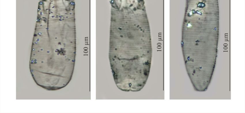
Description (Table 1, Figures 1 and 2)Animals 2021, 11, x FOR PEER REVIEW 4 of 11
Animals 2021, 11, 2712 3 of 10
Figure 1. Demodex crocidurae sp. nov.: female, dorsal view (A), female, ventral view (B), male, dorsal
view (C), gnathosoma, female, ventral view (D), gnathosoma, female, dorsal view (E), claw on the
Figureleg1. Demodex crocidurae
(F), supracoxal spine,sp. nov.:
dorsal female,
view dorsal view
(G), supracoxal (A),lateral
spine, female, ventral
view view (B),(I);
(H), aedeagus male, dorsal
a: vulva,
view (C), gnathosoma, female, ventral view (D), gnathosoma, female, dorsal view (E), claw
b: aedeagus, c: spines on palps, d: seta v”F, e: subgnathosomal seta (seta n), f: pharyngeal bulb,
on the
leg (F),g:supracoxal spine, dorsal view (G), supracoxal spine, lateral view (H), aedeagus (I); a: vulva,
supracoxal spine (seta elc.p).
b: aedeagus, c: spines on palps, d: seta v”F, e: subgnathosomal seta (seta n), f: pharyngeal bulb, g:
supracoxal spine (seta elc.p).Animals 2021, 11, 2712 4 of 10
Table 1. Measurements of morphological features (in micrometers) for adults of Demodex crocidurae
sp. nov.
Morphologic Features Males (n = 32) Females (n = 61)
Length of gnathosoma 17 (15–20), SD 1 17 (15–20), SD 1
Width of gnathosoma (at base) 12 (11–16), SD 1 13 (10–16), SD 1
Length of podosoma 45 (40–53), SD 3 49 (40–55), SD 3
Width of podosoma 23 (19–28), SD 2 24 (20–30), SD 2
Length of opisthosoma 96 (80–120), SD 10 87 (73–110), SD 9
Animals 2021, 11, x FOR PEER REVIEW Width of opisthosoma 30 (24–35), SD 3 29 (23–35), SD 53 of 11
Aedeagus 25 (22–30), SD 2 –
Vulva – 6 (5–10), SD 1
Total body length 158 (139–182), SD 11 153 (138–176), SD 9
Figure 2. Demodex talpae, female (A) and Demodex crocidurae sp. nov., female, holotype (B), male (C).
Figure 2. Demodex talpae, female (A) and Demodex crocidurae sp. nov., female, holotype (B), male (C).
FEMALE (holotype and 60 paratypes): Body 153 (138–176) long and 29 (23–35) wide
(holotype, and×location
Infection150 27). Distinctly separated
in the host. Demodex gnathosoma, rectangular,
crocidurae sp. longerin
nov. was found than wide; on
all exam-
dorsal surface in anterior part of basal (coxal) segment, parallel to base, pair of massive
ined lesser white-toothed shrews (100%), with a mean intensity of 11.7 and intensity range
boomerang-shaped supracoxal spines (setae elc.p) present, ca. 5.0 long (holotype, 5.0).
of 3–26 individuals per host: 93 individuals in total (32 males, 61 females). The demodecid
Palps 3-segmented, terminating in two claw-like, large spines on tibio-tarsus, ca. 2.0 long
mites were found on the hairy skin of the body (head—31 individuals, abdomen—30,
(holotype, 2.0) and two small, conical spines; conical setae v”F near external edge of middle
back—28, and genital-anal area—4). The observed mites did not cause any lesions in ex-
segment (trochanter-femur-genu) present. On ventral surface, horseshoe-shaped pharyn-
amined shrews.
geal bulb with pair of conical subgnathosomal setae (setae n) situated on either side of and
Etymology. The specific epithet crocidurae refers to the specific name of the host.
slightly posterior to anterior limit. Podosoma trapezoidal, widening posterior end; four
pairs of short legs, with coxae integrated into ventral idiosomal wall and five free, over-
3.2. Demodex talpae Hirst, 1921
lapping segments (trochanter–tarsus); two forked claws, ca. 3.0 long (holotype, 3.0) with
Redescriptions
large, (Table 2, Figures
pointed subterminal 2 and
spur, and one3)solenidion (ω) on each tarsus. Epimeral plates
(coxal fields) distinctly sclerotized; pairslong
FEMALE (n = 8): Body 143 (134–154) and 38 (35–43)
I-II trapezoidal, wide.
III-IV Distinctly
pairs separated
rectangular; posterior
gnathosoma, trapezoidal, base width longer than length; on dorsal surface in
edges of pair IV indented archwise; anterior end of vulva between incision. Podosomalanterior part
of basal
shield(coxal)
reachingsegment,
level ofdirected
legs III;posteromedially,
posterior edge of pair
thisofshield
massive, wedge-shaped
is convex. su-
Opisthosoma
pracoxal spines (setae elc.p) present, ca. 5.5 long. Palps 3-segmented, terminating in two
claw-like, large spines on tibio-tarsus, ca. 3.0 long and one small, conical spine. On ventral
surface, horseshoe-shaped pharyngeal bulb with pair of conical subgnathosomal setae (se-
tae n) situated exactly at level of anterior margin on both sides. Podosoma trapezoidal,
widening posterior end; four pairs of short legs, with coxae integrated into ventral idio-Animals 2021, 11, 2712 5 of 10
constitutes 57% (53−63%) of body length (holotype, 57%); widens to 3/4 of opisthosoma
length and then becomes narrower. Whole opisthosoma distinctly annulated; annulation
reaches level of legs III dorsally; annuli relatively wide, ca. 1.5−2.0 µm. Opisthosomal
organ absent. Vulva 6 (5−10) long (holotype, 8.0), located between and behind incision of
IV epimeral plate.
MALE (32 paratypes): Slightly longer and slender than female, 158 (139−182) long,
30 (24−35) wide. Distinctly separated gnathosoma, rectangular, longer than wide. Pharyn-
geal bulb and morphological details of gnathosoma similar to those in females. In addition,
podosoma and legs shaped similar to those in females; however, posterior edge of epimeral
plate IV lacks incision. Opisthosoma constitutes 61% (56−66%) of body length; opistho-
soma, similar to females, distinctly annulated; annuli relatively wide, ca. 1.5−2.0 µm.
Opisthosomal organ absent. Aedeagus 25 (22−30) long, on dorsal surface, located between
epimeral plates II and IV. Genital opening located on dorsal surface, at level of anterior
margin of epimeral plate II.
Type material: Female holotype (reg. no. UGDIZPSCSDDc13f) from Crocidura suave-
olens (reg. no. MSSCs01/2017), Słomowo, Wielkopolska Voivodeship, Poland, August
2017, parasites coll. K. Cierocka, J.N. Izdebska, host coll. J.N. Izdebska, L. Rolbiecki;
60 female paratypes (reg. nos. UGDIZPSCSDDc01-12f, UGDIZPSCSDDc14-61f) and
32 male paratypes (reg. nos. UGDIZPSCSDDc01-32m) from Crocidura suaveolens (reg.
nos. MSSCs01/2017, MSSCs02/2018, MSSCs03-05/2017, MSSCs06-08/2018), Słomowo,
Wielkopolska Voivodeship, Poland, August 2017, August 2018, same collectors.
Type material deposition: Whole type material (mounted microscope slides with
the demodecid mites) is deposited in scientific collections within the framework of the
Collection of Extant Invertebrates in Department of Invertebrate Zoology and Parasitology,
University of Gdańsk, Poland.
Infection and location in the host. Demodex crocidurae sp. nov. was found in all exam-
ined lesser white-toothed shrews (100%), with a mean intensity of 11.7 and intensity range
of 3–26 individuals per host: 93 individuals in total (32 males, 61 females). The demodecid
mites were found on the hairy skin of the body (head—31 individuals, abdomen—30,
back—28, and genital-anal area—4). The observed mites did not cause any lesions in
examined shrews.
Etymology. The specific epithet crocidurae refers to the specific name of the host.
3.2. Demodex talpae Hirst, 1921
Redescriptions (Table 2, Figures 2 and 3)
Table 2. Measurements of morphological features (in micrometers) for adults of Demodex talpae.
Morphologic Features Males (n = 6) Females (n = 8)
Length of gnathosoma 13 (10–15), SD 2 14 (13–15), SD 1
Width of gnathosoma (at base) 18 (17–20), SD 1 18 (17–21), SD 2
Length of podosoma 42 (38–50), SD 6 48 (45–53), SD 3
Width of podosoma 30 (27–33), SD 2 33 (30–38), SD 3
Length of opisthosoma 74 (68–88), SD 7 81 (73–88), SD 6
Width of opisthosoma 34 (33–40), SD 3 38 (35–43), SD 3
Aedeagus 19 (18–22), SD 2 –
Vulva – 9 (8–10), SD 1
Total body length 128 (118–150), SD 13 143 (134–154), SD 6Animals 2021, 11, x FOR PEER REVIEW 7 of 11
Animals 2021, 11, 2712 6 of 10
Figure 3. Demodex talpae: female, dorsal view (A), female, ventral view (B), male, dorsal view (C),
gnathosoma, female, ventral view (D), gnathosoma, female, dorsal view (E), claw on the leg (F),
Figure 3. Demodex
supracoxal talpae:
spine (G), female, dorsalorgan,
opisthosomal view (A), female,
female ventral view
(H), aedeagus (B),
(I); a: male,b:dorsal
vulva, viewc:(C),
aedeagus, spines
gnathosoma, female, ventral view (D), gnathosoma, female, dorsal view (E), claw on the leg (F),
on palps, d: subgnathosomal seta (seta n), e: pharyngeal bulb, f: supracoxal spine (seta elc.p),
supracoxal spine (G), opisthosomal organ, female (H), aedeagus (I); a: vulva, b: aedeagus, c: spines
g: opisthosomal organ.
on palps, d: subgnathosomal seta (seta n), e: pharyngeal bulb, f: supracoxal spine (seta elc.p), g: opis-
thosomal organ.
FEMALE (n = 8): Body 143 (134–154) long and 38 (35–43) wide. Distinctly separated
gnathosoma, trapezoidal, base width longer than length; on dorsal surface in anterior
Infection and location in the host. Demodex talpae was noted in 30.0% of the ten exam-
part of basal (coxal) segment, directed posteromedially, pair of massive, wedge-shaped
ined European moles, with a mean intensity of 4.7 and intensity range of 2.0–8.0 individ-
supracoxal spines (setae elc.p) present, ca. 5.5 long. Palps 3-segmented, terminating in
uals per host: 14 individuals in total (six males, eight females). Mites were found in the
two claw-like, large spines on tibio-tarsus, ca. 3.0 long and one small, conical spine. On
hairy skin of the body (back—six individuals; abdomen—eight). The observed mites did
ventral surface, horseshoe-shaped pharyngeal bulb with pair of conical subgnathosomal
not setae
cause(setae
any lesions in the
n) situated examined
exactly European
at level moles.
of anterior Infestations
margin on both were
sides.not associated
Podosoma trape-
withzoidal,
skin lesions or other symptoms.
widening posterior end; four pairs of short legs, with coxae integrated into ventral
idiosomal wall and five free, overlapping segments (trochanter–tarsus); two forked claws,
ca. 3.0 long with large, pointed subterminal spur. Epimeral plates (coxal fields) distinctlyAnimals 2021, 11, 2712 7 of 10
sclerotized; I-IV pairs trapezoidal; posterior edges of pair IV weakly sclerotized, form trian-
gular incision, which almost surrounding vulva. Podosomal shield reaching level of legs
III; posterior edge of this shield is concave. Opisthosoma constitutes 57% (53−60%) of body
length; widens towards end; widest at end. Opisthosoma distinctly and densely annulated;
annulation also reaches dorsal podosoma side (level of legs III); annuli relatively wide at
ca. 1.0–1.5 µm. Opisthosomal organ present, tubular; opisthosomal pore oval, 1.5–2.0 in
diameter. Vulva 9 (8−10) long, located between and behind incision of IV epimeral plate.
MALE (n = 6): Shorter than female, 128 (118−150) long, 34 (33−40) wide. Distinctly
separated gnathosoma, trapezoidal, base width longer than length. Pharyngeal bulb and
morphological details of gnathosoma similar to those in female. Shape of podosoma and
legs similar to those in female; however, only posterior edge of epimeral plate IV without
incision. Opisthosoma constitutes 58% (55−60%) of body length; widens towards posterior
end. Opisthosoma, similar to females, distinctly annulated; annuli relatively wide, ca.
1.0−1.5 µm. Opisthosomal organ present, similar to that in female. Aedeagus 19 (18−22)
long, on dorsal surface, located between epimeral plates III and IV. Genital opening located
on dorsal surface, on border between epimeral plates II and III.
Material deposition: Mounted microscope slides with specimens were stored in
scientific collections within the framework of the Collection of Extant Invertebrates in
Department of Invertebrate Zoology and Parasitology, University of Gdańsk, Poland. Eight
females (reg. no. UGDIZPTTeDDt01-08f) and six males (reg. no. UGDIZPTTeDDt01-06m)
from Talpa europaea (reg. no. MSTTe01/2016, MSTTe02/2019, MSTTe03/2018), Lublewko,
Pomeranian Voivodeship, Słomowo, Wielkopolska Voivodeship, Pszczółki, Pomeranian
Voivodeship, Poland, October 2016, August 2018, May 2019, parasites coll. K. Cierocka, J.N.
Izdebska, host coll. J.N. Izdebska, L. Rolbiecki.
Infection and location in the host. Demodex talpae was noted in 30.0% of the ten exam-
ined European moles, with a mean intensity of 4.7 and intensity range of 2.0–8.0 individuals
per host: 14 individuals in total (six males, eight females). Mites were found in the hairy
skin of the body (back—six individuals; abdomen—eight). The observed mites did not
cause any lesions in the examined European moles. Infestations were not associated with
skin lesions or other symptoms.
3.3. Differential Diagnosis
Regarding the known Demodecidae, D. crocidurae sp. nov. appears to be closely related
to D. talpae described from the European mole, this being a representative of another family
within the order Soricomorpha. However, D. talpae is smaller and has different body
proportions; it is relatively wider, with shorter opisthosoma; in addition, D. talpae males
and females have similar body size and proportions, whereas D. crocidurae males are typically
slightly longer (with longer opisthosoma) than the females (Tables 1–3, Figures 1–3).
The gnathosoma in D. crocidurae is rectangular, narrow, and longer than wide; in
D. talpae, it is trapezoidal, shorter than wide at the base. The supracoxal spines in both
species are large and massive (ca. 5.0 um in length); however, they are boomerang shaped
in D. crocidurae and wedge-shaped in D. talpae. Additionally, they are directed parallel to
the base of the gnathosoma in D. crocidurae and are directed posteromedially in D. talpae.
In addition, there is a process on the supracoxal spines; this is located in the center of the
spine in D. crocidurae and at the widest part in D. talpae. The terminal segments of the
palpi are equipped with two large and two small spines in D. cocidurae, while there are two
large and one small spines in D. talpae. The subgnathosomal setae are located on either
side of and slightly posterior to the anterior margin of the pharyngeal bulb in D. crocidurae,
and exactly at the level of anterior margin of the pharyngeal bulb in D. talpae. The leg
tarsi are equipped with forked claws differing in shape between both demodecid species.
Differences also exist between the epimeral plates: in D. crocidurae, I–II pairs are trapezoidal
and III–IV are rectangular, while in D. talpae, all epimeral plates are trapezoidal. Moreover,
the posterior edge of epimeral plates IV has an arched shape in D. crocidurae females but
a triangular shape in D. talpae females. In addition, the posterior edge of the podosomalAnimals 2021, 11, 2712 8 of 10
shield is convex in D. crocidurae and concave in D. talpae. The opisthosoma in D. crocidurae
is narrower and longer, widens to 3/4 of its length and then narrows, while in D. talpae it is
wider and shorter, widening posteriorly, with the widest part at the end of the body. In
addition, the opisthosomal organ is absent in D. crocidurae, but it is present in both sexes in
D. talpae. Furthermore, in D. crocidurae males, the aedeagus is longer (22–30 µm in length)
and located at epimeral plates II–IV with the genital opening at the level of anterior margin
of epimeral plates II; in D. talpae males, the aedeagus is shorter (18–22 µm in length) and
located at the border between epimeral plates III and IV, with the genital opening on the
border between epimeral plates II and III.
Table 3. Morphometric comparison between Demodex crocidurae sp. nov. and Demodex talpae.
Feature/Species Demodex crocidurae sp. nov. Demodex talpae
Source Present Study Present Study Hirst [9]
Sex Males Females Males Females Males Females
Sample Size (n = 32) (n = 61) (n = 6) (n = 8) (n = *) (n = *)
Body total length 158 (139–182), SD 11 153 (138–176), SD 9 128 (118–150), SD 13 143 (134–154), SD 6 126 128–130
Body total width 30 (24–35), SD 3 29 (23–35), SD 3 34 (33–40), SD 3 38 (35–43), SD 3 37 34–41
5.3:1 (4.3–6.1:1), SD 5.4:1 (4.3–7.0:1), SD 3.7:1 (3.5–3.9:1), SD 3.8:1 (3.2–4.1:1), SD
Body length to width ratio 3.4:1 **
0.5:1 0.6:1 0.1:1 0.3:1
Opisthosoma length to
61 (56–66), SD 3 57 (53–63), SD 3 58 (55–60), SD 2 57 (53–60), SD 2 57 **
body length ratio (%)
Aedeagus length 25 (22–30), SD 2 – 19 (18–22), SD 2 – 22 –
Vulva length – 6 (5–10), SD 1 – 9 (8–10), SD 1 – –
* The author gives no information about the number of examined mites; only “several specimens” are given. ** Taken from measurements
of Hirst [9].
4. Discussion
Demodex crocidurae sp. nov. clearly differs from all known Demodecidae species.
However, it resembles D. talpae in general body shape, and certain characteristics, namely,
the shape of the supracoxal spines and leg claws, which are also observed in several
demodecid mites of the Muridae, e.g., D. musculi Oudemans, 1897, D. apodemi Hirst, 1918, D.
corniculatus Izdebska, 2012, D. bandicotae Izdebska, Rolbiecki, Morand & Ribas, 2017 [20–23].
Although both the two host species, under discussion, belongs to the order Soricomorpha,
they are representatives of two separate families, Soricidae and Talpidae, respectively with a
doubtful level of affinity [11]. In addition, the lesser white-toothed shrew and the European
mole have different environmental preferences and different biologies. Crocidura suaveolens
is a small mammal (weight in the 3–7 g range), found in Central Europe, Israel, Saudi
Arabia, Middle East, Caucasus, Kyrgyzstan, north-east China and Korea, where it primarily
inhabits bushes, forest communities, as well as parks and gardens and buildings [24]. It
forms a nest among grass or in abandoned rodent burrows. It is active throughout the
year and day and night and has short activity and rest cycles. Conversely, T. europaea is
considerably larger (up to 120 g) and has a considerably wider distribution range, including
the Northern Hemisphere, being found in Europe, Asia and North America. It is also
active throughout the year during both day and night, but it has longer activity and rest
cycles. The mole is also a subterranean mammal, constructing underground nests and
tunnels [12,25]. Thus, it is difficult to expect that the same Demodecidae species will be
found in both host species.
Demodex crocidurae and D. talpae, although morphologically similar (Tables 1–3, Figures 1–3)
and found in comparable microhabitats on their hosts (hairy skin), were found to demon-
strate different infestation levels. Demodex crocidurae was common in all examined shrews,
despite originating from different populations and seasons. In contrast, D. talpae, demon-
strates low infestation parameters; only 14 demodecid mites were recorded in only three
mole specimens in the present study (30.0% of the examined).
Previously, D. talpae was only known from a single record from the United Kingdom,
in which Hirst [9] described the species based on several specimens obtained from a single
mole in 1919. However, D. talpae was recorded from several hosts in recent studies on theAnimals 2021, 11, 2712 9 of 10
mole parasitofauna conducted in Poland [10]; but, due to the different study methodology
(determined by parasite analyses from different groups and locations), and poor preserva-
tion state of the specimens, it was not possible to use all specimens for the redescription.
Knowledge of the distribution of Demodex species is still fragmentary the distribution
of both species may be wider than presently known. In the present study, D. crocidurae
was found in lesser white-toothed shrews from an area located near the northwestern
boundary of their distribution in Europe; the shrew is found relatively rarely in this area,
and considering its solitary behavior (apart from the breeding season), this likely does not
facilitate the spread of this parasite. Despite this, D. crocidurae was well represented on all
individuals of all the host populations. The considerably lower D. talpae infestation level
observed in European mole is probably linked to its subterranean and extremely solitary
behavior. However, its records from distant localities (the United Kingdom and Poland)
over the period of 100 years clearly suggests that it may be present in more areas of its
host distribution.
5. Conclusions
To date only six species (Apodemodex cornutus Bukva, 1996, D. foveolator, D. neomydis
Bukva, 1995, D. soricinus Hirst, 1918, D. talpae, Soricidex dimorphus Bukva, 1982) of Demod-
ecidae parasitic mites are recorded in the Soricomorpha. Here, we describe the seventh
species, D. crocidurae from C. suaveolens which indicates that the diversity of these mites in
this most primitive group of placental mammals is probably greater and requires further
detailed research.
Author Contributions: Conceptualization, J.N.I., K.C. and L.R.; methodology, J.N.I., L.R. and K.C.;
investigation, K.C. and J.N.I.; resources, K.C., J.N.I. and L.R.; writing—original draft preparation,
J.N.I., K.C. and L.R.; writing—review and editing, K.C. and L.R.; supervision, J.N.I. All authors have
read and agreed to the published version of the manuscript.
Funding: This research received no external funding.
Institutional Review Board Statement: Ethical review and approval were waived for this study, due
to the use of only dead animals (specimens found in the field, dead from natural causes).
Conflicts of Interest: The authors declare no conflict of interest.
References
1. Izdebska, J.N.; Rolbiecki, L. The biodiversity of demodecid mites (Acariformes: Prostigmata), specific parasites of mammals with
a global checklist and a new finding for Demodex sciurinus. Diversity 2020, 12, 261. [CrossRef]
2. Bukva, V. Soricidex dimorphus g. n., sp. n. (Acari, Demodicidae) from the common shrew, Sorex araneus. Folia Parasitol. 1982,
40, 343–349.
3. Bukva, V. Redescription of Demodex soricinus (Acari: Demodecidae) from the sebaceous glands of Sorex araneus (Insectivora).
Folia Parasitol. 1993, 40, 243–248.
4. Bukva, V. Demodex neomydis sp. n. (Acari: Demodecidae) from the hair follicles of the Mediterranean water shrew, Neomys anomalus
(Insectivora: Soricidae). Folia Parasitol. 1995, 42, 299–306.
5. Bukva, V. Apodemodex cornutus gen. n. et sp. n. (Acari: Demodecidae): New genus and new species of the hair follicle mite from
the Mediterranean water shrew, Neomys anomalus (Insectivora: Soricidae). Folia Parasitol. 1996, 43, 312–316.
6. Izdebska, J.N. Species of Demodecidae (Acari, Actinedida), new for the fauna of Poland, in common shrew (Sorex araneus L.).
Zool. Pol. 2004, 49, 47–51.
7. Bukva, V. Demodex foveolator sp. n. (Acari: Demodicidae), a new epidermis-dwelling parasite of Crocidura suaveolens (Pallas, 1821).
Folia Parasitol. 1984, 31, 42–52.
8. Cierocka, K.; Izdebska, J.N.; Rolbiecki, L. Demodex foveolator (Acariformes: Demodecidae) from Crocidura suaveolens (Soricomorpha:
Soricidae)–the second observation worldwide, and a checklist of the demodecid mites of soricomorphs. Ann. Parasitol. 2019,
65, 329–332.
9. Hirst, S. On some new or little-known Acari, mostly parasitic in habit. Proc. Zool. Soc. Lond. 1921, 91, 357–378. [CrossRef]
10. Izdebska, J.N.; Rolbiecki, L. New data on parasites of mole Talpa europaea (Mammalia, Insectivora) in northern Poland. Wiad. Parazytol.
2003, 49, 97–98.Animals 2021, 11, 2712 10 of 10
11. Wilson, D.E.; Reeder, D.M. (Eds.) Mammals Species of the World. A Taxonomic and Geographic Reference, 3rd ed.; The Johns Hopkins
University Press: Baltimore, Maryland, 2005. Available online: http://www.departments.bucknell.edu/biology/resources/
msw3/ (accessed on 6 June 2021).
12. Pucek, Z. (Ed.) Keys to Vertebrates of Poland. Mammals; Polish Scientific Publishers: Warszawa, Poland, 1981; pp. 72–75, 99–101.
13. Bukva, V. Sexual dimorphism in the hair follicle mites (Acari: Demodecidae): Scanning electron microscopy of Soricidex dimorphus.
Folia Parasitol. 1993, 40, 71–79.
14. Izdebska, J.N. Demodex spp. (Acari: Demodecidae) in brown rat (Rodentia: Muridae) in Poland. Wiad. Parazytol. 2004, 50, 333–335.
15. Nutting, W.B. Hair follicle mites (Demodex spp.) of medical and veterinary concern. Cornell Vet. 1976, 66, 214–231. [PubMed]
16. Bochkov, A.V. New observations on phylogeny of cheyletoid mites (Acari: Prostigmata: Cheyletoidea). Proc. Zool. Inst. RAS 2008,
312, 54–73.
17. Izdebska, J.N.; Rolbiecki, L. A new genus and species of demodecid mites from the tongue of a house mouse Mus musculus:
Description of adult and immature stages with data on parasitism. Med. Vet. Entomol. 2016, 30, 135–143. [CrossRef] [PubMed]
18. Taxonomic Information System (ITIS). Available online: http://www.itis.gov (accessed on 6 June 2021).
19. Margolis, L.; Esch, G.W.; Holmes, J.C.; Kuris, A.M.; Schad, G.A. The use of ecological terms in parasitology (report of an ad hoc
committee of the American Society of Parasitologists). J. Parasitol. 1982, 68, 131–133. [CrossRef]
20. Izdebska, J.N. A new Demodecidae species (Acari) from the yellow-necked mouse Apodemus flavicollis (Rodentia, Muridae)–
description with data on parasitism. J. Parasitol. 2012, 98, 1101–1104. [CrossRef]
21. Izdebska, J.N.; Rolbiecki, L. A new species of Demodex (Acari, Demodecidae) with data on topical specificity and topography
of demodectic mites in the striped field mouse Apodemus agrarius (Rodentia, Muridae). J. Med. Entomol. 2013, 50, 1202–1207.
[CrossRef]
22. Izdebska, J.N.; Rolbiecki, L. Two new species of Demodex (Acari: Demodecidae) with a redescription of Demodex musculi and data
on parasitism in Mus musculus (Rodentia: Muridae). J. Med. Entomol. 2015, 52, 604–613. [CrossRef]
23. Izdebska, J.N.; Rolbiecki, L.; Morand, S.; Ribas, A. A new species and new host record of Demodecidae (Acariformes: Prostigmata)
associated with the bandicoot rat (Rodentia: Muridae) from Lao PDR with data on parasitism and a checklist of the demodecid
mites of rodents. Syst. Appl. Acarol. 2017, 22, 1910–1923. [CrossRef]
24. Cichocki, J.; Kościelska, A.; Piłacińska, B.; Kowalski, M.; Ważna, A.; Dobosz, R.; Nowakowski, K.; Lesiński, G.; Gabryś, G.
Occurrence of lesser white-toothed shrew Crocidura suaveolens (Pallas, 1811) in Poland. Zesz. Nauk. Uniw. Szczec. Acta Biol. 2014,
21, 149–168.
25. Hutchins, M.; Kleiman, D.G.; Geist, V.; McDade, M.C. (Eds.) Grzimek’s Animal Life Encyclopedia, 2nd ed.; Gale Group: Farmington
Hills, HI, USA, 2003; pp. 285–286.You can also read