Dermoscopic Features of Keloid versus Hypertrophic Scar
←
→
Page content transcription
If your browser does not render page correctly, please read the page content below

The Egyptian Journal of Hospital Medicine (January 2018) Vol. 70 (4), Page 622-624

          Dermoscopic Features of Keloid versus Hypertrophic Scar
                           Mahmoud Abdallah¹, Marwa Yassin¹, Noha Saber².
                    ¹Dermatology Department, Faculty of Medicine, Ain Shams University,
                               2
                                 Dermatology Department, El Talaba Hospital
                 *Corresponding author: nohasaber, drnohasaber@gmail.com. Tele: 0201117549990

ABSTRACT
Background: Hypertrophic scars and keloids can be described as variations of typical wound healing.
Aim of this study was to find out the dermoscopic differentiating features between keloids and hypertrophic
scars in their recent phases of development in Egyptian population.
Patients and Methods: An observational cross-sectional design of 30 cases that assigned according to
histopathological analysis into two groups; Keloids and Hypertrophic scars. Then the polarized non-contact
mode of a Dermlite© DL3 “Gen, USA” at a magnification of 10 × was used to capture dermoscopic images
for both groups. The surface morphology and the dermoscopic criteria were recorded directly from the
patients and included vascular structures (Arborizing, linear and comma shaped vessels), erythematous or
white patches. Results: Statistical analysis revealed that patients with keloids are more likely to be associated
with the presence vascular structures on dermatoscopy. The analysis of types of vascular structures showed
that arborizing vessels, in particular, were significantly related to keloids. In contrast, the dominant
dermoscopic feature in Hypertrophic scarring was the presence of scarring as presented in the form of
erythematous or white patches with scanty or absent vascularization. Conclusions: Dermatoscopy should be
considered as a routine investigation of any case with abnormal scarring for a better differentiation between
keloids and hypertrophic scars, and hence a better evaluation and treatment of each type.
Keywords: Keloid, Hypertrophic scar, vascular structures, handheld dermatoscopy.

INTRODUCTION
  Wound healing is a natural restorative response to            to the high cost and lengthy preparation time;
a tissue injury. It is the interaction of a complex             studied the dermoscopic features of keloids and
cascade of cellular events that generates                       hypertrophic scars to differentiate them based on
resurfacing, reconstitution, and restoration of the             their characteristic dermoscopic features and they
tensile strength of injured skin1. Hypertrophic scars           could demonstrate distinctive dermoscopic features
and keloids can be described as variations of typical           especially in keloids where vascular structures
wound healing. As the scar matures, it becomes                  including arborizing, linear irregular and comma
hyperemic and it may be thickened; however, it                  shaped vessels were frequently seen5.
tends to subside gradually until a flat, white,
pliable, possibly stretched, and mature scar                    MATERIALS AND METHODS
develops. When an imbalance during the healing                    This study was conducted on 30 patients with
process occurs, more collagen is produced than is               recent scars who were selected from the Outpatient
degraded, and the scar grows in all directions2. A              Dermatology clinic of Ain Shams University
keloid is an abnormal proliferation of the scar tissue          Hospitals during the period of September 2015 to
that forms at the site of cutaneous injury. It does not         February 2017. Inclusion criteria were a clinical
regress and grows beyond the original margins of                and histological diagnosis of Keloids and
the scar3. On the other hand, the hypertrophic scar             hypertrophic scars. We excluded all cases who had
is a widened or unsightly scar that does not extend             received previous treatment and patients with old
beyond the original boundaries of the wound.                    scars (more than 6 month). The subjects were
Unlike keloids, the hypertrophic scar reaches a                 classified into 2 groups (15 subjects with keloid
certain size and subsequently stabilizes or                     scars and 15 subjects with hypertrophic scars)
regresses4. Dermoscopy is a widely used non-                    according to histopathology. Then dermoscopic
invasive diagnostic technique which provides up to              images had been captured with a Dermlite© DL3
a ten times greater magnification than the unaided              “Gen, USA” at a magnification of 10 × for both
eye and can show the structure of the upper layer of            keloids and hypertrophic scar groups. The
the dermis, and therefore yielding many                         dermoscope used in the polarized non-contact mode
diagnostically relevant findings5. Because of the               because, the blood vessels that are located in the
difficult distinction between keloids and                       dermis collapse easily by the pressure applied when
hypertrophic scars clinically and the unpractical               performing contact dermoscopy. This causes
application of histopathological differentiation due            blanching of the lesion and loss of important

                                                          621
                   Received: 19/10/2017                             DOI: 10.12816/0043814
                   Accepted: 29/10/2017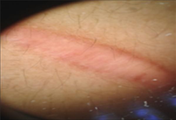
Mahmoud Abdallah et al.
vascular criteria. All participants gave their                   The study included 30 patients with scars
informed consent. Patient demographics and                 (keloid and hypertrophic scar), the age of the
dermoscopic creteria were recorded for both                patients ranged from 4 to 50 years with a mean age
groups.                                                    of 22.6. They were 13 males and 17 females. The
                                                           patients were classified into two groups. Patients
Statistical analysis                                       with keloids (15 patients) were 7 males and 8
  The collected data was revised, coded, tabulated,        females and their ages ranged from 4 to 40 with a
and introduced to a PC using Statistical package for       mean of 20.33 (±9.78 SD). The other group,
Social Science (SPSS 15.0.1 for windows; SPSS              patients with hypertrophic scars, included 15
Inc, Chicago, IL, 2001). Data was presented, and           patients, 6 males and 9 females, and their ages
suitable analysis was done using student T Test,
                                                           ranged from 6 to 50 with a mean of 24.27(SD±
was used to assess the statistical significance of the
                                                           12.68) (table 1). All the scars were recent scars
difference between two study group means, Chi-             (with a maximum scar age of 6 months). The scar
Square test was used to examine the relationship           duration ranged from 3 to 6 months for patients
between two qualitative variables and Fisher’s             with keloids with a mean of 4.93, whereas
exact test used to examine the relationship between        hypertrophic scars were 4 to 6 months with a mean
two qualitative variables when the expected count          of 5.07. Regarding the skin phototype, all the
is less than 5 in more than 20% of cells. The study        enrolled patients were skin phototype III, and IV
was approved by the Ethics Board of Ain Shams              with predominance of skin type III (table 1). No
University.                                                statistical significant difference was observed
RESULTS                                                    between the two study groups as regard the skin
                                                           phototype.
Table (1): Age, sex and skin phototype description among the study groups
                                           The studied groups
                                                                                  P-value          Sig.
                                           Keloids (n=15)       HS (n=15)
      Age (Mean ± SD)                      20.33 ± 9.78         24.27 ± 12.68     0.350‡           NS
                     Male (n %)            7 (46.7%)            6 (40.0%)
      Sex                                                                         0.713*           NS
                     Female (n %)          8 (53.3%)            9 (60.0%)
      Skin           III (n %)             13 (86.7%)           14 (93.3%)
                                                                                  1.0**            NS
    phototype        IV (n %)              2 (13.3%)            1 (6.7%)
SD; standard deviation, n; number of patients, sig.; significance, NS; non-significant, HS; hypertrophic scar, ‡Student t
test, *Chi-square tests, **Fisher exact test
      The presence of different vascular structures in a background of a white scarring patch was the most
characteristic dermoscopic findings in keloidal scarring (Fig.1). In contrast, the dominant dermoscopic feature
in HS scarring was the presence of scarring as presented in the form of erythematous or white patches with
scanty or absent vascularization (Fig.2). Many vascular structures were detected in 80% of the keloidal cases
in comparison to only 20% of hypertrophic scar cases. Keloids showed a significantly higher rate of vascular
structure detection on dermocopic examination in comparison to hypertrophic scars (P value = 0.001) (table
2). The arborizing vessels, linear and comma shaped vessels comprise the three vascular patterns observed in
dermoscopy of keloids with a mean of 22.50 (SD ±9.19) and hypertrophic scars with a mean of 12.67 (SD
±7.02). The rate of detection of arborizing vessels was significantly higher in keloids than in hypertrophic
scars, while the rate of detection of coma shaped vessels and linear vessels
didn’t show statistical difference between keloids and hypertrophic scars (table 2).

                                                                            Fig. 1: A keloid on neck with a
                                                                            dermoscopy showing show
                                                                            arborizing vessels (arrows)
Dermoscopic Features of Keloid versus Hypertrophic Scar
Fig. 1: A keloid on neck with a
Fig. 2: A hypertrophic scar on the arm
Dermoscopically, a white patch with no
vascularization was seen.
Table (2): Comparison between keloids and hypertrophic scars regarding the dermoscopic detection of
vascular structures
The study groups
Keloid HS
P value Sig.
N=15 N=15
No % No %
The prevalence of dermoscopic
12 80 3 20 0.001* H.Sig
Dermoscopic
vascular structures
Features
structure
vascular
Types of
Comma shaped vessels 3 20 1 6.7 0.598** NS
Arborizing vessels 12 80 2 13.3 0.001* H.Sig
Linear vessels 5 33.3 2 13.3 0.390** NS
NO; number of patients, sig.; significance, NS; non-significant, HS; hypertrophic scar, H.Sig.; Highly significant, *Chi-
Square Tests **Fisher exact test.
DISCUSSION
Attempts to clinically differentiate the keloids Macdonald and Deitch7 who reported the
from hypertrophic scars are difficult especially in high incidence of abnormal scarring occurrence in
their early phases of formation. Both types of young
abnormal scarring can be firm, raised, itchy and individuals between 10 to 30 years of age. This was
painful, however; hypertrophic scars are generally attributed to the more liability to trauma in young
confined to the original wound borders, whereas individuals whose skin possesses more elastic
keloids extend beyond the boundaries of the fibers, and hence a greater skin tension. Besides
original lesion6. this, the rate of collagen synthesis is much higher in
Because of the different prognosis of the two young population.
adverse scars, and hence the need for more Regarding the dermoscopic features in keloids
aggressive measures in treatment of keloids; and hypertrophic scars, the most significant
dermatoscopy appears to provide the privilege of difference was the dermoscopic abundance of the
being a non-invasive diagnostic tool for vascular structures in keloids more than in
differentiation between keloids and hypertrophic hypertrophic scars as we could detect vascular
scars in their early phases of formation. In the view structures in 80% of keloidal cases , while only
of the few studies investigating the dermoscopic 20% of hypertrophic scars showed the presence of
characteristics of abnormal scarring conditions, we vessels on dermoscopy (p=0.001).
conducted this study aiming to find out the Yoo and Kim, dermoscopically examined 30
characteristic dermoscopic features of both types of cases of keloids and 11 cases of hypertrophic scars
scars. and they detected the presence of arborizing,
Our study included 30 patients, most of them comma shaped and linear irregular vessels in 90%
were young with a mean age of 22.6 and there was of keloids, whereas only 27% of hypertrophic scars
no significant difference between the keloidal group expressed vascular structures5. This difference is
and the hypertrophic scarring group as regards the probably attributed to the histological differences
age. This finding is consistent with the results of between keloids and hypertrophic scars as regards
their vasculature. The blood vessels in keloids are
623Mahmoud Abdallah et al.
small numerous and aggregating just beneath the REFERENCES
epidermis in keloids while they are vertically 1. Simon PE, Romo T and Al Moutran H (2016): Skin
oriented around the collagen nodules in wound healing. Medscape. Available on http://
hypertrophic scars8. As the dermoscope provides a emedicine. medscape. com/ article/ 884594-
horizontal view of the lesions, the vasculature in the overview#a7.
2. Berman B, Andrea D, Martha H et al. (2016): Keloid
keloids is much easier to be detected in keloids than
and hypertrophic scar. Medscape. Available on http://
in hypertrophic scars. Moreover, the vasculature emedicine. medscape. com/article/1057599-
could be richer in keloids than in hypertrophic overview#a6.
scars. The microvasculature was found to be 3. Wilhelmi BD, Weiner LJ and Polk HC (2015):
associated with luminal occlusion by the Widened and Hypertrophic Scar Healing. Medscape; Jul
endothelial cells in keloids9,10. This impaired 22. http://emedicine.medscape.com/article/1298541-
blood supply and the resultant hypoxia within the overview#a10.
keloid tissue is believed to encourage the 4. Kokoska MS and Prendiville S (2016): Hypertrophic
production of vascular endothelial growth factor Scarring and Keloids. Otolaryngology and Facial Plastic
(VEGF) and hence the enhanced vascularity11. Surgery. Med scape. Available on http:// emedicine.
medscape. com/ article/ 876214-overview#a5.
Our study showed that arborizing vessels are
5. Yoo MG and Kim IH (2014): Keloids and
more reliable than other types of vessels for the Hypertrophic Scars: Characteristic Vascular Structures
dermoscopic differentiation between keloids and Visualized by Using Dermoscopy. Ann Dermatol., 26:
hypertrophic scars because the presence of 603-609.
arborizing vessels was much more in keloids (80%) 6. Lenie JV, Limandjaja GC, Niessen FB et al. (2014):
than in hypertrophic scars (13.3%), the same result Human hypertrophic and keloid scar models: principles,
detected by Jin et al., 2017 reported that the most limitations and future challenges from a tissue
common dermoscopic vascular structure in keloid engineering perspective. Exp. Dermatol., 23: 382–386.
and hypertrophic scars was arborizing, followed by 7. Mcdonald WS and Deitch EA (1987): Hypertrophic
linear irregular and comma-shaped vessels. skin grafts in burned patients: A prospective Analysis of
variables. J. Trauma, 27: 147-150.
The current study emphasized the findings of the
8. Hunasgi S, Koneru A, Vanishree M et al. (2013):
previous study of Yoo and Kim (2014)5 regarding Keloid: A case report and review of pathophysiology and
the dermoscopic characteristic features of keloids differences between keloid and hypertrophic scars. J.
and hypertrophic scars. Thus, based on the distinct Oral Maxillofac. Pathol., 17: 116- 120.
dermoscopic characteristics of keloids and 9. Beer TW, Baldwin HC, Goddard JR et al. (1998):
hypertrophic scars, we believe that dermatoscopy Angiogenesis in Pathological and Surgical Scars. Human
could be a reliable and a valid diagnostic tool that Pathology; 29: 1273-1278.
can distinguish between keloids and hypertrophic 10. Bux S and Madaree A (2010): Keloids show regional
scars in the clinical settings. distribution of proliferative and degenerate connective
However, as the present study is limited by the tissue elements. Cells Tissues Organs, 191: 213-234.
11. Butler PD, Wang Z, Daphne P et al. (2011):
small number of patients, further studies on a larger
Unfolded Protein Response Regulation in Keloid Cells.
scale are encouraged. J. Surg. Res.,167: 151–157.You can also read