Anterior femoral cut in total knee arthroplasty: a classification proposal - Grupo HPA ...
←
→
Page content transcription
If your browser does not render page correctly, please read the page content below
International Journal of Research in Orthopaedics
Medeiros F et al. Int J Res Orthop. 2020 Jan;6(1):7-11
http://www.ijoro.org

                                                   DOI: http://dx.doi.org/10.18203/issn.2455-4510.IntJResOrthop20195792
 Original Research Article

Anterior femoral cut in total knee arthroplasty: a classification proposal
                    Filipe Medeiros*, António Duarte, Bruno Correia, Maria Carvalho,
                             João Vide, Ana Paula Fontes, João Paulo Sousa

  Department of Orthopaedics, Hospital Particular do Algarve, Portugal

  Received: 09 August 2019
  Revised: 17 October 2019
  Accepted: 25 October 2019

  *Correspondence:
  Dr. Filipe Medeiros,
  E-mail: filipejmedeiros@gmail.com

  Copyright: © the author(s), publisher and licensee Medip Academy. This is an open-access article distributed under
  the terms of the Creative Commons Attribution Non-Commercial License, which permits unrestricted non-commercial
  use, distribution, and reproduction in any medium, provided the original work is properly cited.

   ABSTRACT

   Background: The cut of the anterior femur (CAF) sets the rotation of the femoral component, which could affect
   patellar tracking, and influence the clinical results on total knee arthroplasty (TKA). The aim of this study was to
   suggest a classification for anterior femoral cut in TKA.
   Methods: Images of anterior femoral cuts were aggregated in different shapes and defined a classification. One-
   hundred femoral image’s cuts were analysed by 5 orthopaedic surgeons, which classified them twice. To analyse inter
   and intra-observer agreement, the Fleiss Kappa test was used.
   Results: The study proposes the following CAF classification, type 1 (one peak) and type 2 (two peaks); subtypes 1
   (a) a central base peak, 1 (b) a lateral base peak, 2 (a) two peaks where the smallest is in the lower half, and 2 (b) two
   peaks where the smallest is in the upper half. In our study, type 2 (a) was the most common type (54.5%), followed by
   type 1 (b). The analysis showed good intra- and inter-observer agreements (mean K of 0.774 and 0.627, respectively).
   The intra and inter-observer concordance was statistically significant in all the analyses.
   Conclusions: The CAF classification system for TKA is considered a reproducible classification. To our knowledge,
   there is no study describing a shape’s classification of this cut. A slight rotation of the femoral cutting guide could
   change the axial rotation and positioning of the femoral component. An undesirable cut could lead to different
   patellofemoral offset and could consequently cause anterior pain and instability.

   Keywords: Total knee arthroplasty, Total knee replacement, Cut of anterior femur

INTRODUCTION                                                           and the native trochlea.3 Concerning the surgery, implant
                                                                       positioning and sizing, soft-tissue balancing and the cut
Total knee arthroplasty (TKA) is surgical procedure for                of anterior femur (CAF) are critical. The anterior femoral
the treatment of end-stage knee arthritis, with an overall             cut sets the rotation of the femoral component, which
high clinical success. However, approximately 20% of                   affects patellar tracking, it is an extremely important step
the patients are not satisfied and patellofemoral pain and             on total knee arthroplasty.4 If the anterior cut is too
instability have been found to be one of the most                      shallow, it will cause overstuffing of the patellofemoral
common reasons for revision.1 The causes of                            joint. If it is too deep, it will cause notching, and may
patellofemoral complications are multifactorial, including             increase risk for periprosthetic fracture. Tayside
improper surgical technique and limitations in implant                 classification studies the relationship between anterior
design.2 Numerous biomechanical studies suggest                        femoral notching and a risk of fracture, but the analysis is
difficulty to restore the physiological patellofemoral                 in lateral view and it is not the most suitable to determine
tracking due to the difference between trochlear implant               the shape of anterior part of femur. Concerning the

                                    International Journal of Research in Orthopaedics | January-February 2020 | Vol 6 | Issue 1   Page 7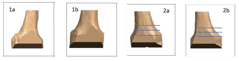
Medeiros F et al. Int J Res Orthop. 2020 Jan;6(1):7-11
morphology of the distal femur, ratio is important, which multiple assessments are analysed, and when the
represents the width of the femur, but new studies evaluation scale presents several nominal or categorical
showed that rectangular/trapezoidal variability of the options. The test was interpreted according to with
distal femur cannot be ignored. The distal femur is Altman as proportional agreement with correction of
considerably more trapezoidal than most femoral chance.10 Kappa is the coefficient of agreement whose
components, and therefore, care must be taken to avoid value varies from +1 (perfect agreement), passes through
anterior prosthetic overhang in TKA.5 Moreover, external 0 (concordance equal to chance) and -1 (complete
rotation may amplify the asymmetry between the medial disagreement). For the interpretation we used the values
and lateral condyles, and exacerbate also prosthetic described by Landis and Koch and the level of
overhang.6 There is a high anatomy variation on distal significance considered was 0.05.
femur condyles that, resulting in different shapes of
trochlear groove and respective cut. Dejour classification RESULTS
is useful to determine the trochlear shape, but insufficient
to predict the frontal cut in arthroplasties.7 To determine Our proposal classification for CAF is type 1 - one peak,
the proper femoral component rotation, several surgical (there is no valley) and type 2 - two peaks, there is one
methods have been utilised, like the transepicondylar valley (at least). The subtypes of type 1 are: 1(a) a central
line, the Whiteside line or the posterior condylar line. base peak and 1(b) a lateral base peak. The subtypes of
These methods have low interindividual reproducibility, type 2 are: 2(a) two peaks where the smallest is in the
and there is no consensus about the best method for lower half and 2(b) two peaks where the smallest is in the
frontal femoral sectioning.8 Only a few previous studies middle or upper half (Figure 1). Our sampling has an
exist on the accuracy of femoral component rotation average age of 68.7±7.9 years old. It is composed by 65%
using Patient Specific Instrumentation in Total Knee of females and 44% of left knees. The type 2(a) was the
Arthroplasty (PSI-TKA). To our knowledge, there is no most common type found in our study (54.5%, 47-67). It
study describing a shape’s classification of this important was followed by type 1b (26.5%, 15-34) and 1(a) (13%,
cut. The aim of this study was to suggest a classification 5-20). The less common type was 2(b) (6%, 3-9). Table 1
for anterior femoral cut in TKA. presents the absolute frequencies for each of the
observers and for each of their evaluations. The results
METHODS for intra-observer agreement are presented in Table 2.
The K mean value was 0.774, with a variation from 0.652
This is a descriptive and comparative study realised in (FM) to 0.886 (MM), whose interpretation is "Good
Hospital Privado do Algarve. After ethical approval, pre- Concordance"; two observers (RD and MM) were found
operative planning magnetic resonance image (MRI) of to have reached an "excellent concordance" value
230 patients who underwent TKA (right and left) during (respectively 0.831 and 0.886). The results for the inter-
2018, in our institution, were retrospectively randomized observer agreement for the first and second moment are
and analysed. We aggregate the different anterior femoral presented in Table 3. The inter-observer agreement was
cuts and define a classification, according its shape. The performed comparing the first reading of each observer,
classification system proposed by the authors was paired two by two, to make all possible combinations and
presented in detail to 5 observers prior to evaluation. One repeating the analysis after the second reading of each
medical student, one orthopaedic resident and three observer. The mean kappa coefficient at first reading was
orthopaedic surgeons voluntarily participated in the 0.563 with a range of 0.431 (FM-JPS) to 0.677 (JV-MM),
study. To attest the classification’s agreement, 100 which analysis reveals a "Moderate Concordance". The
femoral MRI image cuts of our initial sample were second reading was a mean kappa of 0.627, with a
randomly selected and analysed by them, twice with a variation of 0.487 (FM-MM) and 0.771 (FM-RD), whose
week of interval. To analyse inter and intra-observer interpretation corresponds to a "Good Concordance". The
agreement, the Fleiss Kappa test was used.9 This test is intra and inter-observer concordance was statistically
the most appropriate when multiple observers and/or significant in all the analyses.
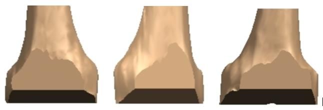
Figure 1: CAF classification: 1 (a) a central base peak, 1 (b) a lateral base peak, 2 (a) two peaks where the smallest
is in the lower half, 2 (b) two peaks where the smallest is in the middle or upper half.
International Journal of Research in Orthopaedics | January-February 2020 | Vol 6 | Issue 1 Page 8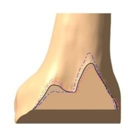
Medeiros F et al. Int J Res Orthop. 2020 Jan;6(1):7-11

         Table 1: Absolute frequencies for each of the observers and for each of their evaluations (n=1000).

                   Observers
                   FM                JPS                 RD                  JV                     MM               Total,
 Classification
                   1st    2nd        1st       2nd       1st       2nd       1st         2nd        1st      2nd     N (%)
                   Evn    Evn        Evn       Evn       Evn       Evn       Ev n        Evn        Evn      Evn
 1a                17     19         11        10        20        21        12          9          5        5       129 (12.9)
 1b                25     28         15        24        24        23        34          31         31       29      264 (26.4)
 2a                49     47         67        60        48        50        48          53         61       62      545 (54.5)
 2b                9      6          7         6         8         6         6           7          3        4       62 (6.2)
Evn: Evaluation.

                              Table 2: Statistical analyses of intra-observer agreement.

                                                                                 CI (95%)
 Observers                          Fleiss’ Kappa index (p value)
                                                                                 Inferior                       Superior
 FM                                 0.652 (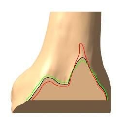
Medeiros F et al. Int J Res Orthop. 2020 Jan;6(1):7-11

native groove line (Figure 3).11 As showed in the image,            Our study has some limitations, namely the classification
6º of external rotation corresponds to type 1b, 6º of               does not cover all the shapes cut. It could be a type 3
internal rotation corresponds to type 2b, and original cut          unclassifiable, but this will be very rare, and it is more
corresponds to type 2(a).                                           beneficial that in doubt, one of the existing types should
                                                                    be chosen rather than losing the classification. This is an
a                  b                     c                          imageology-based study, without clinical testing.
                                                                    Therefore, the authors are developing a clinical study
                                                                    based on this classification. The final limitation is about
                                                                    the influence on instability patellofemoral issues, because
                                                                    other cuts could also influence them.

                                                                    CONCLUSION

 Figure 2 (a-c): Image cuts with higher disagreement                This new classification system for anterior femoral cut in
  on classification that must be classified like 1 (b).             TKA showed adequate inter-observer and intra-observer
                                                                    agreement. Therefore, we recommend applying the
                                                                    proposed classification system in clinical practice when
         a                     b                                    TKA is being done. This classification system could help
                                                                    to understand part of these problems, but more research is
                                                                    needed.

                                                                    Funding: No funding sources
                                                                    Conflict of interest: None declared
                                                                    Ethical approval: The study was approved by the
                                                                    institutional ethics committee
 Figure 3: Changes on classification’s type with axial
(a) and sagittal (b) rotation cut. 6º of external rotation          REFERENCES
     (dark blue line) corresponds to type 1(b), 6º of
  internal rotation (orange line) corresponds to type               1.    Baker PN, van der Meulen JH, Lewsy J. The role of
2(b), and original cut (black line) corresponds to type                   pain and function in determining patient satisfaction
2(a). Lines: black–original; cyan-3° external rotation;                   after total knee replacement: data from the National
    dark blue-6° external rotation; pink-3° internal                      Joint Registry for England and Wales. J Bone Joint
    rotation; orange-6° internal rotation; red-3° of                      Surg. 2007;89-B:893–900.
             extension; green-3° of flexion.                        2.    Varadarajan K, Rubash HE, Li G. Are current total
                                                                          knee arthroplasty implants designed to restore
An internal rotation between 3°-6° tends to be tolerable                  normal trochlear groove anatomy? J Arthroplasty.
and an external rotation up to 8° may not cause clinical                  2011;26:274-81.
problems, it is unknow why some patients are more                   3.    Kulkarni SK, Freeman MA, Poal-Manresa JC. The
symptomatic than others.12 Before applying femoral                        patellofemoral joint in total knee arthroplasty: is the
implants, we should be aware if a type 1(a) or 2(b) exists,               design of the trochlea the critical factor? J
because it could not simulate the native trochlea. It could               Arthroplasty. 2000;15:424–9.
be an anatomic variation, but it also could be a slight             4.    Gujarathi N, Putti AB, Abboud RJ. Risk of
rotation of the femoral cutting guide, resulting in                       periprosthetic fracture after anterior femoral
different type on our classification. Only a few previous                 notching. Acta Orthopaedica. 2009;80(5):553–6.
studies exist on the accuracy of femoral component                  5.    Bonnin M, Saffarini M, Bossard N. Morphometric
rotation using patient specific instrumentation-TKA (PSI-                 analysis of the distal femur in total knee arthroplasty
TKA). This method was recently found to be effective in                   and native knees. Bone Joint J. 2016;98-B(1):49-57.
significantly reducing outliers of optimal rotational               6.    Bonnin M, Saffarini M, Nover L. External rotation
femoral component alignment.13,14 PSI-TKA have the                        of the femoral component increases asymmetry of
advantage on these cuts when comparing with                               the posterior condyles. Bone Joint J. 2017; 99-
conventional cuts, whereas the guide rotation is more                     B(7):894-903.
frequent and can lead easily to an undesirable cut and              7.    Dejour D, Reynaud P, Lecoulte B. Douleurs et
consequently to different patellofemoral offset. Further                  instabilite rotulienne: Essai de classification. Med
investigations comparing the image of anterior cut                        Hyg. 1998;56:1466–71.
planned for PSI-TKA and the real shape of the cut                   8.    Cho Y, Lee MC. Rotational alignment in total knee
intraoperatively are critical for understanding the                       arthroplasty. AP-SMART: Asia-Pacific Journal of
influence of this cut on decreasing patellofemoral pain                   Sports Medicine, Arthroscopy, Rehabilitation and
and maltracking after TKA. The current authors are                        Technology. 2014;9(4):113-8.
presently conducting a study that addresses this point.

                                International Journal of Research in Orthopaedics | January-February 2020 | Vol 6 | Issue 1   Page 10
Medeiros F et al. Int J Res Orthop. 2020 Jan;6(1):7-11
9. Landis KG, Koch GG. The measurement of 13. Heyse T, Tibesku CO. Improved femoral
observer agreement for categorical data. component rotation in TKA using patient-specific
Biometrics.1977;33(1):159-74. instrumentation. Knee. 2014;21(1):268-71.
10. Altman DG. Practical statistic for medical research. 14. Schotanus MGM, Thijs E, Heijmans M. Favourable
3rd edition. 1995: 403-409. alignment outcomes with MRI-based patient-
11. Cho KJ, Erasmus PJ, Muller JH. The effect of axial specific instruments in total knee arthroplasty. Knee
rotation of the anterior resection plane in Surg Sports Traumatol Arthrosc.
patellofemoral arthroplasty. Knee. 2016;23(5):895– 2018;26(9):2659-68.
9.
12. Ghosh KM, Merican AM, Iranpour F. The effect of Cite this article as: Medeiros F, Duarte A, Correia
femoral component rotation on the extensor B, Carvalho M, Vide J, Fontes AP, et al. Anterior
retinaculum of the knee. J Orthop Res. femoral cut in TKA: classification proposal. Int J Res
2010;28(9):1136-41. Orthop 2020;6:7-11.
International Journal of Research in Orthopaedics | January-February 2020 | Vol 6 | Issue 1 Page 11You can also read