Double papilla repositioned flap procedure for the treatment of single tooth recession - A case report
←
→
Page content transcription
If your browser does not render page correctly, please read the page content below
International Journal of Scientific and Research Publications, Volume 11, Issue 3, March 2021                                                         528
ISSN 2250-3153

          Double papilla repositioned flap procedure for the
         treatment of single tooth recession – A case report
                        Anchal saini1, Sachit anand arora2, Rupali kalsi3 , kumar saurav4 , Fatima gilani5

                                                3rd year Postgraduate student of Department of periodontics1
                                                       Principal & HOD ,Department of periodontics2,
                                                          Professor of Department of periodontics3,
                                                           Reader of Department of periodontics4,
                                                3rd year Post Graduate student of Department of periodontics5
                           1,2,3,4,5Department of Periodontics, I.T.S Dental College, Hospital & Research Centre, Greater Noida

                                                           DOI: 10.29322/IJSRP.11.03.2021.p11175
                                                     http://dx.doi.org/10.29322/IJSRP.11.03.2021.p11175

Abstract- Recession is apical displacement of gingival margin to                     recession. Initially there is normal or subclinical inflammation,
Cemento-enamel junction which is associated with certain                             later on there is proliferation of epithelial rete pegs. In later stage
etiological factors like vigorous tooth brushing habit, Aberrant                     increased epithelial proliferation resulting in loss of CT resulting
frenum, Periodontitis, tooth position etc. Various procedures can                    in separation and recession of the gingival tissues due to loss of
be done to achieve root coverage by many procedures like Double                      nutritional supply.[3] Sullivan and Atkins (1968) Classified
papilla, Free gingival graft, connective tissue graft, pedicle grafts.               recession into according to their morphology but it was not useful
Cohen and Ross described Double papilla procedure with success                       to predict outcome of root coverage procedure thus later on
rate of 80% in covering denuded root surface. It is mostly used for                  Miller’s (1985) gave a classification which is based on two things
single tooth recession to increase width of attached gingiva along                   Firstly it describes extent of gingival recession defect, and another
with root coverage. This procedure is little sensitive as two                        is extent of soft & hard tissue loss. [4]
adjacent interdental papilla need to be joined on mid surface of                            Interdental bone loss is more resistant than radicular bone,
denude root to make it one flap.                                                     clinical predictability is good, good colour tissue match are the
                                                                                     main advantages of Double papilla. [5]
Index Terms- Gingival recession, Root Coverage, Double papilla
                                                                                     Case Report
                                                                                            A male patient name Munir, 37 years age came to our
                               I. INTRODUCTION                                       Department of Periodontics, I.T.S Dental College & Hospital
                                                                                     Greater Noida with a chief complaint of recession of gum in
G    ingival recession is apical displacement of gingival margins
     to Cemento-enamel junction (CEJ) . It can be localized or
generalized and associated with factors like inadequate tooth
                                                                                     relation to the Upper left maxillary lateral. (Figure1,2)
                                                                                            On examination lymph nodes were non palpable, lips were
brushing, aberrant frenum pull, periodontitis, high muscle                           competent and face was bilaterally symmetrical. On intraoral
attachment, iatrogenic factors, and smoking. [1]                                     examination, the tooth showed gingival marginal recession of
      Baker and Seymour (1976) [2] described pathogenesis of                         4mm. On the buccal aspect with loss of interdental papilla between
recession and explained different stages in development of                           central and lateral incisor teeth. Recession was classified as class-
                                                                                     Class-II gingival recession according to millers classification

 This publication is licensed under Creative Commons Attribution CC BY.
 http://dx.doi.org/10.29322/IJSRP.11.03.2021.p11175                                                                                      www.ijsrp.org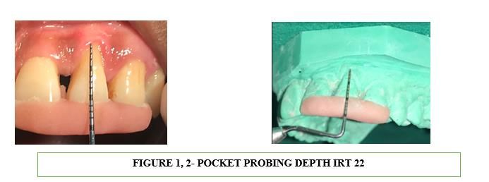
International Journal of Scientific and Research Publications, Volume 11, Issue 3, March 2021 529
ISSN 2250-3153
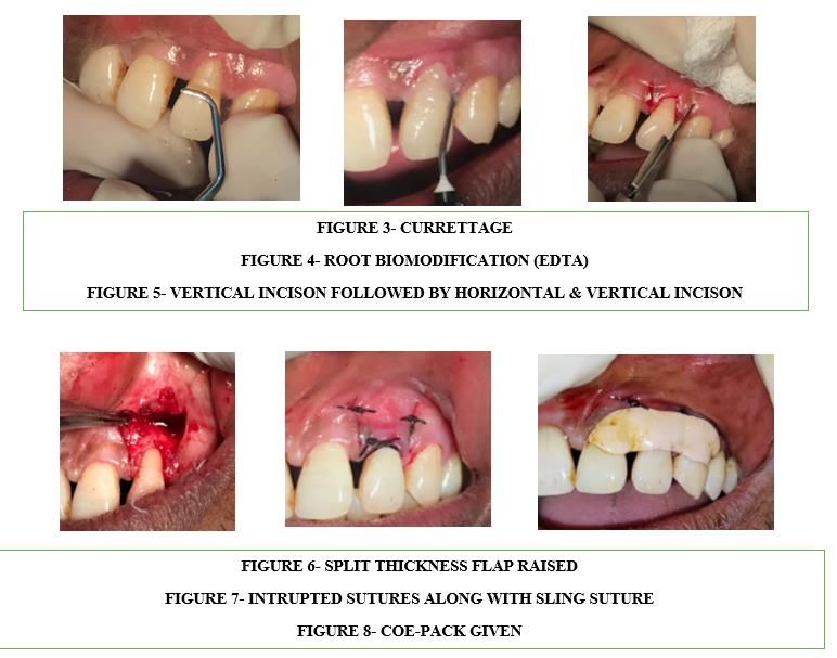
Interrupted with sling sutures were used to achieve proper
II. SURGICAL PROCEDURE stabilization on the mesial and distal papilla using a silk suture
Firstly curettage was done to plane exposed root surface, (figure 7). Pressure for 5 min applied with gloved finger for
followed by root bio modification with EDTA (figure 3,4). Then homeostasis followed by periodontal dressing. Post-surgical
Blade no. 15 was used to make a V-shaped incision on the instructions were given to the patient (figure 8). The patient was
recession tooth. Horizontal followed by Vertical incision was told not to brush the operated area and was advised to use
given to the mesial and distal interdental papilla(figure 5). Then chlorhexidine gluconate mouth wash of 0.2% twice daily for two
partial-thickness pedicle flap elevated until the tissue is mobile so weeks.
we can be suture it on new desired position. (figure 6)
This publication is licensed under Creative Commons Attribution CC BY.
http://dx.doi.org/10.29322/IJSRP.11.03.2021.p11175 www.ijsrp.org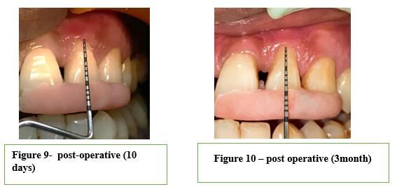
International Journal of Scientific and Research Publications, Volume 11, Issue 3, March 2021                                                    530
ISSN 2250-3153

                                                                                     along with root coverage gain of 1mm. Patient is recalled on 3, 6
                                 III. RESULTS                                        month for follow upto 1 year. Oral hygiene instruction was given.
      Sutures and coe-pack was removed after 10 days followed                        (Figure 9,10)
by clinical examination. Surgical site showed Complete healing                             On 3 month surgical site showed complete healing, with
                                                                                     increase width of keratinized along with root coverage of 1mm.

 This publication is licensed under Creative Commons Attribution CC BY.
 http://dx.doi.org/10.29322/IJSRP.11.03.2021.p11175                                                                                 www.ijsrp.org
International Journal of Scientific and Research Publications, Volume 11, Issue 3, March 2021 531
ISSN 2250-3153
with other techniques as this procedure is mainly done to increase
IV. DISCUSSION width of keratinized gingiva. [10]
Gingival recession can cause aesthetic or hypersensitivity
problems thus necessitate the need for root coverage [6]. Various V. CONCLUSION
techniques can be done to cover recession defects [7] such as Free Clinician should be aware of the procedure to be done for
gingival graft (FGG), Laterally positioned flap (LPG), Double particular type of gingival recession. From this case report it is
papilla flap, Guided tissue regeneration(GTR), and allograft. The concluded that Double papilla flap procedure when done for single
selection of the procedure depends on degree of recession, teeth tooth recession showed predictable root coverage along with good
involved, width of attached gingiva and postoperative colour colour matching with adjacent tissue when treated for class II
harmony. recession
To regenerate periodontal tissue first connective tissue
attachment to root is achieved by doing root bio modification, REFERENCES
generally by EDTA. It is a chelating agent which exposes the [1] [1] Mythri S, Arunkumar SM, Hegde S, Rajesh SK, Munaz M, Ashwin D.
collagen to enhance connective attachment to root surface and Etiology and occurrence of gingival recession - An epidemiological study. J
shows enhanced wound healing when compared with other agents. Indian Soc Periodontol. 2015;19(6):671-5.
Kassab MM (2006) [8] study showed significant root coverage [2] [2] Baker DL, Seymour GJ. The possible pathogenesis of gingival recession.
A histological study of induced recession in the rat. J Clin Periodontol. 1976
when EDTA was applied before regenerative procedure, whereas Nov;3(4):208-19.
Modica et al (2000) [9] showed no significant changes when [3] [3] Ravipudi S, Appukuttan S, Prakash P.S.G., Victor D.J. Gingival
EDTA was applied prior to surgical procedure. Recession: Short Literature Review on Etiology, Classifications and Various
This case report describes double papillae pedicle graft Treatment Options. J. Pharm. Sci. & Res. Vol. 9(2), 2017, 215-220
surgical technique for the treatment of single tooth marginal tissue [4] [4] P.D. Miller. A classification of marginal gingival recession, Int. J.
recession, and to increase width of attached gingiva, It was Periodont. Restor. Dent. 5 (1985) 9
introduced by Cohen and ross (1968) [10] in which two [5] [5] Cohen D W, Ross SE. The double papillae repositioned flap in
periodontal therapy, J. Periodontol. 39 (1968) 65–70.
interproximal papilla were joined on mid surface of teeth to cover
[6] [6] Cmargo PM, Melnick PR, Kenney EB. The use of free gingival grafts
recession in areas of insufficient gingiva. In this study Double for aesthetic purposes. Periodontol 2000. 2001;27 (1): 72-96
papillae pedicle graft showed excellent root coverage when it was [7] [7] Cohen ES. Atlas of cosmetic & reconstructive periodontal surgery.
done correctly, following all the indication of this technique. Philadelphia, Williams & Wilkins, 2nd Ed; 65-135.
Acunzo R et al (2015) [11] study showed 88% root coverage with [8] [8] Kassab MM, Cohen RE, Andreana S, Dentino AR. The effect of EDTA
increase keratinized tissue when Double papilla flap procedure in attachment gain and root coverage. Compend Contin Educ Dent. 2006
Jun;27(6):353-60
was done for isolated tooth defect, Similar results was shown in a
[9] [9] Modica F, Del Pizzo M, Roccuzzo M, Romagnoli R. Coronally
study done by Manisundar N et al (2014) [12]. advanced flap for the treatment of buccal gingival recessions with and
Other procedures can also be combined with double without enamel matrix derivatives. J Periodontol. 2000;71:1693–8
papilla for better results. Harris RJ (2002) [13] and Sunil S et al [10] [10] Kumar PM, Reddy NR, Kumar SS, Chakrapani S. Double papilla flap
(2017) [14] and Benjamin Tanet al (2003) [15] treated recession technique for dual purpose. J Orofac Sci 2012;4:75-8.
defect with connective tissue graft along with double papilla, [11] [11] Acunzo R, Pagni G, Fessi S, Rasperini G. Modified double papillae
results showed root coverage along with increased amount of flap technique: a new surgical approach for the treatment of isolated gingival
recession defects. A case series. Int J Esthet Dent. 2015;10:258–68
keratinized tissue (3mm vs 1.8mm). There are few limitations of
[12] [12] Manisundar N, Paddmanaban P, Ramya V , Bhuvaneswarri J and
this procedure such as technique sensitive as it is difficult to join Hemalatha V.T. Double Papillary Flap - A Treatment for Gingival Recession.
the two adjacent papilla on the mid surface of tooth to make it one World Journal of Medical Sciences 2014; 10 (2): 117-121
flap, Complete Root coverage difficult to obtain unless combined
This publication is licensed under Creative Commons Attribution CC BY.
http://dx.doi.org/10.29322/IJSRP.11.03.2021.p11175 www.ijsrp.orgInternational Journal of Scientific and Research Publications, Volume 11, Issue 3, March 2021 532
ISSN 2250-3153
[13] [13] Harris RJ. Double pedicle flap-predictability and aesthetics using Dental College, Hospital & Research Centre, Greater Noida,
connective tissue. Periodontology 2000 1996;11:39-48
prin.dntl.gn@its.edu.in
[14] [14] Sunil S, Babu HM. Root Coverage using Double Papilla with
Connective Tissue Graft: A 13-month Report of a Successful Case. Journal
Third Author – Rupali kalsi, Professor of Department of
of Health Sciences & Research, 2017;8(2):77-79 periodontics, Department of Periodontics, I.T.S Dental College,
[15] [15] Benjamin Tan, Phay Yew Ming. Partial thickness double papilla flap Hospital & Research Centre, Greater Noida,
with connective tissue graft- case reports. Singapore Dental Journal 2003 ; drrupalikalsi.dntl.gn@its.edu.in
25(1) : 95-100. Fourth Author – kumar saurav, Reader of Department of
periodontics, Department of Periodontics, I.T.S Dental College,
Hospital & Research Centre, Greater Noida,
AUTHORS dr.kumarsauravsingh@its.edu.in
First Author – Anchal saini, 3rd year Postgraduate student of Fifth Author – Fatima gilani, 3rd year Post Graduate student of
Department of periodontics, Department of Periodontics, I.T.S Department of periodontics, Department of Periodontics, I.T.S
Dental College, Hospital & Research Centre, Greater Noida, Dental College, Hospital & Research Centre, Greater Noida,
dranchalsaini94@gmail.com fatimagilani_mds18_21_gn@its.edu.in
Second Author – Sachit anand arora, Principal & HOD
,Department of periodontics, Department of Periodontics, I.T.S Corresponding Author: Saini Anchal
dranchalsaini94@gmail.com
This publication is licensed under Creative Commons Attribution CC BY.
http://dx.doi.org/10.29322/IJSRP.11.03.2021.p11175 www.ijsrp.orgYou can also read