Dynamics in the hemomicrocirculatory bed of the cattle honeycomb bag
←
→
Page content transcription
If your browser does not render page correctly, please read the page content below
E3S Web of Conferences 273, 02023 (2021)                                  https://doi.org/10.1051/e3sconf/202127302023
INTERAGROMASH 2021

         Dynamics in the hemomicrocirculatory bed of
         the cattle honeycomb bag
         Valentina Shpygova1,*, Olga Dilekova1, Viktor Mikhaylenko1, Vladimir Meshcheryakov1,
         and Nikolai Agarkov1
         1
         Stavropol State Agrarian University, Zootekhnichesky lane, 12, Stavropol, 355017, Russia

                       Abstract. The study was conducted on 60 heads of black-and-white cattle
                       of six age groups: 1 day, 30 days, 3 months, 6 months, 18 months and
                       cows 4-5 years old. Injection, morphological and morphometric methods
                       were used. It was found that the hemomicrocirculatory bed of the
                       honeycomb bag has typical links. The diameter of arterioles in the first-
                       order ridges increases by 30.5% during the study period, but significant
                       changes are observed only between the age groups of 3 months – 6 months,
                       6 months-18 months, these differences are 8.3% and 10.7%, respectively.
                       In the capillary link, significant changes are observed between the age
                       groups of 6 months-18 months and 18 months-4-5 years, which is 10.0%
                       and 24.8%, respectively. In the link of postcapillary venules, the diameter
                       of the vessels during the study period increases by 16.9%. In the link of
                       collecting venules, significant changes (p≤0.01) in the diameter of vessels
                       were observed between the age groups of 3-6 months and 6-18 months,
                       which is 22.9% and 8.2%, respectively. In lactating cows (4-5 years old),
                       in addition to wide capillaries, we found postcapillary venules with a
                       diameter of 14.80 to 27.20 µ in the papillae of the lamina mucosa propria.

         1 Introduction
             The uniqueness of ruminants is in their ability to convert roughages into products with
         high nutritional value. The peculiarities of feed intake, digestion, distribution, role in the
         ecosystem, as well as the phylogenetic relationship of ruminant species do not cease to
         interest scientists who seek to use the new information for increasing their productivity and
         find ways to improve ruminants’ genotype [1,2]. The variety of existing
         morphophysiological systems of ruminants can be considered as a pool from which some
         traits can be selected as targets for breeding programs for domestic ruminants [3,4,5,6]. The
         use of industrial technologies in dairy farming involves the thorough grinding of feeds, that
         helps to improve the efficiency of digestion, but affects the relief of the mucous membranes
         of the pre-ventricles, especially the honeycomb bag that performs the sorting function
         [7,8,9]. The mucosa of the adult animals’ honeycomb bag forms first-order ridges and may
         have from four to eight facets where lower second- and third-order ridges are formed. On
         the bottom and in the ridges of the cells there is a large number of conical shaped papillae.
         To date, it is a priority opinion that the mechanism of sorting the contents of the

         *
             Corresponding author: spygova@yandex.ru

   © The Authors, published by EDP Sciences. This is an open access article distributed under the terms of the Creative
   Commons Attribution License 4.0 (http://creativecommons.org/licenses/by/4.0/).
E3S Web of Conferences 273, 02023 (2021)                             https://doi.org/10.1051/e3sconf/202127302023
INTERAGROMASH 2021

         honeycomb bag is based on the physical separation of its contents, including the processes
         of flotation and sedimentation [10, 11]. In the mesh, the musculature of the mucosa plays
         an important role in the contraction of the cell ridges. The contraction of the ridges and the
         honeycomb bag walls are synchronous. Synchronism in the contraction of ridges is due to
         the specific location of their muscles [1]. The similarity in the macroscopic anatomy of the
         honeycomb bag was revealed in different species of wild ruminants, which differ in the
         type of feeding.
             Herbivores face the dilemma that the level of feed intake is negatively related to factors
         such as the thoroughness of grinding feed while chewing and keeping it in the digestive
         tract. [11,12,13]. In addition to the sorting function, the honeycomb bag of ruminants has
         the function of absorbing nutrients, but to a much less extent than the rumen [14]. The
         absorption of nutrients occurs in the metabolic link of the hemomicrocirculatory bed of the
         mucous membrane. The sources of blood supply in the multicameral stomach and the blood
         supply of the chambers are described in a large number of ruminants [15,16], and the
         relationship between the microcirculatory bed of the lamina mucosa propria and the deep
         layers of the pre-ventricular epithelium is abundant in the papillae of the rumen,
         honeycomb bag and omasum [1]. The study of age-related morphometric features of the
         microcirculatory bed has become the aim of our research.

         2 Materials and Methods
         The material for the research was 60 heads of black-and-white cattle of six age groups: 1
         day, 30 days, 3 months, 6 months, 18 months and cows 4-5 years old. The slaughter of
         calves aged from 1 day to 3 months was carried out for scientific and production purposes
         in the conditions of slaughterhouses. The slaughter of animals of older age groups was
         carried out in order to obtain livestock products. All manipulations on selection of
         experimental material were carried out according the Directive 2010/63/EU of the
         European Parliament and the Council of the European Union 2010/09/22 “On Protection of
         Animals Used for Scientific Purposes”.
             To detect the submucosal plexus of the blood bed of the honeycomb bag, the vessels
         were injected with a contrast suspension prepared according to the prescription: glycerin -
         100 ml, barium sulfate (BaSO4) - 100 g, aqua destillata – 400-500 ml. The contrast
         suspension was injected at a pressure of 110-120 mm through a device with a pressure
         gauge. For injection into the veins, a white suspension was used, and before pouring into
         the arteries, a red pigment was added to it. After settling of the suspension particles and its
         solidification, the organ (mesh) was subjected to stratification into the mucosal and
         submucosal, muscular and serous layers. Histological sections were prepared in the
         classical way, stained with hematoxylin-eosin, iron hemnoxylin-picrofuxin according to
         Van Gieson, resorcinol-fuchsin according to Weigert, phosphor-tungsten hematoxylin
         according to Mallory. Histological preparations were examined using an OLYMPUS-BX
         43 light microscope (Japan) and an OLYMPUS C 300 camera (Japan). Vascular
         morphometry was performed in the program "Video Test - master Morphology 4.0" for
         Windows (Russia).

         3 Results
         The data obtained during the study of the hemomicrocirculatory bed of the honeycomb bag
         wall indicated its typical structure and age-related features.
             Intrahepatic arteries of the muscular type, which supply blood to the mesh, passing into
         the submucosal layer, successively give branches to the serous membrane. There they form

                                                       2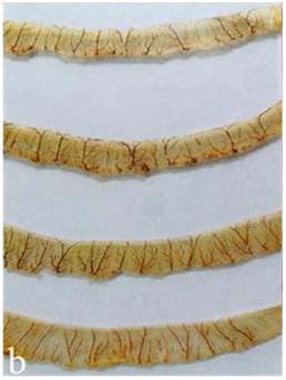
E3S Web of Conferences 273, 02023 (2021)                               https://doi.org/10.1051/e3sconf/202127302023
INTERAGROMASH 2021

         the subsurface plexus. Then, in the submucosal layer an arterial plexus is formed, which
         supplies blood to the muscle and mucous membranes. The hemomicrocirculatory bed of the
         serous membrane represents repeating geometric figure of polygonal shape with a diameter
         of arterioles from 20 to 40 µ, where precapillary arterioles, or metarterioles, depart. The
         collateral arteriole often passes with an arteriole of a larger caliber. In this paper we do not
         provide complete data on the morphometry of the vessels of the microcirculatory bed of the
         serous membrane, but it should be noted that its main age characteristic is an increase in the
         density of the capillary link per square unit of area, thus, in adult animals, the capillary link
         of the module prevails.
             From the submucosal plexus, arterial vessels of various calibers are directed to the
         muscle membrane and form an intermuscular plexus in it, where all five links of the
         hemomicrocirculatory bed can be distinguished (fig. 1).

         Fig. 1. Dynamics in the vessels diameters of the hemomicrocirculatory bed of the honeycomb bag
         muscular membrane.
             An increase in the number of arterioles with a large diameter is observed in the groups
         of calves of three-month and six-month age. The average diameter of arterioles
         significantly (p < 0.01) increases by 54.3% and 12.3% respectively, compared to the
         previous age groups of calves. In older age groups, there is a statistical stability in this
         parameter. During the study period, there were no significant changes in the link of
         precapillary arterioles. Their diameters within the variation series have a minimum
         maximum value from 12.3 to 18.7 µ. In the capillary link, there are significant changes
         (p≤0.01) only between groups of calves aged 1 day and 1 month, that is 34.1%. However,
         in adult animals (4-5 years old) compared to newborns, the diameter of the capillaries is
         43,0% larger. The minimum diameter is noted in newborns of 3.6 µ, the maximum in cows
         of 4-5 years – 8.7 µ, with average statistical values of 5.02±0.15 µ and 7.18±0.17 µ,
         respectively. In the link of postcapillary venules, significant changes (p≤0.01) were also
         observed only between groups of calves aged 1 day and 1 month, which is 9.5%. However,
         over the entire study period, this morphometric parameter increases by 16.9%. In the link of
         collecting venules, significant differences (p≤0.01) were observed between the age groups
         of 1 month – 3 months, 3 months - 6 months and 6 months-18 months. These differences
         are 9.4%, 55.2% and 10.8% respectively.

                                                         3
E3S Web of Conferences 273, 02023 (2021) https://doi.org/10.1051/e3sconf/202127302023
INTERAGROMASH 2021
In the submucosal plexus organ artery branched to the third order at the age of 1 month,
to the fourth order at the age of 3 months, up to the fifth order at the age of 6 months, up to
the sixth order at the age of eighteen months and older. The vessels form polygonal
networks, oriented mainly along the base of the cells. Vessels detected by contrast
suspension filling have a diameter of up to 50 µ (fig. 2).
Fig. 2. a) submucosal arterial plexus of the honeycomb bag; b) arterial vessels with different levels of
division in the ridges of the first-order reticulum cells, male, age 6 months.
In newborns and animals aged 1 month, at the base of the first-order cells ridges, there
are muscle-type arteries with a diameter of 100 to 150 µ. In newborns at the age of 1 day,
the second-order ridges are nourished from arterioles with a diameter of 48 to 100 µ that
pass parallel to each other, across the ridge of the cells at their base. The vessels form
polygonal networks, oriented mainly along the base of the cells. Vessels detected by
contrast suspension filling have a diameter of up to 50 µ. In animals aged six to eighteen
months and older, muscle-type arteries with a diameter of 300 to 600 µ pass at the base of
the first-order cells ridges. Arterioles of various calibers are directed into the mucosa of the
honeycomb bag from the vessels of the submucosal plexus, which are divided into
capillaries in their lamina mucosa propria. In newborn animals, the papillae include 1-2
papillary arterioles, in adult animals there are 5-7 of them. During the study period, the
diameter of arterioles of the first-order ridges increases by 30.5%, but significant changes
are observed only between the age groups of 3 months – 6 months, 6 months-18 months,
and these differences are 8.3% and 10.7%, respectively (fig. 3). Arterioles give off
precapillary arterioles, or metarterioles, along the entire length, which then disintegrate
into metarterioles and capillaries. The arteriole itself also passes into the metarteriole. Due
to the many anastomoses of arterioles with muscle-type arteries, metarterioles, and
capillaries, the conditions for blood delivery to the capillary pool are created, and the
disruption of the functioning of each vessel can be compensated by the work of neighboring
arterioles due to the redistribution of blood through the anastomoses. There were no
significant changes in the diameter of the precapillary arterioles between the age groups,
but during the study period, the average values of this parameter increased by 11.7%.
Precapillary arterioles are divided in the lamina mucosa propria of the honeycomb bag
ridges cells of various levels, the cone-shaped and spherical papillae of the bottom and in
4E3S Web of Conferences 273, 02023 (2021) https://doi.org/10.1051/e3sconf/202127302023
INTERAGROMASH 2021
the interstitial spaces to capillaries with a diameter of 6.56±2.78 µ in newborn calves of one
day age and a diameter of 7.70±1.29 µ in adult animals of 4-5 years old.
Fig. 3. Dynamics in vessel diameters of the hemomicrocirculatory bed of the mucous membrane in
the honeycomb bag.
In the capillary link, significant changes were observed between the age groups of 6
months – 18 months and 18 months-4-5 years, that is 10.0% and 24.8%, respectively (fig.
4). Animals of 4-5 years old were lactating cows.
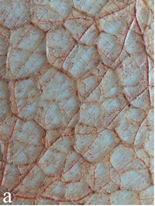
Fig. 4. Capillaries in the papillae of the lamina mucosa propria in the first-order cells’ plica, where: a)
Staining with hematoxylin and eosin; b) staining by Van Gieson; cow, age 4 years. Zoom × 400.
In the link of postcapillary venules, the vessel diameters do not significantly differ
between age groups, but during the study period this morphometric parameter increases by
5E3S Web of Conferences 273, 02023 (2021) https://doi.org/10.1051/e3sconf/202127302023
INTERAGROMASH 2021
16.9%. Total number of vessels per unit area increases as well. In the link of collecting
venules, significant changes (p≤0.01) in the diameter of vessels were observed between the
age groups of 3-6 months and 6-18 months, that is 22.9% and 8.2%, respectively.
4 Сonclusions
The hemomicrocirculatory bed of the honeycomb bag has typical links: arterioles,
precapillaries, capillaries, postcapillaries, collecting venules. Morphofunctional changes in
each layer of the honeycomb bag take place most significantly during the period of
intensive growth of epithelial connective tissue formations (ridges) of the honeycomb bag
mucosa, so the dynamics of morphometric parameters of the hemomicrocirculatory bed in
this mucous membrane is most noticeable. During the study period, the diameter of
arterioles in the first-order ridges increases by 30.5%, but significant changes are observed
only between the age groups of 3 months – 6 months, 6 months-18 months, these
differences are 8.3% and 10.7%, respectively. In animals of different age groups, there are
no significant changes in the diameter of precapillary arterioles, but during the study period,
the average values of this parameter in cows of 4-5 years old are 11.7% higher than in
newborn calves. In the capillary link, significant changes are observed between the age
groups of 6 months-18 months and 18 months-4-5 years, that are 10.0% and 24.8%,
respectively. In the link of postcapillary venules, the vessel diameters between the age
groups did not significantly differ, but during the study period, this morphometric
parameter increased by 16.9%. In the link of collecting venules, significant changes
(p≤0.01) in the diameter of vessels were observed between the age groups of 3-6 months
and 6-18 months, which is 22.9% and 8.2%, respectively. Cattle honeycomb bag performs
the function of absorbing nutrients in a smaller volume than the rumen. Perhaps this
explains the fact that in the papillae of the lamina mucosa propria in lactating cows, besides
wide capillaries, postcapillary venules with a diameter of 14.80 to 27.20 µ have been found.
Subepithelialally, postcapillaries are formed in the mucosa when several capillaries merge,
as well as 2-4 collecting venules in young animals and 4-10 or more collecting venules in
adult animals.
References
1. W. Perez, H.E. Koenig, H. Jerbi et al., Vertebrate zoology 66(3), 419–425 (2016)
2. M. Clauss, R.R. Hofmann, Ecology, evolution and behaviour of wild cattle:
implications for conservation (2014) DOI:
https://doi.org/10.1017/CBO9781139568098.008
3. K.H. Baker, H.W.I. Gray, V. Ramovs et al., Heredity 119(1), 16-26 (2017) DOI:
10.1038/hdy.2017.11
4. M. Clauss, J. Fritz, A. Tschuor et al., Journal of animal physiology and animal
nutrition 101(1), 61-69 (2017) DOI: 10.1111/jpn.12505
5. J. Wang, H. Li, L. Zhang, Y. Zhang, M. Yue, B. Shao, J. Wang, Int. J. Morphol. 32(3),
871-881 (2014) DOI: http://dx.doi.org/10.4067/S0717-95022014000300021
6. W. Pérez, S. Erdogan, R. Ungerfeld, Anat. Histol. Embryol 44(1), 43–49 (2015) DOI:
10.1111/ahe.12106
7. Franco, J. Masot, A. García, E. Redondo, Anat. Histol. Embryol 41(5), 362-73 (2012)
DOI: 10.1111/j.1439-0264.2012.01146.x
8. H. Jerbi, M. Bayoudh, M. Clauss, W. Pérez, Int. J. Morphol. 34(4), 1266–1270 (2016)
http://dx.doi.org/10.1590/S1806-92902017000700008
6E3S Web of Conferences 273, 02023 (2021) https://doi.org/10.1051/e3sconf/202127302023
INTERAGROMASH 2021
9. E. Redondo, A. Garcia, C. Ortega, F.J. Pena, A. Gazquez, J. Masot, Animal science
journal 91(1), e13319 (2020) DOI: 10.1111/asj.13319
10. A. Garcia, P. Rodriguez, J. Masot, A. Franco, E. Redondo, Animal Science Journal
85(11), 951-962 (2014) https://doi.org/10.1111/asj.12231
11. M. Clauss, J. Hummel, Revista Brasileira de Zootecnia 46(7), 606–613 (2017)
http://dx.doi.org/10.1590/S1806-92902017000700008
12. P. Hejcmanová, S. Ortmann, L. Stoklasová, M. Clauss, Comparative Biochemistry and
Physiology Part A: Molecular & Integrative Physiology 246, 110720 (2020)
https://doi.org/10.1016/j.cbpa.2020.110720
13. J. Hummel, S. Hammer, C. Hammer, J. Ruf, M. Lechenne, M. Clauss, Comp.
Biochem. Physiol. A 182, 22-26 (2015) https://doi.org/10.1016/j.cbpa.2014.12.006
14. R. Rackwitz, F. Dengler, G. Gabel, Animals 10(12), 2198 (2020) DOI:
10.3390/ani10122198
15. W. Pérez, N. Vazquez, R. Ungerfeld, Anat. Histol. Embryol 45(3), 240–245 (2016)
DOI: 10.1111/ahe.12192
16. V.M. Shpygova, O.V. Dilekova, V.V. Mikhaylenko, V.A. Meshcheryakov, N.A.
Pisarenko, Research Journal of Pharmaceutical, Biological and Chemical Sciences
9(6), 1234–1238 (2018)
7You can also read