Evaluation of glycemic abnormalities in children and adolescents with β- thalassemia major - Egyptian Pediatric Association ...
←
→
Page content transcription
If your browser does not render page correctly, please read the page content below
M. Diab et al. Egyptian Pediatric Association Gazette                 (2021) 69:9
https://doi.org/10.1186/s43054-021-00052-4
                                                                                                                                           Egyptian Pediatric
                                                                                                                                          Association Gazette

 RESEARCH                                                                                                                                               Open Access

Evaluation of glycemic abnormalities in
children and adolescents with β-
thalassemia major
Aliaa M. Diab1*, Ghada S. Abdelmotaleb1, Khaled Abdel-Azim Eid1, Enas Sebaey S. Mostafa2 and
Eman Sabry Ahmed1

  Abstract
  Background: The quality of life of B-thalassemia major (β-TM) patients has improved with the use of frequent
  blood transfusions. However, this leads to chronic iron overload with its sequelae, as prediabetes and diabetes
  mellitus. This study aimed to assess insulin resistance and glucose abnormalities in a sample of B-thalassemia major
  patients in Benha, Egypt.
  Results: This case-control study included 40 B-thalassemia major patients on regular blood transfusion and iron
  chelation. Their ages ranged from 8 to 16 years, and 30 normal age and sex-matched controls. Thorough clinical
  examination was performed including weight (kg), height (m), body mass index (BMI) (kg/m2), and liver and spleen
  size. Laboratory investigations were done in the form of complete blood count, liver enzymes, serum ferritin, fasting
  plasma insulin, and fasting, and 2 h postprandial plasma glucose. Insulin resistance (IR) was calculated using the
  Homeostasis Model Assessment of insulin resistance (HOMA-IR) index. Insulin resistance was found in 27.5% of
  thalassemic patients; 18.2% of them had diabetes, 72.7% were prediabetics (with impaired fasting glycemia), and
  9.1% had normal fasting and 2 h postprandial plasma glucose level. Insulin resistance increased significantly with
  increased blood transfusion duration, serum ferritin, liver enzymes, fasting plasma insulin, fasting plasma glucose,
  and 2 h postprandial plasma glucose (ROC). The curve analysis showed that the duration of blood transfusion,
  serum ferritin, fasting plasma insulin, fasting, and 2 h postprandial plasma glucose could significantly predict insulin
  resistance at a certain cut-off point.
  Conclusion: Our data show that HOMA-IR can be used to detect insulin resistance in β-TM patients on long-term
  blood transfusions, especially patients with high serum ferritin and impaired liver enzymes.
  Keywords: Glycemic abnormalities, Diabetes, Insulin resistance, Thalassemia major, B-Thalassemia

Background                                                                                 overload. The precise mechanism of iron-induced dia-
B-Thalassemia major (β-TM) patients’ life span and                                         betes is still unknown, but these three mechanisms are
quality are highly dependent on regular blood transfu-                                     most likely to occur: insulin deficiency, insulin resistance
sion, which comes with the cost of iron overload [1].                                      (IR), and liver dysfunction. Evidence suggests that the
Despite advances in iron-chelating agents, iron overload                                   most crucial factor in the pathogenesis of the disease’s
remains a significant challenge in managing β-TM pa-                                       clinical complications is oxidative stress (caused by iron
tients [2]. Diabetes mellitus (DM) is one of the import-                                   accumulation) [3]. Both IR and impaired insulin secre-
ant endocrinal disorders that happen due to iron                                           tion lead to impaired glucose tolerance and type 2 dia-
                                                                                           betes mellitus (T2DM) [4].
* Correspondence: aliaadiab@yahoo.com                                                        It is said that iron excess and its related oxidative
1
 Pediatric Department, Benha University Hospital, Benha, Egypt
Full list of author information is available at the end of the article                     stress can mediate pancreatic islet cell apoptosis

                                           © The Author(s). 2021 Open Access This article is licensed under a Creative Commons Attribution 4.0 International License,
                                           which permits use, sharing, adaptation, distribution and reproduction in any medium or format, as long as you give
                                           appropriate credit to the original author(s) and the source, provide a link to the Creative Commons licence, and indicate if
                                           changes were made. The images or other third party material in this article are included in the article's Creative Commons
                                           licence, unless indicated otherwise in a credit line to the material. If material is not included in the article's Creative Commons
                                           licence and your intended use is not permitted by statutory regulation or exceeds the permitted use, you will need to obtain
                                           permission directly from the copyright holder. To view a copy of this licence, visit http://creativecommons.org/licenses/by/4.0/.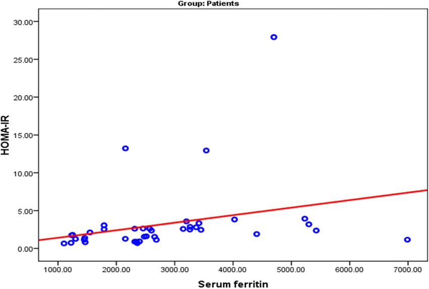
M. Diab et al. Egyptian Pediatric Association Gazette   (2021) 69:9                                                   Page 2 of 7

resulting in reduced insulin secretory capacity [5]. Islet            aspartate aminotransferase (AST) (performed by Biosys-
cells are also very susceptible to oxidative damage due to            tem A 15 autoanalyzer), and serum ferritin (performed
an almost exclusive dependence on mitochondrial glu-                  by AIA 15 fluorescence, chemiluminescent immunoassay
cose metabolism to secrete glucose-induced insulin and                system, TOSOH Corporation, Tokyo, JAPAN). More-
have a flawed system of antioxidant defense [6]. Re-                  over, fasting plasma glucose was obtained after 8 h of
cently, McClain and colleagues have demonstrated a                    fasting (3 ml of venous blood (done by Glucose TR,
high prevalence of irregular homeostasis of glucose in                SPINREACT), and at the same time, fasting plasma insu-
patients with hemochromatosis as well as impaired insu-               lin level was collected (measured by DRG® Insulin
lin secretion and insulin resistance [7].                             ELISA, USA (EIA-2935)), then after taking a meal, 2 h
   In several pathophysiological states, IR broadly occurs.           postprandial plasma glucose (2 h pp plasma glucose) was
Several researchers have indicated that IR is already                 obtained (2 ml of venous blood (done by Glucose TR,
present in diabetic patients before blood glucose irregu-             SPINREACT)).
larities. Hyperinsulinemia and IGT, in other words, are                  DM and prediabetes diagnosis is based on the Ameri-
both T2DM reserve forces. Even in subjects with normal                can Diabetes Association criteria [10]:
glucose tolerance [8], hyperinsulinemia and IR are dan-                  -Prediabetes (impaired fasting glycemia (IFG)) was di-
gerous. Homeostasis Model Assessment (HOMA), which                    agnosed if fasting plasma glucose was 100-125 mg/dL
involves the measurement of only fasting plasma insulin               (5.6-6.9 mmol/L).
and fasting plasma glucose, is the easiest and most                      -Diabetes was diagnosed if fasting plasma glucose was
widely used marker in clinical practice to measure IR                 > 126 mg/dL (11.1 mmol/L).
[9].                                                                     Evaluation of the IR index was done using the Homeo-
                                                                      stasis Model Assessment (HOMA-IR) = (fasting plasma
Aim of the study                                                      glucose (mmol/L) X fasting plasma insulin (μu/mL))/
We aimed in this study to evaluate IR and glucose ab-                 22.5). HOMA-IR value of ≥ 2.7 was considered to be an
normalities in a sample of β-TM patients in Benha,                    indicator of IR [11].
Egypt.
                                                                      Statistical methods
Methods                                                               The collected data were tabulated and analyzed using
Subjects                                                              SPSS version 16 software (SpssInc, Chicago, ILL Com-
This case-control study was conducted on 40 children                  pany). Categorical data were presented as numbers and
and adolescents with β-TM (27 males and 13 females),                  percentages, chi-square (χ2) and Fisher’s exact tests were
aged between 8 and 16 years and 30 normal, age and                    used to analyze them. Quantitative data were tested for
sex-matched controls. The diagnosis of the patients was               normality using the Shapiro-Wilks test, assuming nor-
confirmed by Hb electrophoresis. They were regularly                  mality at P > 0.05. Normally distributed variables were
transfused at the hematology units of the Pediatric De-               expressed as mean ± standard deviation and analyzed by
partments, at Benha University Hospitals and Benha                    St. “t” for two independent groups. Simultaneously, non-
Children’s Hospital. Informed consent was taken before                parametric data were presented as a median and inter-
conducting the study from parents of both cases and                   quartile range (IQR) and analyzed by Mann Whitney U
controls.                                                             test (ZMWU) test. Spearman’s correlation coefficient
  This research was approved by the ethical committee,                (rho) was used to assess the correlation between non-
Faculty of Medicine, Benha University.                                parametric variables. Receiver operator characteristic
  Patients suffering from any acute illness, liver disease,           curve (ROC) curves were constructed to detect the stud-
or those previously diagnosed with DM were excluded                   ied parameters’ cut-off values to early diagnose IR
from this study.                                                      among β-TM patients. P value ≤ 0.05 was considered
                                                                      significant(s), P > 0.05 was non-significant (NS), P ≤
Methods                                                               0.001 is highly significant (HS) [12].
All cases were subjected to detailed history taking re-
garding blood transfusion duration and the chelation                  Results
therapy details. Then, clinical examination including                 The patient group included forty thalassemic patients
measurement of weight (kg), height (m), body mass                     with a mean age of 11 ± 2.86 ys (range, 8-16 years);
index (BMI) (kg/m2), and liver and spleen size was per-               67.5% were males (27 patients), and 32.5% were females
formed for all the subjects. Laboratory investigations                (13 patients). The control group consisted of thirty
were done for all the subjects, including complete blood              healthy subjects with a mean age of 10.8 ± 2.49 years
picture (CBC) (performed by Sysmex XS-8001 cell coun-                 (range, 8-16 years); 60.0% were males (18 patients), and
ter), liver enzymes; alanine aminotransferase (ALT) and               40% were females (12 patients), with no statistically
M. Diab et al. Egyptian Pediatric Association Gazette (2021) 69:9 Page 3 of 7
significant difference between the two groups (P = 0.51) and serum ferritin and a highly significant positive cor-
(Table 1). relation with the 2 h pp plasma glucose. There was a sig-
All patients were on iron chelating therapy (34 pa- nificant positive correlation between the 2 h pp plasma
tients were on Desferasirox, four patients were on Defer- glucose and serum ferritin (Table 3).
iprone, and two patients were on combined therapy ROC curve analysis showed that the duration of blood
(deferasirox and deferoxamine)). Two of our patients transfusions (mean, 10.2 years), serum ferritin (mean,
were splenectomized. 3173 ng/ml), fasting insulin, fasting, and 2 h pp plasma
Thalassemic patients had significantly higher serum glucose can significantly predict IR at the shown cut-off
ferritin, ALT, fasting, 2 h pp plasma glucose levels, and values (Table 4) (Fig. 2).
HOMA-IR than controls (Table 1).
A total of 27.5% of the thalassemic patients (11 pa- Discussion
tients) had IR, while none of the controls group had IR. Regular and frequent blood transfusions usage in pa-
Patients with IR included two patients with DM (18.2%), tients with β-TM has improved patients’ lifespan and
eight patients with high fasting plasma glucose (72.7%), quality of life. However, it contributes to chronic iron
and one patient with regular fasting and 2 h pp plasma overload, sometimes causing endocrine issues, mainly
glucose levels (no glycemic abnormality) (9.1%). DMD [13].
Patients with IR had a significantly longer duration of In our study, 27.5% of the thalassemic patients (11 pa-
blood transfusions, higher serum ferritin, ALT, fasting tients) had IR. Patients with IR included two patients
plasma insulin, fasting, and 2 h pp plasma glucose than with DM (18.2%), eight patients with high fasting plasma
those with no IR (Table 2). glucose (72.7%), and one patient with regular fasting and
HOMA-IR showed a significant positive correlation 2 h pp plasma glucose levels (no glycemic abnormality)
with blood transfusion duration, ALT, serum ferritin (9.1%).
(Fig. 1), fasting plasma glucose, and 2 h pp plasma glu- Patients with IR had a significantly longer duration of
cose, and a highly significant positive correlation be- blood transfusions and higher serum ferritin, ALT, fast-
tween HOMA-IR and fasting plasma insulin. There was ing plasma insulin, fasting, and 2 h pp plasma glucose
a significant positive correlation between serum ferritin compared with those with no IR.
and fasting plasma insulin, fasting, 2 h pp plasma glucose This study showed that fasting plasma glucose and 2 h
levels, HOMA-IR, and ALT level. There was a significant pp plasma glucose were significantly higher in thalasse-
positive correlation between fasting plasma insulin and mic patients than the control group (P = 0,002 and P <
serum ferritin and fasting and 2 h pp plasma glucose. 0.001, respectively), similar to the study of Metwally and
There was a significant positive correlation between fast- El-Said [14]. Moreover, IR was significantly higher in pa-
ing plasma glucose and duration of blood transfusions tients than in controls (P = 0.015). This is consistent
Table 1 Comparison between patient and control groups
Variable Patients (N = 40) Controls (N = 30) St. “t” P
Mean ± SD Range Mean ± SD Range
Age (years) 11.0 2.86 8-16 10.8 2.49 8-16 0.31 0.75
Sex (no., %) Male 27 (67.5%) 18 (60%) χ (0.42)
2
0.51
Female 13 (32.5%) 12 (40%)
BMI (kg/m2) 20.1 5.47 13.6-40.4 19.6 2.98 15.4-24.8 0.43 0.66 (NS)
Hb% 8.49 1.15 6.3-10.0 10.68 1.34 8.4-13.0 7.34 < 0.001 (HS)
ALT (U/L) 55.5 15.4 35-96 48.3 4.22 41-59 2.47 0.016 (S)
AST (U/L) 51.8 12.3 35-95 43.9 5.0 34-53 3.31 0.002 (S)
S ferritin (ng/ml) 2808.1 1344.0 1103-6989 20 3.0 30-18 4.05 < 0.001 (HS)
Fasting plasma glucose (mg/dl) 104.7 21.0 70-151 90.4 11.9 74-108 3.1 0.002 (S)
Fasting plasma insulin (μU/ml) 10.9 14.8 2.62-74.9 8.0 7.1 2.98-43.7 0.99 0.32
2 h pp plasma glucose (mg/dl) 157.0 32.2 99-225 114.5 10.5 95-130 St. “t” = 6.94 < 0.001 (HS)
HOMA-IR% 3.21 4.77 0.66-27.9 1.51 0.49 0.55-2.39 2.43 0.015 (S)
IR (no., %) No 29 (72.5%) 30 (100%) 0.002 (S)
Yes 11 (27.5%) 0 (0%)
St. “t” Student “t” test, S Significant, HS highly significant, ALT alanine aminotransferase, AST aspartate aminotransferase, HOMA-IR Homeostasis Model Assessment
of insulin resistance index, IR insulin resistanceM. Diab et al. Egyptian Pediatric Association Gazette (2021) 69:9 Page 4 of 7
Table 2 Comparison between thalassemic patients with and without IR according to different variables
No IR (N = 29) IR (N = 11) St. P
“t”
Mean ± SD Range Mean ± SD Range
Age (years) 10.8 2.44 8-16 12.5 3.09 8-16 1.82 0.076
2
BMI (kg/m ) 19.6 4.69 13.6-30.6 21.4 7.25 16.3-40.4 0.94 0.35 (NS)
Liver span 8.75 3.98 5-19 10.00 4.07 5-15 0.76 0.44 (NS)
Spleen size 3.32 2.38 0-7 4.30 3.78 0-10 0.68 0.49 (NS)
Duration of bl transfusion (years) 9.38 2.50 6-15 12.55 2.75 7.9-15.6 3.47 =0.001 (HS)
Hb% 8.43 1.18 6.3-10.0 8.62 1.11 6.5-10.0 0.45 0.65 (NS)
ALT(U/L) 52.0 13.5 35-93 64.7 17.1 37.5-96 2.46 0.018 (S)
AST(U/L) 49.7 8.54 35-65 57.4 18.47 36.2-95 1.83 0.075
Serum ferritin (ng/ml) 2447.2 1044.4 1103-5426 3759.7 1617.7 1234-6989 2.63 0.008 (S)
Fasting plasma glucose 98.4 18.94 70-125 121.5 17.40 88-151 3.17 0.002 (S)
2 h pp plasma glucose 146.4 26.8 99-199 184.7 29.3 144-225 3.02 0.003 (S)
Fasting plasma insulin 7.06 3.69 2.62-20.66 21.34 25.64 3.34-74.9 2.67 0.008 (S)
St. “t” Student t test, IR insulin resistance, ALT alanine aminotransferase, AST aspartate aminotransferase
with that reported in a study on Chinese children The incidence of DM in our study was higher than the
with β-TM, where IR was significantly higher in pa- overall prevalence of diabetes in Chinese thalassemic
tients than controls [15]. In this study, 5% of thalas- children under 18 years, which was 2% [15]. Our results
semic patients had fasting plasma glucose in the were also higher than the reported incidence in the
range of the provisional diagnosis of diabetes. This is study conducted by Bhat and Periasamy, where diabetes
similar to the Egyptian study conducted by Metwal- was not diagnosed in any of the β-TM patients included
ley and El-Saied, who reported that the incidence of in their study [17].
diabetes was 5% among studied β-TM cases [14]. Our study showed that there was a significant positive
This is also nearly similar to a recent study that correlation between HOMA-IR in β-TM patients and
found that the prevalence of DM in β-TM patients duration of blood transfusion and serum ferritin, in
was 6.54% [16]. agreement with the study of Hafez et al., who reported
Fig. 1 Scatter graph showing significant positive correlation between HOMA-IR and serum ferritinM. Diab et al. Egyptian Pediatric Association Gazette (2021) 69:9 Page 5 of 7
Table 3 Correlations between serum glycemic parameters and various parameters in the patient group
With Serum ferretin (n = Fasting plasma Fasting insulin (n 2 h pp plasma HOMA-IR (n = 40)
40) glucose (n = 40) = 40) glucose (n = 40)
Rho P Rho P Rho P Rho P Rho P
Age 0.375 0.017 (S) 0.169 0.29 0.069 0.67 0.029 0.86 0.291 0.068
Duration of blood transfusion 0.181 0.26 0.361 0.022 (S) 0.192 0.23 0.252 0.11 0.481 0.002 (S)
BMI (kg/m2) 0.206 0.20 0.018 0.91 −0.064 0.69 0.096 0.55 0.005 0.97
Liver size 0.008 0.96 0.157 0.33 −0.088 0.59 0.046 0.78 0.031 0.85
Spleen size 0.293 0.075 0.071 0.67 −0.023 0.89 0.033 0.84 −0.051 0.76
Hb 0.019 0.91 0.147 0.36 0.166 0.31 0.244 0.13 0.146 0.37
ALT 0.519 =0.001 (HS) 0.225 0.16 0.149 0.35 0.200 0.21 0.391 0.013 (S)
AST 0.251 0.12 0.120 0.46 0.02 0.90 0.156 0.34 0.124 0.44
Serum ferritin 0.361 0.022 (S) 0.425 0.006 (S) 0.393 0.012 (S) 0.396 0.012 (S)
Fasting plasma glucose 0.361 0.022 (S) 0.315 0.048 (S) 0.473 0.002 (S)
Fasting plasma insulin 0.425 0.006 (S) 0.707 < 0.001 (HS)
2 h pp plasma glucose 0.393 0.012 (S) 0.837 < 0.001 (HS) 0.347 0.028 (S) 0.383 0.015 (S)
HOMA-IR 0.396 0.012 (S)
ALT alanine aminotransferase, AST aspartate aminotransferase, HOMA-IR Homeostasis Model Assessment of insulin resistance index
that there was a positive correlation between serum fer- ferritin and liver enzymes; AST (r = 0.978, P ˂0.001), and
ritin and HOMA IR [18], and in agreement with Bhat ALT (r = 0.98, P ˂ 0.001) [21]. In our study, 11 patients
and Periasamy, who found that there was a progressive had IR, two of them had DM, eight patients had IFG,
increase in IR with the increase in the number of units and one patient did not have any glycemic abnormality,
transfused and age [17]. Also, in agreement with Ansari suggesting that IR precedes the glycemic abnormalities
et al., who found that the association between serum fer- (prediabetes and DM). This was similar to the study of
ritin values and HOMA-IR index value was highly statis- Soliman et al., who reported that three of their adoles-
tically significant (P < 0.001) [19]. cents with B-TM showed IR state, one of them had DM,
In this study, HOMA-IR significantly increased with one had prediabetes, and the third one did not have any
the increase in ALT level. This is similar to a study done glycemic abnormality [20]. This state of IR may over-
by Liang et al., which reported a significant positive cor- work the beta-cell function and, in addition to iron tox-
relation between HOMA-IR, the gold standard marker icity, leads to prediabetes and DM later.
of IR, and age, serum ferritin, and ALT levels, suggesting In our study, there was a significant increase in DM,
that the degree of iron overload and hepatic dysfunction and prediabetes with the increase of the duration of
were responsible for the IR [15]. This study demon- blood transfusion, ALT, 2 h pp plasma glucose, and
strated a significant positive correlation between serum HOMA-IR than those with no glycemic abnormality,
ferritin and fasting plasma insulin, fasting plasma glu- and there was a highly significant increase in serum fer-
cose and 2 h pp plasma glucose, and HOMA-IR. This ritin of DM and IFG (prediabetic) than those with no
was similar to the study, which reported that the ferritin glycemic abnormality.
level was positively correlated with the fasting plasma The only patient in our study who had IR with no gly-
glucose and 2 h pp plasma glucose [20]. This study cemic abnormality had the youngest age, the least dur-
showed a highly significant positive correlation between ation of blood transfusion, and the least serum ferritin.
serum ferritin in thalassemic patients and ALT level (P This might show that IR precedes prediabetes and DM,
= 0.001). This was in agreement with Ezzat et al., who which may develop after that with age progression and
found a significant positive correlation between serum increased blood transfusion duration. This highlights the
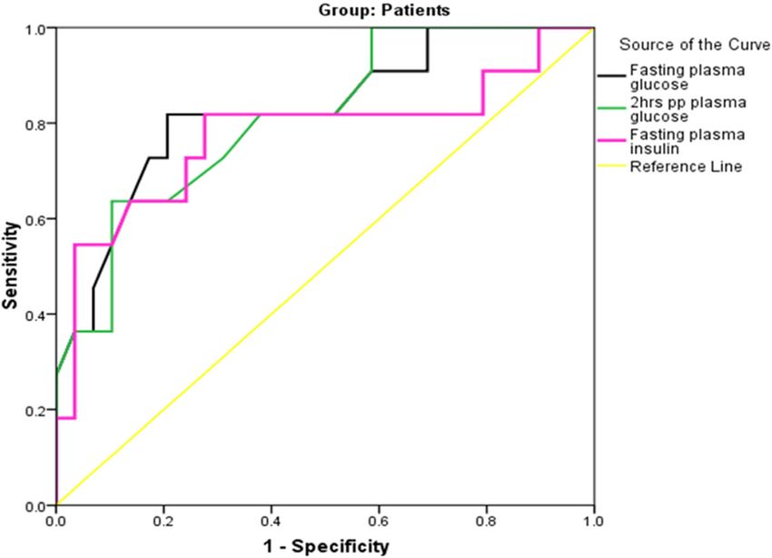
Table 4 ROC curve analysis for the performance of glucose parameters in early diagnosis of IR
Variable Sensitivity Specificity PPV NPV AUC 95% CI P
Fasting plasma g ≥ 115.5 mg/dl 81.8% 79.3% 60% 92% 0.828 0.68-0.98 0.002 (S)
2 h pp plasma glucose ≥ 162.5 mg/dl 81.8% 62.1% 45% 90% 0.812 0.66-0.96 0.003 (S)
Fasting insulin ≥ 9.13μU/ml 81.8% 72.4% 52.9% 91.3% 0.776 0.58-0.96 0.008 (S)
PPV positive predictive value, NPV negative predictive value, AUC area under the curve, CI confidence intervalM. Diab et al. Egyptian Pediatric Association Gazette (2021) 69:9 Page 6 of 7
Fig. 2 ROC curve for the performance of glucose parameters in early diagnosis of insulin resistance
importance of good observation and close monitoring of unlike other variables such as serum ferritin or blood
glycemic parameters of these patients. However, to con- transfusion duration [22].
firm our results, this requires more extensive longitu-
dinal studies rather than cross-sectional studies. This Conclusion
was in agreement with Metwally and El-Saied. They Our results show that HOMA-IR can be used to detect
found that fasting and 2 h pp glucose, fasting insulin, IR in (β-TM) patients on long-term transfusions, espe-
HOMA-IR, ALT, and serum ferritin levels showed a sig- cially patients with high serum ferritin and liver enzymes
nificant increase in thalassemic patients with DM and (ALT).
prediabetes than patients with standard glucose toler-
ance [14].
Recommendation
In this study, there was no significant difference be-
Adolescents and children with β-TM on long-term
tween patients with IR and those with no IR regarding
transfusions should be periodically monitored with gly-
liver span and spleen size; this was in agreement with
cemic indices and serum ferritin levels for early detec-
Bhat and Periasamy, who found that the size of the liver
tion of DM. More efficient therapeutic strategies that
and spleen did not correlate with any of the parameters
could ameliorate insulin resistance should be considered
like IR, age, ferritin, number of transfusions or glycemic
in treating transfusion-dependent thalassemic patients.
indices, like fasting glucose and fasting insulin [17].
ROC curve analysis in our study showed that the dur- Abbreviations
ation of blood transfusion at a cut-off value ≥ 10.2 years β-TM: B-Thalassemia major; HOMA-IR: Homeostasis Model Assessment of
and serum ferritin at a cut-off value ≥ 3173 ng/dl could insulin resistance index; IR: Insulin resistance; IFG: Impaired fasting glycemia;
DM: Diabetes mellitus; T2DM: Type 2 diabetes mellitus; PP: Postprandial;
significantly predict IR, with a sensitivity of 63.6% and BMI: Body mass index; ALT: Alanine aminotransferase; AST: Aspartate
81.8% and a specificity of 69% and 82.8%, respectively. aminotransferase; ROC curve: Receiver operator characteristic curve;
Also, ROC curve analysis showed that fasting plasma HS: Highly significant; NS: Non-significant; CBC: Complete blood count
glucose at a cut-off value ≥ 115.5 mg/dl, 2 h pp glucose
Acknowledgements
at a cut-off value ≥ 162.5 mg/dl, and fasting plasma insu- The authors thank all included patients.
lin at a cut off value ≥ 9.13 could significantly predict IR,
with a sensitivity of 81.8% and a specificity of 79.3%, Authors’ contributions
62.15%, and 72.4%, respectively. This was in disagree- All authors have read and approved the manuscript. AD: Writing this
manuscript and collecting data. GA: Share in writing this manuscript and
ment with Ghergherehchi and Habibzadeh, where they revising data. KE: Collecting data and revising data. EM: Laboratory step. EA:
found that only ALT could predict DM occurrence, Writing this manuscript and collecting dataM. Diab et al. Egyptian Pediatric Association Gazette (2021) 69:9 Page 7 of 7
Funding 15. Liang Y, Bajoria R, Jiang Y et al (2017) Prevalence of diabetes mellitus in
This study had no funding from any resource. Chinese children with thalassaemia major. Trop Med Int Health 22(6):716–
724
16. He L, Chen W, Yang Y, Xie Y et al (2019) Elevated prevalence of abnormal
Availability of data and materials
glucose metabolism and other endocrine disorders in patients with B-
Collected from outpatient clinics pediatric department
thalassemia major: a meta-analysis. Biomed Res Int 2019:1–13
17. Bhat KG, Periasamy PK (2016) Effect of long-term transfusion therapy on the
Ethics approval and consent to participate glycometabolic status and pancreatic beta cell function in patients with
This research accepted by the Research Ethics Committee (REC) of the beta thalassemia major. J Fam Med Primary Care. 3:119–123
Faculty of Medicine, Benha University (chairman: Prof/ Nermeen Adly 18. Hafez M, Yousry I, Hamed FA et al (2009) abnormal glucose tolerance in B-
Mahmoud), Ethics committee reference number RC 1.11.2020. thalassemia major. Hemoglobin 33(2):101–108
All procedures performed in studies involving human participants were in 19. Ansari AM, Bhat K G, Dsa SS et al (2018) Study of insulin resistance in
accordance with the ethical standards of the institutional and/or national patients with B-thalassemia major patients and validity of TYG index. J
research committee and with the 1964 Helsinki Declaration and its later Pediatr Hematol Oncol 40(2):128–131
amendments or comparable ethical standards. A written informed consent 20. Soliman AT, Yasin M, El-Awwa A, De Sanctis V (2013) Detection of glycemic
was obtained from each patient after explaining all steps of this study. abnormalities in adolescents with beta thalassemia using continuous
glucose monitoring and oral glucose tolerance in adolescents and young
adults with β-thalassemia major: pilot study. Indian J Endocrinol Metab
Consent for publication 17(3):490–495
All patients or their parents have been consented for taking their laboratory 21. Ezzat AM, Abdelmotaleb GS, Shaheen AM, Ismail YM, Diab AM (2016)
results for scientific researches Peroxidative stress and antioxidant enzymes in children with b-thalassemia
major. Medical Research Journal 15:57–62
Competing interests 22. Ghergherehchi R, Habibzadeh A (2015) Insulin resistance and beta cell
The authors declare no conflict of interest. function in B-thalassemia major patients. Hemoglobin 39(1):69–73
Author details
1
Pediatric Department, Benha University Hospital, Benha, Egypt. 2Clinical
Publisher’s Note
Springer Nature remains neutral with regard to jurisdictional claims in
Pathology Department, Benha University Hospital, Benha, Egypt.
published maps and institutional affiliations.
Received: 11 August 2020 Accepted: 25 January 2021
References
1. Musallam KM, Cappellini MD (2012) AT Wood Taher. Iron overload in non-
transfusion-dependent thalassemia: a clinical perspective. Blood reviews. 26:
16–S9
2. Koohi F, Kazemi T, Miri-Moghaddam E (2019) Cardiac complications and
iron overload in beta thalassemia major patients—a systematic review and
meta-analysis. Annals of hematology. 98(6):1323–1331
3. Swaminathan S, Fonseca VA, Alam MG et al (2007) The role of iron in
diabetes and its complications. Diabetes Care. 30:1926–1933
4. Praveen EP, Sahoo J, Khurana ML et al (2012) Insulin sensitivity and -cell
function in normoglycemic offspring of individuals with type 2 diabetes
mellitus: impact of line of inheritance, Indian. Journal of Endocrinology and
Metabolism 16(1):105–111
5. American Diabetes Association (2004) Diagnosis and classification of
diabetes mellitus. Diabetes Care. 27:S5–S10
6. Tiedge M, Lortz S, Drinkgern J et al (1997) Relation between antioxidant
enzyme gene expression and antioxidative defense status of insulin-
producing cells. Diabetes. 46:1733–1742
7. McClain DA, Abraham D, Rogers J et al (2006) High prevalence of abnormal
glucose homeostasis secondary to decreased insulin secretion in individuals
with hereditary haemochromatosis. Diabetologia. 49:1661–1669
8. Rubins HB, Robins SJ, Collins D et al (2002) Diabetes, plasma insulin, and
cardiovascular disease: subgroup analysis from the Department of Veterans
Affairs High-density Lipoprotein Intervention Trial (VA-HIT). Arch Intern Med
162(22):2597–2604
9. Govindarajan G, Gill H, Rovetto M et al (2006) what is insulin resistance?
Heart Metab 30:30–34
10. American Diabetes Association, Diabetes Care 2020; 43 (Supplement 1): 14–
S31 https://doi.org/10.2337/dc20-S002.
11. Isiklar A, Karsidag K (2018) Association between thalassemia trait and insulin
resistance. Acta Medica Mediterranea. 34:323–327
12. Khothari CR (2004) Research methodology: methods and techniques. New
Age International, New Delhi
13. Delvecchio M, Cavallo L (2010) Growth and endocrine function in
thalassemia major in childhood and adolescence. J Endocrinol Invest 33:61–
68
14. Metwalley KA, El-Saied AA (2014) Glucose homeostasis in Egyptian children
and adolescents with β-Thalassemia major. Indian J Endocr Metab 18:333–
339You can also read