FELINE CARDIAC DIAGNOSTIC SCHEME ABCDs OF FELINE CARDIOMYOPATHY - UPDATED APRIL 2021
←
→
Page content transcription
If your browser does not render page correctly, please read the page content below

FELINE CARDIAC DIAGNOSTIC SCHEME
 ABCDs OF FELINE CARDIOMYOPATHY
                    UPDATED APRIL 2021
© 2021 Cardiac Education Group                                                               cardiaceducationgroup.org

    A                    CARDIOMYOPATHY STAGES
                         Cats that are predisposed to cardiomyopathy1 but currently
                         have no clinical evidence of myocardial disease.

                                  DIAGNOSTICS
                                     • Patient history
                                     • Yearly auscultation2
                                     • Screening echocardiography for predisposed breeds
                                     • Genetic tests are available for Main Coon and Ragdoll breeds
                                     • Elevated NT-proBNP concentrations may identify cats that may benefit
                                       from further diagnostic evaluation. See NT-proBNP Testing in Cats.

                                  CEG RECOMMENDATIONS
                                     • No treatment
                                     • Client education
                                     • Annual re-evaluation

                 KEY:     Red text: High priority               Black text: Lower priority

                           FOOTNOTES

                           1. Predisposed breeds include Maine Coon, Ragdoll,
                              British Shorthair, Persian, Bengal, Sphynx, Norwegian
                              Forest cat, and Birman breeds. Additional “at risk”
                              breeds are likely to be identified and additional breed-
                              specific genetic testing may become available in
                              future years.
                           2. The absence of a heart murmur does not exclude the
                              possibility of preclinical cardiomyopathy.
© 2021 Cardiac Education Group cardiaceducationgroup.org
B CARDIOMYOPATHY STAGES
Cats with suspected cardiomyopathy that do not have
clinical signs3
Stage B cats are divided into Stage B1 or B2 based on risk of imminent
congestive heart failure (CHF) or aortic thromboembolism (ATE).
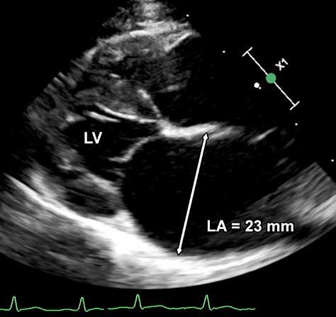
• B1: low risk of imminent CHF or ATE based primarily on minimal left
atrial (LA) enlargement.
• B2: increased risk of imminent CHF or ATE based on more severe LA
enlargement (for example, LA diameter ≥ 20 mm on long axis, LA:Ao ≥
1.8) or presence of other risk factors4.
DIAGNOSTICS: STAGE B1 & B2
• Patient history
• Cardiac and pulmonary auscultation5
• Echocardiography6 7
IVSd: 6.99 mm
• Blood pressure
LVIDd: 10.09 mm
• Resting serum thyroxine concentration (cats ≥ 6 years of age)
LVWd: 8.61 mm
• NT-proBNP8
• Thoracic radiographs9
• Electrocardiogram (ECG) when cardiac arrhythmia is evident during
clinical examination
• Clinical lab tests: serum biochemistries, PCV/TS (or CBC) and urinalysis
(prior to initiating any therapy in B2 patients)
KEY: Red text: High priority Black text: Lower priority
FOOTNOTES
3. Cardiomyopathy may be suspected when a murmur, 6. Echocardiographic findings allow diagnosis of specific 8. Abnormal SNAP® NT-proBNP or Cardiopet NT-proBNP
gallop or arrhythmia is detected on physical examination. type of cardiomyopathy, including hypertrophic quantitative results (Idexx Laboratories, Inc.) are an
4. Other risk factors: presence of arrhythmia, extreme LV cardiomyopathy (HCM), restrictive cardiomyopathy indication for echocardiography. See NT-proBNP Testing
hypertrophy, spontaneous echo contrast/thrombus, (RCM), dilated cardiomyopathy (DCM) or non-specific in Cats.
regional wall motion abnormalities, LV systolic cardiomyopathic phenotypes as well as degree of severity 9. Radiographic findings are not diagnostic for
dysfunction of changes. See ACVIM Feline CM Consensus Statement. cardiomyopathy but may be used to track progressive
5. Abnormal auscultation findings indicate that further 7. Point-of-Care (POC) exams may be used to document cardiomegaly or document evidence of respiratory
evaluation is warranted, but are not diagnostic degree of LA enlargement and/or presence of disease, and may be used as baseline information for B2
for cardiomyopathy. Conversely, some cats with spontaneous echo contrast or intracardiac thrombus in cats.
cardiomyopathy may have normal auscultation findings. cats without clinical signs.
© 2021 Cardiac Education Group                                                                                     cardiaceducationgroup.org

         B                              CARDIOMYOPATHY STAGES
                                        Cats with suspected cardiomyopathy that do not have
                                        clinical signs

                                                CEG RECOMMENDATIONS - B1 & B2

                                                      • No specific dietary changes or exercise restrictions at this stage
                                                      • Manage systemic hypertension if present
                                                      • Manage hyperthyroidism if present

                                                      Stage B1
                                                      • No treatment10, 11

                                                      Stage B2
                                                      • Echocardiographic identification of cardiomyopathic phenotype and
                                                        severity of anatomic changes including degree of LA enlargement is
                                                        recommended.

                                                      • Thromboprophylactic therapy12 recommended when risk factors for ATE
                                                        are present, primarily moderate to severe LA enlargement (for example,
                                                        LA diameter ≥ 20 mm on long axis, LA:Ao ≥ 1.8)13 or other identified risk
                                                        factors.

                                                      • Owner monitoring of resting or sleeping respiratory rate strongly
                                                        recommended (See Monitoring Your Pet’s Respiratory (Breathing) Rate).
                                                      • Treat ventricular tachycardia or frequent ventricular ectopy14 or atrial
                                                        fibrillation15 when present; consider consultation with a cardiologist.
                                                      • Additional therapies16 are controversial in Stage B2 disease.
                                                        Consultation with a cardiologist may be helpful.

                            KEY:         Red text: High priority              Black text: Lower priority

FOOTNOTES
10. Atenolol therapy for preclinical cats with          12. Clopidogrel recommended. Feline Formulary.           sotalol) most often first choice for therapy of
    dynamic left ventricular outflow obstruction is     13. Other risk factors for ATE include spontaneous       ventricular ectopy.
    controversial. Consultation with a cardiologist         echo contrast (“smoke”), visible intracardiac    15. Diltiazem recommended.
    may be helpful.                                         thrombus, left atrial (LA) decreased             16. For example, ACEi or spironolactone.
11. Beta-blocker therapy (e.g. atenolol or                  systolic function (LA FS%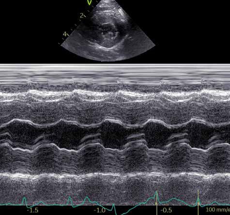
© 2021 Cardiac Education Group                                                                                                  cardiaceducationgroup.org

              C                                  CARDIOMYOPATHY STAGES
                                                 Cats with past or current clinical signs17 of congestive heart
                                                 failure or aortic thromboembolism.

                                                           DIAGNOSTICS

                                                           • Patient history
                                                           • Cardiac and pulmonary auscultation
                                                           • Echocardiography for definitive diagnosis of underlying structural heart
Left atrial size                                             disease; this test may need to be delayed until the patient is clinically
assessment using
POC ultrasound                                               stable.
                                                           • Thoracic radiographs18 or POC19 ultrasound to identify pulmonary edema,
                       Ao
                                                             left atrial enlargement or pleural effusion
                                                           • Cageside SNAP® NT-proBNP might help discriminate respiratory disease
                                                             causes of acute clinical signs versus congestive heart failure20 in cats with
                  LA                                         respiratory distress.21
                                                           • Blood pressure
                                                           • Electrocardiogram (ECG) when cardiac arrhythmia is evident during
                                                             clinical examination.
Detection of pleural                                       • Clinical lab tests:
effusion using POC
ultrasound                                                         • Serum biochemistries, PCV/TS (or CBC) and urinalysis (prior to initiating any
                                                                     therapy and to monitor for renal and electrolyte abnormalities after therapy).
                   LV Wall
                                                                   • Thyroid testing can be submitted if current thyroid status is not known (cats ≥
                             Pleural                                 6 years of age).
                             Effusion

                                    KEY:          Red text: High priority                  Black text: Lower priority

     FOOTNOTES
     17. Clinical signs may include general signs of illness         thoracocentesis should be performed to stabilize             blood sample or a pleural effusion sample that has
         (e.g. hiding, inappetence), congestive heart                patients with severe pleural effusion.                       been diluted 1:1 with saline.
         failure signs (e.g. tachypnea, respiratory distress,    19. Point-of-Care (POC) ultrasound may be used to            21. "Normal" SNAP NT-proBNP results make congestive
         hypothermia), signs of arrhythmia (e.g. syncope) and/       document cardiac disease as the cause of dyspnea             heart failure UNLIKELY to be the cause of the patient's
         or signs of aortic thromboembolism (e.g. paresis,           by identifying left atrial enlargement in the presence       respiratory distress. A strong "Abnormal" result
         paralysis).                                                 of pleural effusion or pulmonary B-lines that may            SUPPORTS a diagnosis of congestive heart failure.
     18. Thoracic radiographs may be too stressful to be             indicate pulmonary edema.                                    A weak "Abnormal" result should be interpreted with
         completed in severely dyspneic patients. Emergency      20. NT-proBNP assessment can be performed using a                caution.
© 2021 Cardiac Education Group                                                                                                 cardiaceducationgroup.org

          C                                   CARDIOMYOPATHY STAGES
                                              Cats with past or current clinical signs of congestive heart failure
                                              or aortic thromboembolism.

                                                        CEG RECOMMENDATIONS

                                                        • Standard Treatment:
                                                              • Acute CHF22: Oxygen supplementation, furosemide, anxiolysis,
                                                                thoracocentesis if needed, supportive care23
                                                              • Chronic CHF22,24: Furosemide, ACEi, clopidogrel if indicated by
                                                                echocardiographic findings:
                                                                        • Most patients: furosemide, clopidogrel25
                                                                        • Renin-angiotensin-aldosterone system blockade
                                                                          recommended (ACEi and spironolactone) if tolerated. 26
                                                              • ATE: analgesia27, anticoagulant therapy28, supportive care (including
                                                                CHF therapy if needed)
                                                              • Treat ventricular arrhythmias or atrial fibrillation as outlined for B2
                                                              • Owner assessment of cat’s home sleeping respiratory rate
                                                                recommended to monitor for recurrence of CHF. (See Monitoring Your
                                                                Pet’s Respiratory (Breathing) Rate)

                                KEY:           Red text: High priority                 Black text: Lower priority

FOOTNOTES
22. If systolic dysfunction is present, pimobendan or           medications should be based on patient tolerance.          for pain control. Concurrent use of butorphanol with
    dobutamine therapy may be helpful.                      25. If clopidogrel is not tolerated, factor Xa inhibitors      these medications may reduce analgesic efficacy.
23. Injectable butorphanol (IM) recommended for                 (apixaban, rivaroxaban) may be considered.              28. Immediate commencement of low molecular weight
    anxiolysis, supportive care includes access to water        Consultation with a cardiologist is recommended.            heparin or unfractionated heparin injections or
    and access to gentle warming for hypothermic            26. May cause facial pruritis in some cats.                     oral dosing of factor Xa inhibitor (e.g. apixaban,
    patients.                                                                                                               rivaroxaban) is recommended. (Feline Formulary)
                                                            27. Potent opioid-based analgesics (such as fentanyl,
24. Although furosemide is almost always needed                 hydromorphone or methadone) are recommended
    to control chronic CHF in cats, addition of other
© 2021 Cardiac Education Group                                                                                   cardiaceducationgroup.org

         D                              HEART FAILURE STAGES
                                        Cats with end-stage disease with clinical signs of CHF refractory
                                        to standard therapy or repeated thromboembolic events.

                                                 DIAGNOSTICS

                                                 • Patient history
                                                 • Cardiac and pulmonary auscultation
                                                 • Thoracic radiographs29 or POC30 ultrasound to identify pleural effusion or
                                                   pulmonary edema
                                                 • Echocardiography for definitive diagnosis of underlying structural heart
                                                   disease31.
                                                 • Blood pressure
                                                 • Clinical lab tests: serum biochemistries, PCV/TS (or CBC) and urinalysis
                                                   (prior to initiating any therapy and to monitor for renal and electrolyte
                                                   abnormalities after therapy). Thyroid status should be reassessed in
                                                   Stage D patients.
                                                 • NT-proBNP might help discriminate between cats with respiratory causes
                                                   of clinical signs or congestive heart failure32.
                                                 • Electrocardiogram (ECG) when cardiac arrhythmia is evident during
                                                   clinical examination.

                                                 CEG RECOMMENDATIONS

                                                 • Standard Treatment: Furosemide, pimobendan, ACEi & spironolactone
                                                 • Clopidogrel33 if indicated by echocardiographic findings
                                                 • Torsemide may be considered in place of furosemide if high doses of
                                                   furosemide not effective for recurrent CHF
                                                 • Atrial fibrillation – diltiazem therapy
                                                 • Ventricular arrhythmias – sotalol therapy
                                                 • Other therapies may be helpful; consultation with a cardiologist is strongly
                                                   recommended.
                                                 • Dietary changes – avoid excessive sodium intake and maintain adequate
                                                   protein and caloric intake. Dietary intake is prioritized over sodium
                                                   restriction.
                                                 • Appetite stimulants may be useful.

                            KEY:         Red text: High priority              Black text: Lower priority

FOOTNOTES
29. Thoracic radiographs may be too stressful to           to identify pleural effusion and to identify        sample that has been diluted 1:1 with saline.
    be completed in severely dyspneic patients.            pulmonary B-lines that may indicate             33. If clopidogrel is not tolerated, factor Xa
    Emergency thoracocentesis may be required to           pulmonary edema.                                    inhibitors (apixaban, rivaroxaban) may be
    stabilize patients with severe pleural effusion.   31. Echocardiography may need to be delayed             considered. Consultation with a cardiologist is
30. Point-of-Care (POC) ultrasound may be used             until patient is stabilized.                        recommended.
    to document cardiac disease as the cause           32. NT-proBNP assessment can be performed
    of dyspnea by identifying LA enlargement,              using a blood sample or a pleural effusion
cardiaceducationgroup.org

                                 ABOUT THE CARDIAC EDUCATION GROUP (CEG)
                                 Founded in 2009, the Cardiac Education Group is a registered not-for-
                                 profit organization of board-certified veterinary cardiologists from both
                                 academia and private practice that offers independent recommendations
                                 for the evaluation and treatment of canine and feline heart disease. The
                                 CEG mission is to improve the lives of dogs and cats with heart disease
                                 by providing resources and information in order to promote detection,
                                 diagnosis and therapy of heart disease and heart failure with greater
                                 accuracy and confidence.

                                                             John D. Bonagura, DVM, MS, Diplomate ACVIM
                                                           Adjunct Professor, North Carolina State University
                                                             Rebecca L. Stepien, DVM, MS, Diplomate ACVIM
                                                                             Clinical Professor of Cardiology,
                                                                           University of Wisconsin-Madison
                                                              Brian A. Scansen, DVM, MS, Diplomate ACVIM
                                                                          Associate Professor of Cardiology,
                                                                 Service Head, Cardiology & Cardiac Surgery,
                                                                                   Colorado State University
                                                                    Barret J. Bulmer, DVM, Diplomate ACVIM
                                                        Tufts Veterinary Emergency Treatment & Specialties,
                                                                                                Walpole, MA
                                                                   Whit M. Church, DVM, Diplomate ACVIM
                                                          Desert Veterinary Medical Specialists, Phoenix, AZ
                                                                 Alan W. Spier, DVM, PhD, Diplomate ACVIM
                                                                    BluePearl Veterinary Partners, Tampa, FL
                                                            Sonya G. Gordon, DVM, DVSc, Diplomate ACVIM
                                                                                  Professor of Cardiology,
                                                                         Eugene Ch’en Chair of Cardiology,
                                                                                    Texas A&M University

   TO LEARN MORE OR SIGN UP
   FOR OUR NEWSLETTER, VISIT
  cardiaceducationgroup.org .

                                                                    The CEG is sponsored by educational grants from
© 2021 Cardiac Education Group
                                                        Boehringer Ingelheim Vetmedica, Inc. and IDEXX Laboratories.
You can also read