Feline sarcoid in a 1-year-old domestic short-haired cat caused by bovine papillomavirus type 14 in Switzerland
←
→
Page content transcription
If your browser does not render page correctly, please read the page content below

Fallberichte | Case reports
Feline sarcoid in a 1-year-old domestic
short-haired cat caused by bovine
papillomavirus type 14 in Switzerland
C. Kiefer1, K. Tobler2, A. S. Ramsauer2, U. Biegel4, N. Kuehn3, M. Ruetten3
1 Tierarztpraxis Stadthof, Wangen a. A., Switzerland, 2 Institute of Virology, Vetsuisse Faculty, University of
Zurich, Switzerland, 3 PathoVet AG, Tagelswangen, Switzerland, 4 Research Institute for Organic Agriculture
(FiBL), Frick, Switzerland
Summary Felines Sarcoid bei einer 1-jährigen https://doi.org/
10.17236/sat00127
europäischen Hauskatze ausgelöst
A 1year old domestic short haired cat, living on a farm durch bovines Papillomavirus Typ 14 Received: 28.12.2016
in Switzerland, was presented to the veterinarian with a Accepted: 09.05.2017
in der Schweiz
5 cm in diameter mass, bulging from her left nostril.
The mass was only incompletely removed because of its
unfavourable location. Histologically, the lesion con Eine einjährige Bauernhofkatze zeigte einen 5 cm gros
sisted of an infiltrative growing spindeloid proliferation sen Knoten an der linken Nasenöffnung, der aufgrund
in close approximation to the epidermis and was diag seiner Lokalisation nur unvollständig entfernt werden
nosed as a feline sarcoid tumour. The presence of Bovine konnte. Hinsichtlich der infiltrativ wachsenden, spin
Papillomavirus type 14 (BPV14) specific DNA could be delförmigen Proliferation wurde histologisch ein felines
identified in the tissue by using two PCR assays. The Sarkoid diagnostiziert. Die aus dem Gewebe isolierte
amplified sequences of 194 and 549 base pairs (bp) were DNA konnte durch zwei PCRs als solche von bovinem
99% and 100% identical with a virus isolated after aut Papillomavirus 14 (BPV14) identifiziert werden. Die 194
opsy, from a cat with feline sarcoid in the USA. The cat und 549 Basenpaare (bp) langen, amplifizierten Sequen
recovered completely after an even incomplete surgical zen waren 99 bzw. 100% identisch mit einer von einem
excision and no recurrence could be observed 10 months in den USA isolierten Virus, welches mit felinem Sar
later. koid in Zusammenhang gebracht wurde. Trotz unvoll
ständiger Exzision, ist bis 10 Monate nach der Opera
Keywords: feline sarcoid, bovine Papillomavirus type 14,
cat, spindeloid neoplasia, surgery tion kein Rezidiv aufgetreten.
Schlüsselwörter: felines Sarkoid, bovines Papillomavirus
Typ 14, Katze, spindelzellige Neoplasie, Chirurgie
Introduction based on the coding sequences of the highly conserved
major capsid protein L1. The majority of PVs only infect
Feline sarcoids (synonym: feline cutaneous fibropapil epithelium and are highly host specific. The bovine
lomas) are rare intradermal fibroblastic proliferations of papillomaviruses (BVPs) of the Deltapapillomavirus
cats living in rural areas. So far, feline sarcoids were genus, however, have the ability to infect both epithelial
reported in North America, New Zealand, England, and mesenchymal cells of different species (Bernard et
Sweden and Australia (Schulman et al., 2001; Munday al., 2010; Joh et al., 2011; Munday et al., 2014). Lesions
et al., 2010). These lesions resemble those described as caused by PVs in domestic cats may be oral papillomas
equine sarcoids (Gumbrell et al., 1998; Schulman et al., (Felis catus (Fca) PV1) (Munday et al., 2015), feline cu
2001; Gross et al., 2005). Equine sarcoids are the most taneous viral plaques, Bowenoid carcinomas (FcaPV2
common skin tumours of horses and are divided into 5 and FcaPV3) (Lange et al., 2009; Munday et al., 2013)
different clinical entities (Martens et al., 2000). In cats, and feline sarcoids. Feline sarcoids are spindeloid sarco
however, these different morphologies are not described. mas of younger cats with close connection to the epi
Sarcoids are caused by small doublestranded DNA pap dermis, which are reported not to metastasize but often
illomaviruses (PVs), which are classified into genera tend to develop recurrences after incomplete surgical
Band 159, Heft 9, September 2017, 487–491, © GST | SVS SAT | ASMV 9 | 2017 487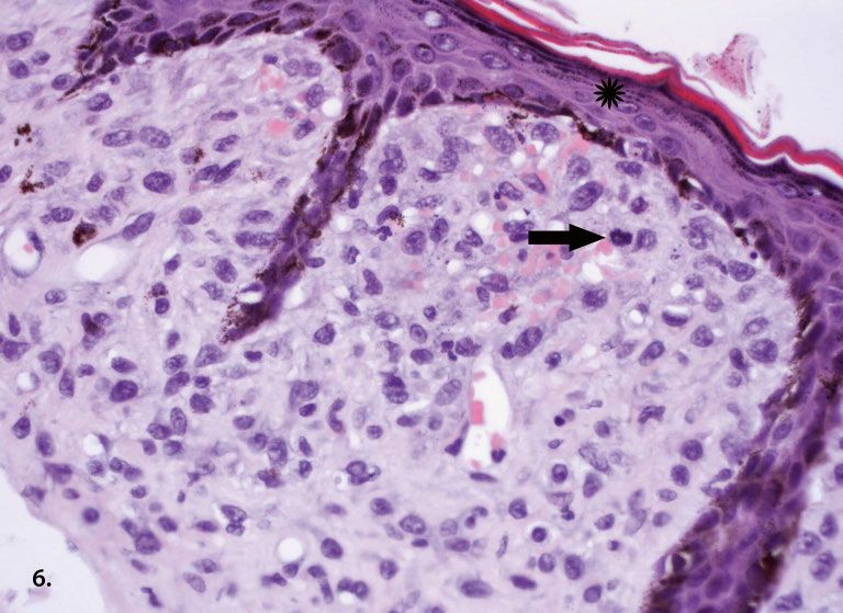
Fallberichte | Case reports
Feline sarcoid in a 1-year- excision. The entire genomic sequence of PVs involved was not possible because of the location next to the left
old domestic short-haired in feline sarcoids (FeSarPV) isolated from cats was re nostril. The wound was closed by a single button suture
cat caused by bovine
papillomavirus type 14 cently published and classified as a Deltapapillomavirus. with Supramid® 3–0, (B. Braun Medical AG, Sempach,
in Switzerland However, since DNA of this virus was also amplified Switzerland). A small suture dehiscence occured 10 days
from samples of normal skin and fibropapillomas of after surgery and was closed again with Supramid®
C. Kiefer et al.
cattle (Munday et al., 2010; daSilva et al., 2012), it seems (Fig. 3). Due to the incomplete excision of the mass,
likely that feline sarcoids are due to crossspecies infec with “close margins”, the cat was given a Viscum Album
tion by BPVs (Schulman et al., 2001). Extract (VAE) as adjuvant treatment (injections with
Iscador® P, Iscador AG, Arlesheim, Switzerland) fol
lowed by oral application with Viscum quercus praepa
Case history ratum 3% Dilaq, (Iscador AG, Arlesheim, Switzerland).
The cat was presented to the veterinarian showing a
slightly bulging mass on her left nostril measuring 1 cm Histology
in diameter. The mass was firm, alopecic and the surface
slightly ulcerated (Fig. 1). A fine needle aspiration (FNA) The excised material was fixed in 4% buffered formalin
was performed under anaesthesia with Domitor®, for 24 h, dehydrated in a 70–95% ethanol series, fol
(Provet AG, Lyssach, Switzerland) and Morphasol®4, lowed by Xylol and paraffin embedding. Sections (2 to
(Dr. E. Gräub AG, Bern, Switzerland) subcutaneously, 3 µm) were mounted on glass slides and stained with
followed by intravenous anaesthesia with Propofol® 2%, HaematoxylinEosin (HE) using standard procedures.
(Provet AG, Lyssach, Switzerland). Unfortunately, the Histologically the mass consisted of spindeloid to stel
FNA was not diagnostic. As a bacterial infection was late cells expanding the dermis and subcutis and was
suspected, an initial treatment with Veraflox®, (Provet intimately associated with to the epidermis. The neo
AG, Lyssach, Switzerland) 5 mg/kg b.w. (once daily) plastic cells were oval to spindeloid with moderate
orally, was given. Six weeks later, the cat was presented amounts of pale basophilic cytoplasm with indistinct
again due to a massive growth of the lesion. The bulging cell borders (Fig. 4) and round to oval nuclei showing a
mass on her left nostril measured now 5 cm in diameter finely stippled chromatin pattern and only slightly vis
(Fig. 2). An excision of this mass seemed inevitable. ible nucleoli, moderate anisocytosis, anisocaryosis and
anisonucleoliosis. The neoplastic cells were embedded
in abundant extracellular matrix (mucopolysacchar
Therapy ides), separating the adnexa and often forming whorls
around small blood vessels or hair follicles. The mitotic
Under anaesthesia with the same protocol as mentioned rate was moderate with 11 mitotic figures in 10 high
above, the mass was surgically removed. A total excision power fields. Within the tumour were small areas of
Figure 1: Cat at first presentation with a Figure 2: The cat at the second presentation Figure 3: Complete healing of the operation
bulging mass measuring 1 cm in diameter. 2 month after the first treatment attempt. field 6 months after the closure of the suture
The mass grew up to 5 cm in diameter. dehiscence.
488 SAT | ASMV 9 | 2017 Band 159, Heft 9, September 2017, 487–491, © GST | SVS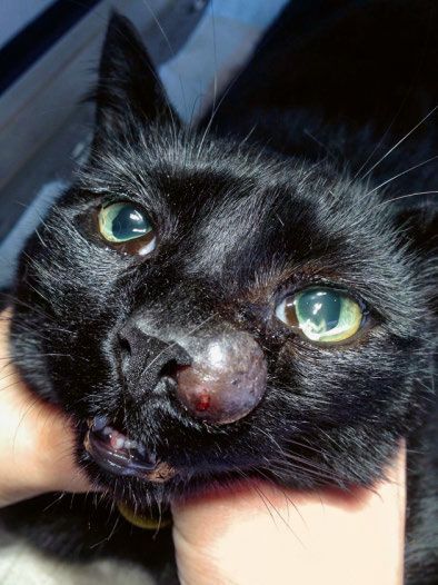
Fallberichte | Case reports
coagulative necrosis randomly distributed visible. The
overlaying epidermis was multifocally ulcerated or if still
intact, hyperplastic, forming high rete ridges and were
strongly pigmented by melanin. In the stratum spino
sum were deposited a moderate amount of keratohyalin
granula (Fig. 5).
Molecular analysis
DNA was extracted from the center of the formalin fixed
tissue using the QIAamp DNA Mini Kit (Qiagen, Hom
brechtikon, Switzerland) according to the manufactur
er’s instructions. For detection of viral DNA of FeS
arPV/BPV14, a previously reported primer set jmpSAF
(5’GGAACAAACCTCACAATCAC3’) and jmpSAR
(5’CCAGTTCTCTAATACTGAGG3’) was used (Mun
day et al., 2010). As these primers just amplify a short
product of 194 bp, additional primers amplifying a 549
bp product in the L1 region (5771 to 6319) of the BPV14 Figure 4: Mesenchymal neoplasia adjacent to the epidermis with the spindeloid to
genome BPV14for (5’TGG TAA AGA GGT GCC CAA stellate cells embedded in abundant extracellular matrix (mucopolysaccharides),
separating the adnexa and forming whorls around small blood vessels. HE staining,
AG3’) and BPV14rev (5’GCT TCC TCA GCC ATT 4x magnification.
TTG AG3’) were designed. PCR was performed with a
reaction mix containing 8 µl water, 2 µl of each forward
and reverse primer (10µM each), 1µl extracted DNA as
template and 12 µl REDTaq ReadyMIX (SIGMA
ALDRICH, Buchs, Switzerland) in a total volume of
25 µl. The cycling program for all PCR assays started
with a denaturation step of 3 min at 94 °C, followed by
40 cycles of 30 sec at 94 °C, 30 sec at 55 °C and 30 sec
at 72 °C. PCR products were separated by agarose elec
trophoresis. Both primer sets for feline sarcoid virus
BPV14 amplified bands of the expected size. The PCR
amplimers were excised from the agarose gel and puri
fied using Zymoclean Gel DNA Recovery Kit (ZYMO
RESEARCH, Irvine, USA) according to the manufac
ture’s protocol. Nucleotide sequences were determined
(Microsynth, Balgach, Switzerland) and compared to
the published reference sequences of BPV14 with the
NCBI Basic Local Alignment Search Tool (“BLAST”)
(http://www.ncbi.nlm.nih.gov/blast/Blast.cgi) (Altschul
et al., 1990). The sequencing results of the shorter and
the longer amplimer showed 99% and 100% identity to Figure 5: Round to oval nuclei with moderate anisocaryosis. A mitotic figure (arrow)
the published BPV14 sequence (Genbank accession is visible just underneath the epidermis. In the stratum spinosum a moderate amount
of keratohyalin granula (star) were deposited. HE staining, 40x magnification.
#KP276343).
Discussion tumour interfered with eating the cat was euthanized
(Munday et al., 2015). Veterinarians tend to classify
Our sequences covering 743 bp (194 bp and 549 bp) were mesenchymal tumours of the skin and subcutis into soft
identical to the L1 of BPV14, which was sequenced and tissue sarcomas and apply then a grading system. Such
classified from a cat in the USA (Munday et al., 2015). grading systems are applied for canine and human tu
Similar to our case, the cat had a rapidly growing mass mours (Coindre et al., 2006; Dennis et al., 2011) but not
at the nose tip. Therapy attempts with intralesional in for those from cats. Adaption and implementation of
jections of cisplatin and surgery were performed twice, these grading systems would classify the herein de
but the mass regrew within only 2 months. When the scribed tumour as soft tissue sarcoma grade II according
Band 159, Heft 9, September 2017, 487–491, © GST | SVS SAT | ASMV 9 | 2017 489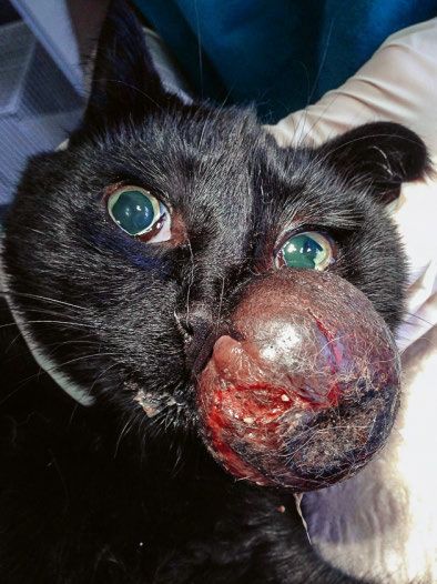
Fallberichte | Case reports
Feline sarcoid in a 1-year- to the morphology of the neoplastic cells, the mitotic owner of the cat reported herein disapproved the radi
old domestic short-haired rate and necrotic area within the tumour. Higher mitot ation because it is extensive, costly and the cat has to
cat caused by bovine
papillomavirus type 14 ic rate is often prognostic for reduced survival time in deal with frequent anaesthesia and possible side effects.
in Switzerland many tumours (Coindre et al., 2006; Esplin et al., 2008; If the VAE treatment had any immunemodulating ef
Dennis et al., 2011; Thompson et al., 2011). and such fect as reported in the literature, remains unanswered
C. Kiefer et al.
grading would imply a short disease free period with a in our case (Bussing, 2006).
risk of metastasis that might have led to euthanasia of
the cat. Although the tumour resembles closely equine In summary, if a radiation therapy is not an option, we
sarcoids, it could readily have been misdiagnosed as suggest it is still worth to attempt surgery even when it
fibrosarcoma, which is another kind of mesenchymal is difficult to excise the mass completely. The cat has
neoplasia with some risk to metastasize (Morrison and now been free of disease for 10 months. Although we
Starr, 2011). Our case taught us the importance to dif cannot assure its cancer free state, the measures have
ferentiate and to recognize these lesions as feline sar prolonged its life and may even lead to complete recov
coids since, feline sarcoids are not reported to metasta ery.
size. The accurate diagnosis of the origin of cells and
aetiology is particularly more important than the clas
sification with a grading system, which could lead to Acknowledgement
wrong prognostic outcome.If the neoplasia cannot be
excised completely, radiation therapy is recommended We kindly thank Prof. Lloyd Vaughan for critically read
to prevent recurrence (Gross and Affolter, 1998). The ing the manuscript and making very helpful suggestions.
References Gross T. L., Ihrke P. J., Walder E. J., Affolter V. K.: Feline Sar-
coid. In: Skin diseases of the dog and cat. 2sd editon, Black-
Altschul, S. F., Gish, W., Miller, W., Myers, E. W., and well publishing, Iowa, USA, 2005, 730–731.
Lipman, D. J.: Basic local alignment search tool. J. Mol. Biol. Gumbrell R. C., Rest J. R., Bredelius K., Batchelor D. J., Wil-
1990, 215: 403–410. liamson J.: Dermal fibropapillomas in cats. Vet. Rec. 1998,
Bernard H. U., Burk R. D., Chen Z., van Doorslaer K., 4: 142(14): 376.
Hausen H., de Villiers E. M.: Classification of papillomavirus- Joh J., Jenson A. B., King W., Proctor M., Ingle A., Sund-
es (PVs) based on 189 PV types and proposal of taxonomic berg J. P., Ghim S.J.: Genomic analysis of the first laborato-
amendments. Virology 2010, 401: 70–79. ry-mouse papillomavirus. J. Gen. Virol. 2011, 92: 692–698.
Bussing A.: Immune modulation using mistletoe (Viscum Lange C. E., Tobler K., Markau T., Alhaidari Z., Bornand V.,
album L) extracts Iscador. Arzneimittelforschung 2006, Stockli R., Trussel M., Ackermann M., Favrot C.: Sequence
56: 508–515. and classification of FdPV2, a papillomavirus isolated from
Clottu CO., Klocke P., Burger D., Straub R., Gerber V.: Treat- feline Bowenoid in situ carcinomas. Vet. Microbiol. 2009,
ment of Clinically Diagnosed Equine Sarcoid with a Mistle- 137: 60–65.
toe Extract (Viscum album austriacus). J. Vet. Intern. Med. Martens A., De Moor A., Demeulemeester J., Ducatelle R.:
2010, 24: 1483–1489. Histopathological characteristics of five clinical types of
Coindre J. M.: Grading of soft tissue sarcomas. Arch. Pathol. equine sarcoid. Res. In Vet. Sci. 2000, 69: 295–300.
Lab. Med. 2006, 130: 1448–1453. Morrision W. B, Starr R. M.: Vaccine-Asssociated feline Sar-
Da Silva M. A., Carvalho C. C., Couthino L. C., Reis M. C., coma Task Force. Vaccine-associated Feline Sarcoma. Task
de Aragao Batista M. V., de Castro R. S., Dos Anjos F. B., Force guidelines. JAVMA 2001, 218:5, 697–701.
de Freitas A. C.: Co-infection of bovine papillomavirus and Munday J. S., Knight C. G., Howe L.: The same papilloma-
feline-associated papillomavirus in bovine cutaneous warts. virus is present in feline sarcoids from North America and
Transbound Emerg. Dis. 2012, 59: 539–543. New Zealand but not in any non-sarcoid feline samples. J.
Dennis M. M., McSporran K. D., Bacon N. J., Schulmann F. Y., Vet. Diagn. Invest. 2010, 22: 97–100.
Foster R. A., Powers B. E.: Prognostic factors for cutaneous Munday J. S., Dunowska M., Hills S. F., Laurie R. E.: Genomic
and subcutaneous soft tissue sarcomas in dogs. Vet. Pathol. characterization of Felis catus papillomavirus-3: A novel
2011, 48: 73–84. papillomavirus detected in a feline Bowenoid in situ car-
Esplin D. G.: Survival of dogs following surgical excision of cinoma. Vet. Microbiol. 2013, 165: 319–326.
histologically well-differentiated melanocytic neoplasms Munday J.S.: Bovine and human papillomaviruses, a com-
of the mucous membranes of the lips and oral cavity. Vet. parative review. Vet. Pathol. 2014, 51: 1063–1075.
Pathol. 2008, 45: 886–896.
Munday J. S., Fairley R. A., Mills H., Kiupel M., Vaatstra B. L.:
Gross T. L., Affolter V. K.: In: Advances in skin oncology Oral papillomas associated with Felis catus papillomavirus
(workshop report). In: Advances in Veterinary Dermatology. type 1 in two domestic cats. Vet. Pathol. 2015, 52: 1187–1190.
Eds. Kwochka K.W., Willemse T., von Tscharner C. Butter-
worth-Heinemann, Oxford, 1998, 382–385.
490 SAT | ASMV 9 | 2017 Band 159, Heft 9, September 2017, 487–491, © GST | SVS
Fallberichte | Case reports

Munday J. S., Thomson N., Dunowska M., Knight C. G.,             Corresponding author                  Feline sarcoid in a 1-year-
Laurie R. E., Hills S.: Genomic characterisation of the feline   Maja Ruetten                          old domestic short-haired
sarcoid-associated papillomavirus and proposed classifi-         PathoVet AG                           cat caused by bovine
cation as Bos taurus papillomavirus type 14. Vet. Microbiol.     Buckstr. 2                            papillomavirus type 14
2015, 177: 289–295.                                              CH-8317 Tagelswangen                  in Switzerland

Schulmann F. Y., Krafft A. E., Janczewski T.: Feline cutane-     Tel: +41 208 9920
                                                                 E-Mail: maja.ruetten@pathovet.ch      C. Kiefer et al.
ous fibropapillomas: clinicopathologic findings and associa-
tion with papillomavirus infection. Vet. pathol. 2001,
38: 291–296.

Thompson J. J., Pearl D. L., Yager J. A., Best S. J., Coomber
B. L., Foster R. A.: Canine subcutaneous mast cell tumor:
characterization and prognostic indices. Vet. Pathol. 2011,
48: 156–168.

Band 159, Heft 9, September 2017, 487–491, © GST | SVS                                                SAT | ASMV 9 | 2017     491
You can also read


