Granulomatous Mastitis Due to Non-Tuberculous Mycobacteria: A Diagnostic and Therapeutic Dilemma - MDPI
←
→
Page content transcription
If your browser does not render page correctly, please read the page content below
Case Report
Granulomatous Mastitis Due to Non-Tuberculous
Mycobacteria: A Diagnostic and Therapeutic Dilemma
Owais Ahmed Patel * , Girish D. Bakhshi, Amogh R. Nadkarni and Zarin S. Rangwala

                                          Grant Government Medical College and Sir JJ Group of Hospitals, Mumbai 400008, India;
                                          gdbakhshi@yahoo.com (G.D.B.); amoghrn@gmail.com (A.R.N.); zarinrangwala@gmail.com (Z.S.R.)
                                          * Correspondence: theowaispatel@gmail.com; Tel.: +91-6362256141

                                          Abstract: Non-tuberculous mycobacterial (NTM) infections of the breast are rare. These infections
                                          present as cellulitis of the breast or breast abscess. Their diagnosis poses a challenge as they manifest
                                          signs of acute inflammation, unlike tuberculous mycobacterial infections which present in a chronic
                                          pattern. However, on aspiration of pus from the site of infection, primary smear may show acid
                                          fast bacilli. This poses a diagnostic dilemma. The present case is that of a 34-year-old woman who
                                          presented with recurrent mastitis. She had history of right breast swelling, for which surgical excision
                                          had been performed three months prior at another facility. Her histopathology had showed cystic
                                          granulomatous neutrophilic mastitis (CNGM). The patient again presented with right breast abscess
                                          which was confirmed on ultrasonography. Incision and drainage along with removal of necrotic
                                          tissue was done. Primary smear of pus showed acid fast bacilli on Ziehl–Neelson staining. Bacterial

                                   culture and line probe speciation revealed non-tuberculous mycobacterium M. abscessus, which
                                          responded well to prolonged anti-microbial therapy. These rapidly growing NTM require prolonged
Citation: Patel, O.A.; Bakhshi, G.D.;
Nadkarni, A.R.; Rangwala, Z.S.
                                          treatment and are quite often recurrent. M. abscessus is a rare cause of CNGM, with this being only
Granulomatous Mastitis due to             the third reported case in literature. A brief case report with a review of literature is presented.
Non-Tuberculous Mycobacteria: A
Diagnostic and Therapeutic Dilemma.       Keywords: breast; mastitis; non-tuberculous mycobacteria; breast abscess
Clin. Pract. 2021, 11, 228–234.
https:// doi.org/10.3390/
clinpract11020034
                                          1. Introduction
Academic Editors:
                                               Cystic neutrophilic granulomatous mastitis (CNGM) remains a little-known benign
Vishwanath Venketaraman and Vipul
                                          breast entity with obscure aetiology first reported in 1972 as a differential mimicking
D. Yagnik
                                          breast carcinoma [1]. Less than 1% of all breast specimens show CNGM. It is commonly
                                          observed in women of childbearing age, often with a history of breast feeding. It is typically
Received: 7 December 2020
Accepted: 25 March 2021
                                          associated with the gram-positive bacillus Corynebacterium kroppenstedtii. A mainstay
Published: 14 April 2021
                                          of therapy in CNGM remains focused on lipophilic antibiotics and surgical excision in
                                          refractory cases. CNGM is rarely associated with non-tuberculous mycobacteria (NTM) [2].
Publisher’s Note: MDPI stays neutral
                                          NTM may cause infections of breast tissue after cosmetic surgery and in immunosuppressed
with regard to jurisdictional claims in
                                          individuals. These mycobacterial species show resistance to conventional antibiotic therapy.
published maps and institutional affil-   We hereby present a case of Mycobacterium abscessus associated with CNGM in India.
iations.
                                          2. Case Presentation
                                                A 34-year-old woman from Maharashtra, India presented with right breast swelling,
                                          generalized malaise, and decreased appetite. She gave prior history of bilateral breast
Copyright: © 2021 by the authors.
                                          abscesses three months back, for which she underwent incision and drainage at another
Licensee MDPI, Basel, Switzerland.
                                          facility. Histopathology reports had demonstrated acute on chronic granulomatous mastitis
This article is an open access article
                                          with the presence of Langhans’ type of giant cells with acid-fast bacilli seen on Ziehl–
distributed under the terms and           Neelson staining. She was started on a course of anti-tubercular drugs at the previous
conditions of the Creative Commons        facility. The patient denied any history of previous trauma to the breast or diabetes mellitus.
Attribution (CC BY) license (https://     She reported no history of breastfeeding during the period when she developed the abscess.
creativecommons.org/licenses/by/                At presentation, she reported a swelling on her right breast associated with throbbing
4.0/).                                    pain and low-grade fever. The left breast showed a healthy scar of previous surgery. Vital

Clin. Pract. 2021, 11, 228–234. https://doi.org/10.3390/clinpract11020034                                https://www.mdpi.com/journal/clinpract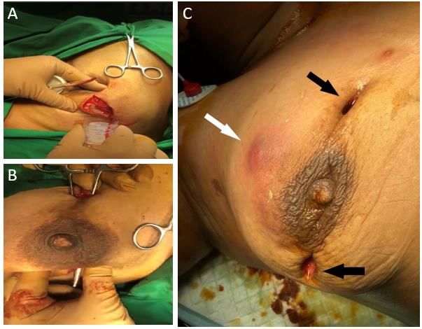
facility. The patient denied any history of previous trauma to the breast or diabetes melli-
tus. She reported no history of breastfeeding during the period when she developed the
Clin. Pract. 2021, 11 229
abscess.
At presentation, she reported a swelling on her right breast associated with throbbing
pain and low-grade fever. The left breast showed a healthy scar of previous surgery. Vital
parameters were
parameters were unremarkable
unremarkable and and local
local examination
examination demonstrated
demonstrated aa tender
tender swelling
swelling
over
over the right breast
breast in the upper and outer aspect. It was about 10 cm × 7 cmsize
in the upper and outer aspect. It was about 10 cm × 7 cm in and
in size
associated
and with with
associated local local
warmth, induration,
warmth, and fluctuation.
induration, Blood tests
and fluctuation. Bloodrevealed a leuco-
tests revealed
acyte count ofcount
leucocyte 18,000/mm
of 18,000/mm 3 with neutrophilia.
3 with neutrophilia. A diagnosisAofdiagnosis
recurrentofbreast abscess
recurrent was
breast
abscess
made. She was made. She
underwent underwent
incision incisionofand
and drainage drainage
the breast underof general
the breast under general
anaesthesia. Intra-
anaesthesia.
operatively an Intra-operatively
indurated massan indurated
with multiple mass with
small multiple
abscesses small abscesses
containing greyishcontaining
green pus
greyish
was seen green pus1A,B).
(Figure was seenAbout(Figure
150 cc 1A,B).
of pus About 150 cc of pus
was evacuated andwas evacuated
all necrotic andwas
tissue all
necrotic tissue was debrided. This pus cavity was in continuity with
debrided. This pus cavity was in continuity with previously drained abscess. Post opera-previously drained
abscess.
tively shePostwas operatively
started on a she wasofstarted
course on a course
intravenous of intravenous
third generation third generation
cephalosporin. Pus was
cephalosporin. Pus wasZiehl–Neelson
sent for Gram staining, sent for Gram staining, Ziehl–Neelson
(ZN) staining and culture.(ZN)
Gram staining andwas
staining culture.
neg-
Gram staining
ative for wasbut
bacteria, negative
primary forZNbacteria, butdemonstrated
staining primary ZN staining
acid-fastdemonstrated
bacilli in fair acid-fast
number,
bacilli in fair
suggestive ofnumber, suggestive
mycobacterial of mycobacterial
infection. Histopathology infection. Histopathology
report was suggestive ofreport was
periductal
suggestive of periductal and perilobular inflammation comprising
and perilobular inflammation comprising lymphocytes, plasma cells, polymorphs, foamy lymphocytes, plasma
cells, polymorphs,
macrophages, foamy and
histiocytes, macrophages,
occasional histiocytes,
Langhans’ type and of
occasional Langhans’
giant cells, along with type of
areas
giant cells, along with areas of micro-abscesses and frank oedema. Further,
of micro-abscesses and frank oedema. Further, cystic spaces surrounded by neutrophilic cystic spaces
surrounded
aggregates were by neutrophilic aggregatestowere
noted corresponding CNGM.notedNo corresponding to CNGM.
micro-calcifications No micro-
or granulomas
calcifications
were reportedor(Figure
granulomas
2). As were reported
per hospital (FigureCartridge-based
protocol 2). As per hospital protocol
Nucleic AcidCartridge-
Amplifi-
based Nucleic Acid Amplification Test for Mycobacterium tuberculosis (GeneXpert ® ) was
cation Test for Mycobacterium tuberculosis (GeneXpert ) was ordered to rule out tubercu-
®
ordered
losis. Thetotest
rulewas
outnegative.
tuberculosis. The test was negative.
Figure 1.
Figure 1. (A):
(A): Abscess
Abscess containing
containing pus
pus (B):
(B): Communicating
Communicating abscesses
abscesses (C):
(C): Actively
Actively draining
draining sites
sites of
of
previous incision (black arrows). New onset induration (white arrow).
previous incision (black arrows). New onset induration (white arrow).Clin. Pract. 2021, 11 230
Clin. Pract. 2021, 12, FOR PEER REVIEW 3
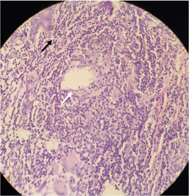
Figure2.2.Histopathological
Figure Histopathologicalphotograph,
photograph,Haematoxylin
Haematoxylin && Eosin
Eosin stain,
stain, 40
40×;
×;white
whitearrow
arrowshowing
showing
epithelioid cells around a granuloma; black arrow showing neutrophilic infiltrate.
epithelioid cells around a granuloma; black arrow showing neutrophilic infiltrate.
Aerobicculture
Aerobic cultureobtained
obtainedafteraftertwo
twoweeks
weeksrevealed
revealedrapidly
rapidlygrowing
growingnon-tuberculous
non-tuberculous
mycobacteria. Line probe speciation further demonstrated Mycobacterium
mycobacteria. Line probe speciation further demonstrated Mycobacterium abscessus abscessussub.
sub.
abscessusasasthe
abscessus thecausative
causativeorganism.
organism.Based Basedon onhistological
histologicalandandmicrobiological
microbiologicalreporting,
reporting,
diagnosisofofcystic
diagnosis cystic neutrophilic
neutrophilic granulomatous
granulomatous mastitis
mastitis secondary
secondary to Mycobacterium
to Mycobacterium ab-
absces-
scessus
sus was made.
was made. Patient
Patient by time
by this this time had developed
had developed a new a new abscess
abscess at 12ato’12 o’ clock.
clock. Previ-
Previous
ousincision
two two incision
sites atsites at 10
10 and 3 o’and 3 o’
clock clock positions
positions were notedwere noted
to be to bedraining
actively activelygreenish
draining
greenish pus (Figure 1C). On incision and drainage, all three sites were
pus (Figure 1C). On incision and drainage, all three sites were noted to be communicating noted to be com-
municating
with with
each other. each
The other.breast
necrotic The necrotic breast
tissue was tissue wasdebrided
aggressively aggressively debrided
extending extend-
up to pec-
ing up
toralis to pectoralis
fascia fascia outer
in the superior in theaspect,
superior outer aspect,
following which following
the patientwhich the patient
was advised was
regular
advised changes.
dressing regular dressing changes. Anti-tubercular
Anti-tubercular therapyBased
therapy was stopped. was stopped. BasedThoracic
on American on Amer-
ican Thoracic Society/Infectious
Society/Infectious Diseases Society Diseases Society
of America of America
guidelines andguidelines and antibiotic
antibiotic sensitivity sen-
report,
combination antimicrobial
sitivity report, combination therapy involvingtherapy
antimicrobial Clarithromycin
involving and Amikacin for and
Clarithromycin two weeks
Amika-
was
cin given,
for twofollowed
weeks was by Clarithromycin
given, followed monotherapy
by Clarithromycinfor two months. Follow
monotherapy for two upmonths.
of six
months
Followhas shown
up of her to be
six months hasdisease
shownand her symptom free.and symptom free.
to be disease
3.3.Discussion
Discussion
CNGM
CNGMisisaa rarerare disease
disease of breast seen
of breast seen in
in around
around1% 1%ofofall
allbreast
breastspecimens.
specimens. It isIt a
isdistinct
a distinct type of granulomatous mastitis with a peculiar histopathological
type of granulomatous mastitis with a peculiar histopathological pattern. There is pattern.
There is however
however a lack
a lack of of consensus
consensus over theover the definition
definition of theofdisease
the disease
withwith
CNGM CNGMoftenoften
used
used interchangeably with Idiopathic Granulomatous Mastitis (IGM) and
interchangeably with Idiopathic Granulomatous Mastitis (IGM) and Granulomatous Lob- Granulomatous
Lobular Mastitis
ular Mastitis (GLM).
(GLM). WuWu et [3]
et al. al. [3] suggested
suggested the the
useuse of ‘Cystic
of ‘Cystic Neutrophilic
Neutrophilic Granu-
Granuloma-
lomatous Mastitis’ over IGM and GLM and proposed to recognise CNGM
tous Mastitis’ over IGM and GLM and proposed to recognise CNGM as a distinct entity. as a distinct
entity. The characteristic feature of the disease is the presence of lobulo-centric granulo-
The characteristic feature of the disease is the presence of lobulo-centric granulomatous
matous mastitis with cystic spaces rimmed by neutrophils and occasionally containing
mastitis with cystic spaces rimmed by neutrophils and occasionally containing Gram-pos-
Gram-positive bacilli.
itive bacilli.Clin. Pract. 2021, 11 231
Mycobacterium abscessus is rapidly emerging nontuberculous mycobacterium that is
notorious for being resistant to standard anti-microbial therapies, thus posing therapeutic
challenges. Literature review showed sixteen cases of M. abscessus associated with infec-
tious mastitis, demonstrated women ranging 20–54 years (Table 1) [4–18]. However only
two cases of CNGM were associated with Mycobacterium abscessus.
Typically seen in parous women of childbearing age, the disease mimics several more
common conditions. CNGM presents as a breast mass with nipple inversion. Breast pain,
sinus formation and abscesses may also be seen. The disease is usually unilateral. However,
8.5% patients present with bilateral disease. In the present case though, the patient had
a past history of surgery for bilateral breast abscesses. However, recurrent breast abscess
was seen only in the right breast.
First noted as a distinct histopathological picture of CNGM in 2002, Taylor et al. were
able to identify Corynebacterium species in a cohort of 34 patients with cystic lipid filled
spaces surrounded by neutrophilic aggregates [19]. Corynebacterium kroppenstedtii was
the most common isolate. However, the exact pathogenic role of coryneforms in CNGM
is unknown. The present case showed acid-fast bacilli on primary smear, however a
Cartridge-based Nucleic Acid Amplification Test (GeneXpert® ) ruled out tuberculosis of
the breast, which is endemic to the region. Bacterial culture revealed rapidly growing NTM
which was M. abscessus.
Postulated aetiologies in soft tissue infections by M. abscessus include direct contami-
nation by material or water in traumatic injuries or surgical wound or colonization and
dissemination in immunocompromised patients. It is known to cause infection of the breast
tissue in immunocompromised patients (or patients who have undergone reconstructive
breast surgery [5–9,12,13,16]. However, in this case there was no such history).
Ultrasonography of the disease is rarely reported in literature with the most common
findings being mass, dilated ducts, oedema, and abscesses [3]. Most of these findings are
assigned a Breast Imaging and Reporting Data System (BIRADS) score of 4 (suspicious of
malignancy). CNGM, therefore presents many diagnostic and therapeutic challenges as
it mimics invasive carcinoma [1,17]. Sonography in present case showed breast abscess
with necrotic tissue. Histopathologic analysis combined with bacterial culture led to the
diagnosis of CNGM secondary to non-tuberculous mycobacterium. However, carcinoma
of the breast and tuberculosis must be ruled out before mainstay therapy is undertaken.
Two prominent subspecies, M. abscessus subsp. abscessus and M. abscessus subsp.
massiliense, have been known to encode different erm-41 gene patterns, which encodes
for macrolide resistance. This poses obvious challenges as the recommended guidelines
suggest Clarithromycin as the gold standard of monotherapy combined with Amikacin
or Cefoxitin. Resistance rates of Clarithromycin ranges up to 20%, while Cefoxitin and
Amikacin yield around 10% and 10% respectively [20]. In the present case, initially drainage
of abscess and debridement of necrotic tissue was done. This was followed by intravenous
amikacin for 14 days and oral Clarithromycin tablets for 10 weeks. A recurrence of mastitis
is another issue associated with M. abscessus. Hence, these patients require prolonged
antibiotic therapy and regular follow up.Clin. Pract. 2021, 11 232
Table 1. Review of literature of reported cases of granulomatous mastitis secondary to Mycobacterium abscessus, including
current case.
Case Report Age Predisposition Presentation Histopathology Treatment Antimicrobial Outcome
Trupiano JK Granulomatous Surgical Antimicrobials No
17 Nipple piercing Breast Mass inflammation recurrence
(2001) [4] with AFB Resection not received
Histiocytic and Clarithromycin x
Breast No
Fox LP (2004) 29 Augmentation giant cell Surgical 24 weeks,
[5] Abscess Drainage recurrence
Surgery reaction, Cefoxitin x
granulation, AFB 3 weeks
Breast Clarithromycin x No
Feldman EM 48 Augmentation Sinus not performed Surgical
(2007) [6] Drainage 24 weeks recurrence
Surgery
Breast
Augmentation
Surgery with Clarithromycin, Clinical
Taylor JL (2006) Surgical Levaquin x
21 Cystic Fibrosis on Sinus Not reported deterioration
[7] Drainage & death
Prednisone, 44 weeks
Azathioprine,
Tacrolimus
Chronic
Autoimmune inflammatory Clarithromycin x
Pasticci (2009) 54 Haemolytic Surgical Recurrence
[8] Anaemia on Abscess reaction with Drainage 10 weeks,
prednisone giant cells with Amikacin
AFB
Breast No
Jackowe DJ 44 Augmentation Sinus Not performed Surgical Not reported
(2010) [9] Surgery Drainage recurrence
Clarithromycin x
Yasar et al. 38 None Breast Mass Aspiration 16 weeks, No
(2011) [10] with sinus Not performed recurrence
Linezolid
8 weeks
Urganci AU Granulomatous Surgical Clarithromycin x No
27 None Breast Mass mastitis with recurrence
(2011) [11] AFB Drainage 6 weeks
Clarithromycin x
Ruegg (2015) Breast 20 weeks, No
39 Augmentation Abscess Not performed Surgical Tigecycline,
[12] Surgery Drainage recurrence
Linezolid,
Amikacin
Baroudi el at. Crohn’s disease, off Micro abscesses Clarithromycin x No
50 Abscess Antimicrobials recurrence
(2016) [13] treatment with mastitis 12 weeks
Rifampin,
Isoniazid,
Chronic Pyrazinamide,
Wankhade AB 30 None Breast Mass Granulomatous Surgical no follow up
(2017) [14] resection Ethambutol,
mastitis Clarithromycin,
duration
unknown
Wang YS (2017) Rifampin,
29 None CNGM Surgical No
Abscess Drainage Isoniazid, recurrence
[15] Pyrazinamide
Breast
Jensen et al. 36 Augmentation Sinus Not performed Antimicrobials Cefalexin x Recurrence
(2018) [16] Surgery 8 weeks
Clarithromycin,
Ramchandra S 33 None Breast Mass CNGM Antimicrobials no follow up
(2019) [17] duration
unknown
Mixed
inflammatory Clarithromycin x
Shaikh A 32 None Breast Mass infiltrate with Surgical 4 weeks, no follow up
(2020) [18] granulomatous resection Amikacin
reaction with fat 4 weeks
necrosis
Clarithromycin,
Present Case 34 None CNGM Surgical No
Abscess Drainage Amikacin x recurrence
8 weeks
NR: Not reported.Clin. Pract. 2021, 11 233
4. Conclusions
CNGM due to non-tuberculous mycobacteria is a rare entity. It is usually seen in
immunocompromised individuals or those who undergo breast reconstruction. Sometimes,
no obvious predisposing factors can be found.
In regions where tuberculosis is endemic, granulomatous inflammation of the breast
is usually assumed to be tubercular in origin. It is imperative that bacterial cultures and
speciation are done to rule out non-tubercular mycobacterial infection to make sure the
patient does not receive inadequate or potentially harmful therapies.
Author Contributions: Conceptualization, O.A.P. and G.D.B.; methodology, O.A.P. and G.D.B.;
validation; O.A.P., G.D.B. and A.R.N.; formal analysis, O.A.P., G.D.B. and A.R.N.; investigation,
O.A.P. and Z.S.R.; resources, Z.S.R.; data curation Z.S.R.; writing—original draft preparation, O.A.P.;
writing—review and editing, O.A.P., G.D.B., A.R.N. and Z.S.R.; visualization, O.A.P., G.D.B., A.R.N.
and Z.S.R.; supervision, G.D.B.; project administration, G.D.B. All authors have read and agreed to
the published version of the manuscript.
Funding: This research received no external funding.
Institutional Review Board Statement: Not applicable.
Informed Consent Statement: Informed consent was obtained from all subjects involved in the study.
Data Availability Statement: Data is contained within the article.
Conflicts of Interest: The authors declare no conflict of interest.
References
1. Kessler, E.; Wolloch, Y. Granulomatous mastitis: A lesion clinically simulating carcinoma. Am. J. Clin. Pathol. 1972, 58, 642–646.
[CrossRef] [PubMed]
2. Wolfrum, A.; Kümmel, S.; Theuerkauf, I.; Pelz, E.; Reinisch, M. Granulomatous Mastitis: A Therapeutic and Diagnostic Challenge.
BRC 2018, 13, 413–418. [CrossRef] [PubMed]
3. Wu, J.M.; Turashvili, G. Cystic neutrophilic granulomatous mastitis: An update. J. Clin. Pathol. 2020, 73, 445–453. [CrossRef]
[PubMed]
4. Trupiano, J.K.; Sebek, B.A.; Goldfarb, J.; Levy, L.R.; Hall, G.S.; Procop, G.W. Mastitis Due to Mycobacterium abscessus after Body
Piercing. Clin. Infect. Dis. 2001, 33, 131–134. [CrossRef] [PubMed]
5. Fox, L.P.; Geyer, A.S.; Husain, S.; Della-Latta, P.; Grossman, M.E. Mycobacterium abscessus cellulitis and multifocal abscesses of
the breasts in a transsexual from illicit intramammary injections of silicone. J. Am. Acad. Dermatol. 2004, 50, 450–454. [CrossRef]
[PubMed]
6. Feldman, E.M.; Ellsworth, W.; Yuksel, E.; Allen, S. Mycobacterium abscessus infection after breast augmentation: A case of
contaminated implants? J. Plast. Reconstr. Aesthet. Surg. 2009, 62, 330–332. [CrossRef] [PubMed]
7. Taylor, J.L.; Palmer, S.M. Mycobacterium abscessus chest wall and pulmonary infection in a cystic fibrosis lung transplant
recipient. J. Heart Lung Transpl. 2006, 25, 985–988. [CrossRef] [PubMed]
8. Pasticci, M.B.; Lapalorcia, L.M.; Antonini, G.; Mencacci, A.; Mazzolla, R.; Baldelli, F. Community-acquired mastitis due to
Mycobacterium abscessus: A case report. J. Med. Case Rep. 2009, 3, 130. [CrossRef] [PubMed]
9. Jackowe, D.J.; Murariu, D.; Parsa, N.N.; Parsa, F.D. Chronic fistulas after breast augmentation secondary to Mycobacterium
abscessus. Plast. Reconstr. Surg. 2010, 126, 38–39. [CrossRef] [PubMed]
10. Yasar, K.K.; Pehlivanoglu, F.; Sengoz, G.; Cabioglu, N. Successfully treated Mycobacterium abscessus mastitis: A rare cause of
breast masses. Indian J. Med. Microbiol. 2011, 29, 425. [CrossRef] [PubMed]
11. Urgancı, A.U.; Yorulmaz, İ.; Karaca, B.Y. Granulomatous mastitis due to mycobacterium abcessus. J. Breast Health 2011, 7, 3.
12. Rüegg, E.; Cheretakis, A.; Modarressi, A.; Harbarth, S.; Pittet-Cuénod, B. Multisite Infection with Mycobacterium abscessus after
Replacement of Breast Implants and Gluteal Lipofilling. Case Rep. Infect. Dis. 2015, 2015, 361340. [PubMed]
13. Baroudi, R.; Flaugher, M.; Hemadeh, O. An Unusual Infection of Breast Tissue. Fed. Pract. 2016, 33, 28–30. [PubMed]
14. Wankhade, A.B.; Ghadage, D.; Bhore, A.V. Breast abscess due to Mycobacterium abscessus: A rare case. Ann. Trop. Med. Public
Health 2017, 10, 447. [CrossRef]
15. Wang, Y.S.; Li, Q.W.; Zhou, L.; Guan, R.F.; Zhou, X.M.; Wu, J.H.; Rao, N.Y.; Zhu, S. Granulomatous Lobular Mastitis Associated
with Mycobacterium abscessus in South China: A Case Report and Review of the Literature. Case Rep. Infect. Dis. 2017, 2017.
[CrossRef] [PubMed]
16. Jensen, E.; Holst-Albrechtsen, S.; Christensen, K.Ø.; Birk-Sørensen, L.; Juel, J. [Mycobacterium abscessus infection after cosmetic
breast surgery in India]. Ugeskr Laeger 2018, 180, V09170655. [PubMed]Clin. Pract. 2021, 11 234
17. Ramachandra, S.; Al Kindi, M.; Al Amri, F.S.S. Granulomatous Mastitis—A Rare Presentation of Atypical Mycobacterial Infection.
J. Pathol. Infect. Dis. 2019, 2, 1–3.
18. Shaikh, A.; Vohra, L.M. Mycobacterium Abscessus: A Rare Cause of Peri-Ductal Mastitis in Endemic Regions. J. Coll. Physicians
Surg. Pak. 2020, 30, 537–540. [PubMed]
19. Taylor, G.B.; Paviour, S.D.; Musaad, S.; Jones, W.O.; Holland, D.J. A clinicopathological review of 34 cases of inflammatory
breast disease showing an association between corynebacteria infection and granulomatous mastitis. Pathology 2003, 35, 109–119.
[PubMed]
20. Naik, M.A.; Korlimarla, A.; Shetty, S.T.; Fernandes, A.M.; Pai, S.A. Cystic Neutrophilic Granulomatous Mastitis: A Clinicopatho-
logical Study With 16s rRNA Sequencing for the Detection of Corynebacteria in Formalin-Fixed Paraffin-Embedded Tissue. Int. J.
Surg. Pathol. 2020, 28, 371–381. [CrossRef] [PubMed]You can also read