SURVEILLANCE OF INFECTIOUS DISEASES IN ANIMALS AND HUMANS IN SWEDEN 2020 - Chapter excerpt - Echinococcosis
←
→
Page content transcription
If your browser does not render page correctly, please read the page content below
SURVEILLANCE OF INFECTIOUS
DISEASES IN ANIMALS AND HUMANS
         IN SWEDEN 2020

          Chapter excerpt -
           Echinococcosis
Editor: Karl Ståhl
Department of Disease Control and Epidemiology
National Veterinary Institute (SVA), SE-751 89 Uppsala, Sweden
Authors: Charlotte Axén, Mia Brytting, Ioana Bujila, Erika Chenais, Rikard Dryselius, Helena Eriksson, Eva
Forsgren, Malin Grant, Gittan Gröndahl, Gunilla Hallgren, Kristina Hammarén Busch, Anette Hansen, Marika
Hjertqvist, Mia Holmberg, Cecilia Hultén, Helena Höök, Cecilia Jernberg, Jerker Jonsson, Oskar Karlsson
Lindsjö, Ulrika König, Elina Lahti, Emelie Larsdotter, Moa Lavander, Mats Lindblad, Anna Lundén, Margareta
Löfdahl, Oskar Nilsson, Maria Nöremark, Anna Ohlson, Ylva Persson, Karin Persson-Waller, Thomas Rosendal,
Karl Ståhl, Lena Sundqvist, Robert Söderlund, Magnus Thelander, Karin Troell, Henrik Uhlhorn, Anders Wal-
lensten, Per Wallgren, Stefan Widgren, Ulrika Windahl, Joakim Wistedt, Beth Young, Nabil Yousef, Siamak
Zohari, Erik Ågren, Estelle Ågren, Elina Åsbjer
Cover: Juvenile mink in hand. Photo: Elina Kähkönen
Copyright of map data: © EuroGeographics for the administrative boundaries

Reporting guidelines: Reporting guidelines were introduced in 2018 for those chapters related to purely
animal pathogens. The guidelines build on experiences from several EU projects, and have been validated by
a team of international experts in animal health surveillance. The aim is to develop these guidelines further
in collaboration within the global surveillance community and they have therefore been made available in
the form of a wiki on the collaborative platform GitHub (https://github.com/SVA-SE/AHSURED/wiki). Feel
free to contribute!

Layout: The production of this report continues to be accomplished using a primarily open-source toolset.
The method allows the source text, produced by authors, to be edited independently of the template for
the layout which can be modified and reused for future reports. Specifically, the chapter texts, tables and
captions are authored in Microsoft Word and then converted using pandoc and R to the LaTeX typesetting
language. Most figures and maps are produced using the R software for statistical computing and the LaTeX
library pgfplots. Development for 2020 has further improved the importing of content from Excel files to
automatically build figures in the pgfplots LaTeX library. The tool is available as an R-package on GitHub
(https://github.com/SVA-SE/mill/). The report generation R-package and process was designed by Thomas
Rosendal, Wiktor Gustafsson and Stefan Widgren. In 2020, final typsetting was done primarily by Wiktor
Gustafsson with contributions from the report authors.

Print: TMG Tabergs AB.

Except where otherwise noted, the reuse of this document is authorised under the Creative Commons At-
tribution 4.0 International (CC BY 4.0) licence. This means that reuse is allowed provided appropriate credit
is given and any changes are indicated. For any use or reproduction of photos or other material that is not
owned by SVA, permission must be sought directly from the copyright holders.

Suggestion citation: Surveillance of infectious diseases in animals and humans in Sweden 2020, National
Veterinary Institute (SVA), Uppsala, Sweden. SVA:s rapportserie 68 1654-7098.
This report may be subject to updates and corrections. The latest version is always available for download
at www.sva.se.

                                                      visiting address. Ulls väg 2B address. 751 89 Uppsala
                                                      telephone. +46 18-67 40 00 fax. +46 18-30 91 62
                                                      e-mail. sva@sva.se web. www.sva.se
Echinococcosis
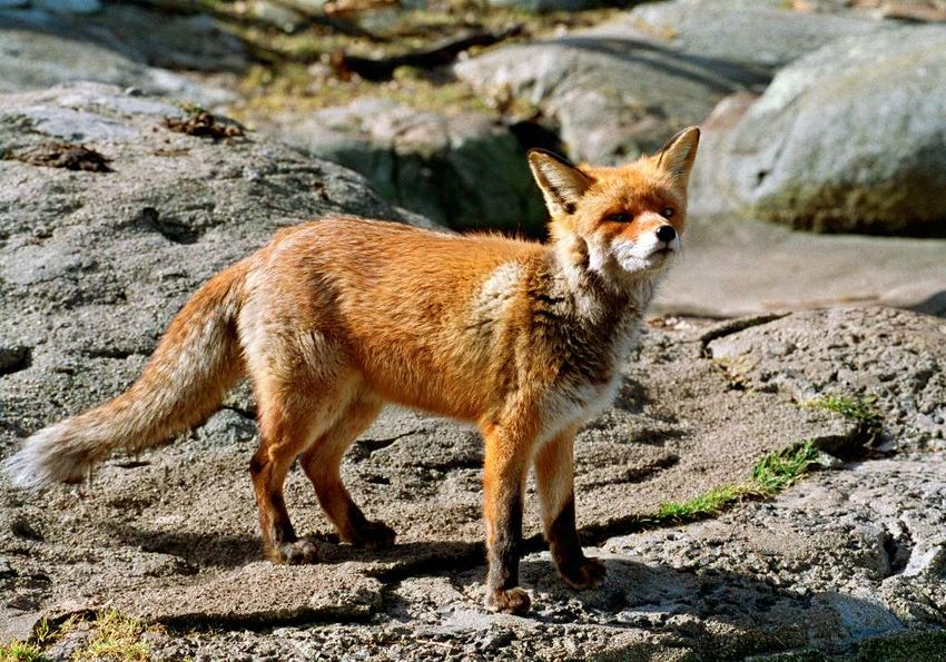
BACKGROUND The definitive hosts of this parasite are mainly foxes, but rac-
Echinococcosis is a common name for different diseases coon dogs, dogs, coyotes and wolves can also act as defini-
in humans caused by tapeworms belonging to the genus tive hosts. Rodents, mainly voles, serve as intermediate
Echinococcus. The genus contains several species, of hosts. Foxes contract E. multilocularis by eating infected
which E. multilocularis is the causative agent of alveolar rodents.
echinococcosis, while cystic echinococcosis (hydatid dis-
ease) is caused by species within the E. granulosus sensu History
lato (s.l.) complex, mainly E. granulosus sensu stricto (s.s.), Prior to 2010, E. multilocularis had not been detected in any
but also other species such as E. canadensis and E. ortleppi. definitive host, and no case of alveolar echinococcosis had
The life cycles of these parasites are similar with carniv- been reported in Sweden. As a response to the finding of
orous definitive hosts and intermediate herbivorous/omnivo- E. multilocularis in foxes in Denmark, an active monitoring
rous intermediate hosts. However, host ranges vary between programme of red foxes (Vulpes vulpes) was implemented
the different Echinococcus species. Humans are dead-end in Sweden in 2000. From 2000 to 2009, a total of 2962 red
hosts and may become infected by accidental ingestion of foxes, 68 raccoon dogs (Nyctereutes procyonoides) and 35
eggs shed by the definitive host. wolves (Canis lupus) were examined for E. multilocularis,
all with negative results. Samples from the majority of foxes
ALVEOLAR ECHINOCOCCOSIS (n=2675) were examined by ELISA (CoproAntigen ELISA)
Background at the Institute for Parasitology, Zurich University, for the
Echinococcus multilocularis is endemic in large parts of Eu- presence of the E. multilocularis coproantigen. The remain-
rope and has a reported increasing geographical range. Al- ing samples and those that were ELISA positive, were exam-
though a rare disease in humans, alveolar echinococcosis is ined using the sedimentation and counting technique (SCT)
of considerable public health concern due to its high mortal- (n=726). All samples from raccoon dogs and wolves were
ity if untreated as well as high treatment costs. examined by SCT.
During 2020, fox scats were collected from three areas in Sweden where Echinococcus multilocularis previously has been found. The results show that the parasite
is still present in two of the areas. Photo: SVA.
DISEASE SURVEILLANCE 2020 39During 2010, 304 foxes were examined for E. multilocularis. cases with clinical symptoms, and both were considered to
A total of 103 were tested by SCT and 201 by egg PCR. One have been infected abroad. No human cases were diagnosed
fox shot in south-west Sweden (county of Västra Götaland) in 2013 to 2015. From 2016 and onwards, there have been
and analysed in 2011 was found to be positive. one to four cases reported yearly.
During the spring of 2011, a national surveillance pro-
gramme was implemented where 2985 hunter-shot foxes Disease
were analysed with the segmental sedimentation and count- Animals
ing technique (SSCT). Three foxes were found positive: one In the definitive animal host, the infection is asymptomatic.
in Västra Götaland, one in Södermanland and one in the The main intermediate hosts, rodents, will usually die from
county of Dalarna. In addition, 119 faecal samples from the infection if not captured by a predator.
hunting dogs collected in the region of the first positive find-
ing were analysed with egg PCR and all were negative. In Humans
the same area 236 rodents were necropsied and all potential In humans, alveolar echinococcosis may develop into a se-
lesions examined by an in-house PCR without any positive rious, potentially fatal disease characterised by infiltrative
finding. tumour-like lesions in the affected organ. The incubation
To obtain a better prevalence estimate in a known in- period for developing alveolar echinococcosis in humans is
fected area, fox scats were collected, by a systematic sam- assumed to be between 5 and 15 years. Because of the long
pling procedure, from a circular area with a diameter of 25 incubation period, the disease is most frequently seen in
km surrounding a positive finding in the county of Söder- adults. The most common site of localisation is the liver
manland. The samples were collected in 2011 and analysed but other organs can also be affected. Symptoms depend on
in 2012, using semi-automated magnetic capture probe- the site and size of the lesion.
based DNA extraction and real-time PCR method (MC-
PCR). Six out of 790 (0.8%) faecal samples were positive. Legislation
A second national screening was initiated in 2012 and Animals
continued in 2013 and 2014. In all, a total of 2779 fox scat Detection of the parasite is notifiable according to Swedish
samples were analysed, and three positive fox scats were legislation (SJVFS 2013:23). Before 2012, all imported
identified, one from Gnesta, one from Katrineholm (both in dogs and cats (except from certain countries) were required
the county of Södermanland) and one from the county of to be de-wormed with praziquantel before entering Sweden
Västra Götaland. as a preventive measure. Because E. multilocularis has been
From the five known infected areas (including one area detected in Sweden, there is presently no legal requirement
in the county of Kronoberg which was identified as infected to deworm pets entering the country. However, as the preva-
in 2014, see below), hunters were asked to submit 30 foxes lence of the parasite in foxes is very low in Sweden compared
from each circular area with a diameter of 40 km. The aim to many European countries, dog owners are still encouraged
was to follow up the positive findings, and to collect para- to deworm their dogs prior to entry to Sweden.
sites from any positive cases, for further subtyping. Sam-
pling was initiated in 2012 and finalized in 2016. In Väs- Humans
tra Götaland two foxes were positive, in Södermanland three Infection with Echinococcus spp. has been notifiable since
foxes from Katrineholm and one from Gnesta were positive, 2004 according to the Communicable Disease Act (SFS
whereas no fox from Dalarna or Kronoberg was positive. In 2004:168) with the amendments of SFS 2013:634. However,
2018, on one single occasion, fox scats were collected in notification at the species level is not required. If cases of E.
Gnesta and 6 of 13 samples tested positive. This showed multilocularis occur in humans, the data will be presented in
that the parasite was still present in this location. the annual report at the website of the Public Health Agency
Within the Emiro research project (finalized in of Sweden (www.folkhalsomyndigheten.se). Before 2004,
2016) and the FoMA Zoonosis monitoring programme Echinococcus spp. was reported on a voluntary basis by the
(www.slu.se/en/environment) at the Swedish University of laboratories.
Agricultural Sciences (SLU), the parasite was found for the
first time in intermediate hosts; voles caught in the county Surveillance
of Södermanland in 2013 (Gnesta/Nyköping). One out of Animals
187 field voles (Microtus agrestis) and eight out of 439 As E. multilocularis does not cause clinical signs in the
water voles (Arvicola amphibius) had metacestode lesions definitive host, surveillance in these species must either be
confirmed by PCR and sequencing. Protoscoleces were active or enhanced passive for example by collection of ma-
demonstrated in the Microtus agrestis and in three out of terials from animals submitted for other reasons. In 2020,
eight Arvicola amphibius. No lesions were found in bank all free-living wolves submitted to necropsy at the National
voles (Myodes glareolus; n=655) or mice (Apodemus spp.; Veterinary Institute were tested with MC-PCR. In addition,
n=285). Within this project, a new infected area was identi- fox scats were collected from the areas in Uddevalla, Gnesta,
fied in 2014 near the town Växjö in the county of Kronoberg. and Katrineholm/Finspång where the parasite has been pre-
In 2012, alveolar echinococcosis was diagnosed in hu- viously found.
mans in Sweden for the first time. There were two human
40 DISEASE SURVEILLANCE 2020Humans Based on the knowledge available today, there is a risk
Surveillance is passive and based on identification of the dis- for occasional cases of alveolar echinococcosis acquired in
ease by a treating physician or by laboratory diagnosis. Both Sweden in the future, but the infection will most likely con-
the physician and the laboratory are obligated to report iden- tinue to be very rare in humans.
tified cases to the regional and national level to enable fur-
ther analyses and adequate intervention measures. CYSTIC ECHINOCOCCOSIS
Background
Results Cystic echinococcosis is caused by Echinococcus granulo-
Animals sus s.l. and domestic dogs and wolves are the most frequent
During 2020, 29 wolves (Canis lupus lupus), four red foxes, definitive hosts. Eggs of the parasite are excreted in fae-
three dogs and one cat were tested with the MC-PCR and ces into the environment where they can infect intermediate
all were negative. However, analysis of fox scats collected hosts such as sheep, pigs, cattle, horses and wild ruminants.
from areas where the parasite has previously been found re- The eggs develop into the larval stage (hydatid cyst) mainly
vealed that it was still present in two of the three areas (12 in the liver but also in other organs of the intermediate host.
of 109 fox scats from Uddevalla, Västra Götaland and 7 of The definitive hosts get the infection when consuming or-
18 from Gnesta, Sörmland were positive), while none of 108 gans containing hydatid cysts.
samples from the area in Katrineholm, Sörmland/Finspång,
Östergötland tested positive. History
Echinococcosis was quite common in reindeer in the north-
Humans ern parts of Scandinavia in the first half of the 20th century.
In 2020, there were three cases of alveolar echinococcosis In the 1990s, single cases of E. granulosus s.l. were de-
reported. One person had probably acquired the infection tected in moose and reindeer in Sweden. Since then, the par-
in his country of origin in Eastern Europe, while it cannot asite has not been detected in any intermediate host. How-
be ruled out for the remaining two cases that they had been ever, in a retrospective study of biobank material from 116
infected in Sweden, but they could also have acquired the wolves submitted to the National Veterinary Institute during
infection while travelling abroad. 2012–2020, two wolves shot in 2012 tested positive with a
MC-PCR detecting E. canadensis genotype 8/10 as well as
Discussion E. ortleppi.
E. multilocularis occurs sporadically in Sweden. It is not
known how and when the parasite was introduced into Disease
the country. The national screening finalised in 2014 can Animals
be used as a baseline estimate of the national prevalence, In animals, the infection is usually asymptomatic.
against which the future trend can be assessed. It is well
known from other countries that the prevalence of this par- Humans
asite varies geographically. Regional screenings have pre- In humans, the main site for cystic echinococcosis is the
viously shown a prevalence of more than 1% in a part of liver. However, it may also be located in the lungs, brain or
the county of Södermanland, and within the Emiro research other tissues. Infected patients may remain asymptomatic
project and FoMA Zoonosis monitoring programme 18 of for years or permanently. Clinical signs of disease depend
80 (20%) fox scats were found to be positive in one of four on the number of cysts, their size, localisation and pressure
investigated small areas. However, the true geographical dis- exerted on surrounding organs or tissues. The incubation pe-
tribution is unknown but so far no positive cases have been riod for developing cystic echinococcosis ranges from one to
found north of Dalarna county. Until now, the infection has several years.
been detected in five different areas. The recent finding of
positive fox scats in two of these areas shows that the para- Legislation
site is still present in these locations. In 2021 a new national Animals
screening to assess the present prevalence in foxes will be Detection of the parasite is notifiable in all animals accord-
initiated, which is expected to run for three years. ing to (SJVFS 2013:23).
E. multilocularis has also been found in intermediate
hosts, for the first time in 2014 within the Emiro research Humans
project. This finding increases our knowledge about in Echinococcosis has been notifiable according to the Com-
which biotypes the life cycle of the parasite can be com- municable Disease Act since 2004 (SFS 2004:168) with the
pleted. It has been suggested that the absence of Microtus amendments of SFS 2013:634. However, notification on
arvalis in Sweden may be a contributing factor to the low species level is not required. If cases of E. granulosus oc-
prevalence of the parasite. However, in some small areas, cur in humans, the data will be presented in the annual re-
prevalence has been reported to be higher and more research port at the website of the Public Health Agency of Sweden
is needed to clarify which intermediate host(s) are most im- (www.folkhalsomyndigheten.se). Before 2004 Echinococ-
portant. cus spp. was voluntarily reported by the laboratories.
DISEASE SURVEILLANCE 2020 41Surveillance This species is considered as less pathogenic, and possibly
Animals with a lower zoonotic potential, than E. granulosus sensu
At slaughter, all livestock are inspected for cysts during rou- stricto that is prevalent in other parts of Europe and identi-
tine meat inspection. Semi-domesticated reindeer are in- fied mainly in a cycle between dogs and farm animals.
spected at slaughter, but not all free-ranging hunted cervids In humans, cystic echinococcosis is a rare disease seen
are inspected. If cysts, that could be hydatid cysts, are found in immigrants or other people who have resided in endemic
in the liver or lung they should be sent to the National Vet- countries. In Sweden, no domestically acquired human
erinary Institute for diagnosis. cases have been reported since the infection became no-
tifiable. In Finland, on the other hand, pulmonary cystic
Humans echinococcosis (E. canadensis) was confirmed in 2015 in
Surveillance is passive and based on identification of the dis- a patient with no history of travelling abroad. The infection
ease by a treating physician or by laboratory diagnosis. Both was presumably transmitted by hunting dogs.
the physician and the laboratory are obligated to report iden-
tified cases to the regional and national level to enable fur- REFERENCES
ther analyses and adequate intervention measures. Isaksson M, Hagström A, Armua-Fernandez M, Wahlström
H, Ågren E, Miller A, Holmberg A, Lukacs M, Casulli
Results A, Deplazes P, Juremalm M (2014) A semi-automated
Animals magnetic capture probe based DNA extraction and real-
E. granulosus s.l. was not detected in any animal sampled time PCR method applied in the Swedish surveillance of
in 2020. In addition to the routine inspection at slaughter, Echinococcus multilocularis in red fox (Vulpes vulpes) fae-
5 wolves and 2 dogs were tested by MC-PCR detecting E. cal samples. Parasit Vectors 7:583
canadensis genotype G8 and G10 as well as E. ortleppi.
Miller, A. The role of rodents in the transmission of
Humans Echinococcus multilocularis and other tapeworms in a low
In 2020, 20 cases of cystic echinococcosis were reported. endemic area. Doctoral Thesis 2016. Faculty of Veteri-
For eleven of these cases, it was not possible with available nary Medicine and Animal Sciences, Swedish University of
laboratory methods to determine which species they were Agricultural Sciences, Uppsala, Sweden
infected with, but the epidemiology suggested that they also
had cystic echinococcosis. Annually around 15–30 cases are Wahlström H, Comin A, Isaksson M, Deplazes P (2016) De-
reported in Sweden. In 2020, the reported cases ranged in tection of Echinococcus multilocularis by MC-PCR: evalu-
age from 8 to 68 years (median 34 years). Eight cases were ation of diagnostic sensitivity and specificity without gold
women and 12 were men. They were all considered to have standard. Infect Ecol Epidemiol 6:30173
been infected abroad in areas where the parasite is endemic.
The most frequently specified countries of infection were Wahlström H, Lindberg A, Lindh J, Wallensten A, Lindqvist
Syria (6 cases) and Iraq (4 cases). R, Plym-Forshell L, Osterman Lind E, Ågren EO, Widgren
S, Carlsson U, Christensson D, Cedersmyg M, Lindström E,
Discussion Olsson GE, Hörnfeldt B, Barragan A, Davelid C, Hjertqvist
E. granulosus s.l. has not been detected in animal inter- M, Elvander M (2012) Investigations and actions taken dur-
mediate hosts in Sweden since the late 1990s, when it was ing 2011 due to the first finding of Echinococcus multilocu-
reported in three reindeer in the northernmost regions of laris in Sweden. Eurosurveillance 17:28
Sweden, bordering to Norway and Finland. However, retro-
spective analysis of biobank samples from 2012–2020 has Davison KR, Lavikainen A, Konyaev S, Schurer J, Miller
revealed that two wolves shot in 2012 were infected with AL, Oksanen A, Skirnisson K, Jenkins E (2016) Echinococ-
genotype G8/10 (or possibly G5). In Finland, the parasite cus across the north: Current knowledge, future challenges.
is present at a low prevalence in wildlife (wolves, moose Food Waterborne Parasitol 4:39
and reindeer) and has been genotyped as E. canadensis.
42 DISEASE SURVEILLANCE 2020You can also read