Hearing Screening & Surveillance - Unit 3 Training Programme - HSE
←
→
Page content transcription
If your browser does not render page correctly, please read the page content below
Training Programme
for Public Health Nurses and Doctors
in Child Health Screening, Surveillance and Health Promotion

Unit 3
Hearing Screening
& Surveillance
February 2005
Acknowledgements

The Programme of Action for Children wishes to thank

    Ms Patricia Barr, Senior Audiological Scientist, HSE, North West

    Ms Teresa Cawley, Child Health Training & Dev Officer, HSE, North West

    Ms Joan Dunne, Audiological Scientist, HSE, Mid West

    Ms Clare Farrell, Public Health Nurse, HSE, Dublin NA
     (“Can Your Baby Hear You” Project)

    Ms Maria Monaghan, Senior Audiological Scientist, HSE, North East

    Dr. Christina McMaster, Regional Coordinator, BHFC, HSE, North West

    Dr Theresa Pitt, Senior Audiological Scientist, HSE, South East

    Ms Deirdre Rooney, Administration, PAC

                                                                        1
CONTENTS PAGE
AUDIOLOGICAL DEFINITIONS AND TERMS........................................... 3
RATIONALE
The importance of screening for hearing in young infants ........................ 6
Outline of Screening Theory .................................................................... 7
Describing Hearing Loss – Terms and Definitions.................................... 8
How the Ear Works.................................................................................. 9
Types of Hearing Impairment................................................................. 10
Causes of Permanent Childhood Hearing Impairment............................. 12
Risk Factors for PCHI ............................................................................ 13
Glue Ear – The Facts.............................................................................. 15
Development of Sound Localisation....................................................... 18
HEARING SCREENING AND SURVEILLANCE
Protocols & Recommendations - Hearing Assessment ........................... 19
Partnership with Parents – “Can your baby hear you”............................. 21
Outline Checklist for Speech/Language/Auditory milestones…………. .23
Updated Recommendations for School Entry Hearing Screening……….24
Distraction Screening Test of Hearing.................................................... 25
Performance of the Distraction Test: Parent’s Role................................. 26
Performance of the Distraction Test: The Distractor’s Role .................... 26
Pitfalls for Distractor.............................................................................. 27
Performance of the Distraction Test: Role of Tester Behind ................... 28
Pitfalls for the Tester Behind.................................................................. 30
SCREENING OUTCOMES AND REFERRAL CRITERIA
Baby Achieves the Required Number of Responses ............................... 31
Baby Does Not Achieve the Required Number of Responses ................. 31
Referral Criteria ..................................................................................... 31
Auditing your Programme Performance ................................ ………….32
PARENT INFORMATION
Before the Screen......................................................................................... 34
After the Screen ........................................................................................... 34
Services for Children with Permanent Childhood Hearing Impairment......... 35
APPENDIX 1 – Can your baby hear you? - Sample Audit Records.....................................................37
APPENDIX 2 – Updated Protocol: Distraction Testing for Screening Purposes ................................43
APPENDIX 3 – Specifications for Hearing Test Room ........................................................................47
APPENDIX 4 – Sample Case History Sheet ..........................................................................................48
APPENDIX 5 – Equipment Contacts .....................................................................................................49
APPENDIX 6 – Scoring Sheet ................................................................................................................50
APPENDIX 7 – Evaluation Sheet ...........................................................................................................51
REFERENCES ................................................................................................ 52
2AUDIOLOGICAL DEFINITIONS and TERMS
ACQUIRED HEARING LOSS
Loss thought to have developed more than a week after birth, through infection, illness, or
genetic causes not manifest at birth (various risk factors are identified).
AUDIOLOGIST
Audiologists have specific training in the science of hearing. Many work with adults in the
community or support ENT clinics in hospitals.
AUDIOLOGICAL SCIENTIST
Audiological scientists have specific post-graduate qualifications in audiology and specialise
mainly in identification and habilitation of hearing loss in babies and children.
AUTOMATED AUDITORY BRAINSTEM RESPONSES
Screening test using small electrode gel pads to pick up electrical activity generated by sound
stimuli; easy to use with newborns.
AVERAGE HEARING LOSS
The amount of hearing loss calculated using the average decibel (dB) detection level for four
sound frequencies: 500, 1000, 2000 and 4000 Hz in better ear. Losses are generally grouped into
Mild (=95) for research and
epidemiological purposes.
COCHLEAR IMPLANT (CI)
Electrode permanently placed in or against the inner ear to stimulate the nerve electrically.
Some sound perception can result, which often leads to speech recognition with suitable
rehabilitation.
CONDUCTIVE HEARING LOSS
Defect or blockage in outer ear canal, eardrum, middle ear space or Eustachian Tube. There are
several main risk factors identified for conductive hearing loss.
CONGENITAL HEARING LOSS
Hearing loss thought to be present from birth (including birth-related causes). Risk factors are
well-defined for congenital hearing loss and can be used to identify about half of all such
children soon after birth.
CRANIO-FACIAL ABNORMALITY (CFA)
A defect in the skull or facial bone structure often causing permanent conductive hearing loss.
dB ‘A’ : A weighted scale, normally used for hearing levels when test is not done under
headphones. It can be ‘corrected’ to dB HL using fixed values.
dB HL : Decibel Hearing Level, the commonest measurement unit as shown on a Pure Tone
Audiogram
3DEGREE OR SEVERITY OF HEARING LOSS
Mild, Moderate, Severe and Profound Hearing Loss are the common categories.
DISTRACTION TESTING (DT) & UNIVERSAL DISTRACTION TESTING (UDT)
Screen or Test involving 2 testers used for children under 18 months.
EARLY DEVELOPMENTAL HEARING SCREEN (EDS)
Generally refers to DT at 7-9 months developmental clinic
EUSTACHIAN TUBE
Narrow tube which connects the nasopharynx with the middle ear space; blockage of the tube
can result in ear symptoms and OME.
GROMMETS
Tiny ventilation tubes put into the eardrums to treat OME; extruded in 4-8 months
MIXED HEARING LOSS
Hearing Loss with conductive and sensori-neural elements.
NORMAL HEARING
Usually taken as all thresholds lower/better than 25dB HL, or equivalent.
OTITIS MEDIA (WITH EFFUSION), OR OME OR ‘GLUE EAR’
Fluid build-up in middle ear space which should be air-filled. OME can cause mild/moderate
conductive hearing loss – usually temporary. ~10% of under-fives may have OME at a given
time; many improve spontaneously within weeks. OME incidence falls greatly after age seven.
OTOACOUSTIC EMISSIONS (OAE)
Signals generated by a healthy cochlea in response to incoming sound; can be utilised as a
hearing screening tool. Either Transient Evoked Otoacoustic Emission (TEOAE) or Distortion
Product Otoacoustic Emission (DPOAE) are measured.
OTOSCOPE
Instrument for examining the ear canals and eardrums
PERMANENT CHILDHOOD HEARING IMPAIRMENT (PCHI)
Permanent Hearing loss averaging >=40dB HL in better ear, present by age 5 (for epidemiology
purposes); includes children with Congenital HI (approx. 1.1 per thousand) and those with early
acquired childhood HI (approx. 0.15 per thousand), but excludes most with OME. Up to 1 per
thousand extra children may acquire PHI of this degree before leaving school.
PREVALENCE RATE
The average number of children for every 1,000 live births, with a given degree of hearing loss
(or other stated condition).
PURE TONE AUDIOMETRY
Hearing test normally carried out with children aged 3 and older wearing headphones and
requiring co-operation – results are recorded on an Audiogram.
SCREENING TEST
A simply administered test which can be passed or not; intended to be applied to an entire,
defined population. It is different to SURVEILLANCE.
4SENSORI-NEURAL HEARING LOSS (SNHL)
Hearing loss involving defects or damage to the inner ear (cochlea) or the nerve pathways from
the cochlea to the brain. Most children with PCHI have SNHL.
SURVEILLANCE
A flexible continuous process that is broader in scope than screening, whereby knowledgeable
professionals perform skilled observations on children throughout all encounters during child
health care.
COMMON ABBREVIATIONS WHICH MAY BE SEEN ON REPORTS:
AABR = Automated Auditory Brainstem Response Screening
AC = Air Conduction (overall hearing loss)
BC = Bone Conduction (measure of SNHL)
BHFC = Best Health for Children, 1999 (Denyer et al, 1999).
Bil. = Bilateral/ Unil. = Unilateral
CHL = Conductive Hearing Loss
CI = Cochlear Implant
CP = Cerebral Palsy OR Cleft Palate
DNA = Did not attend
ENT = Ear, Nose and Throat (clinic or Surgeon)
ET = Eustachian Tube
FH = Family History
Gr/T/A = Grommets/Tonsils/Adenoid surgery
HA = Hearing Aid
M (on audiograms) = Masking used
NAD = Nothing abnormal detected
NFA = No further appointments
NH = Normal Hearing
NHSP = Newborn Hearing Screening Programme
OAE = Otoacoustic Emissions
(TEOAE: Transient Evoked OAE; DPOAE: Distortion Product OAE)
O/E = On examination
OME = Otitis Media with Effusion
Perf. = Perforation of the eardrum
PTA = Pure Tone Audiogram/Audiometry
SFA = Sound Field Audiometry
SLM = Sound Level Meter
SNHL = Sensori-Neural Hearing Loss
TM = Tympanic Membrane
UNHS = Universal Newborn Hearing Screening
WL = Waiting List
WNL = Within Normal Limits
5RATIONALE
THE IMPORTANCE OF SCREENING FOR HEARING IN YOUNG INFANTS
Congenital permanent childhood hearing impairment (PCHI) affects 1-1.3 babies per 1000 births
(Davis et al, 1997; NRB, 1996), making it commoner than all other screenable birth disorders
combined (White, 2003). PCHI is a handicap of such significant effect as to warrant our effort to
identify it early. There is no medical cure for PCHI.
Early identification is the first step in lessening the effects of hearing impairment.
Early identification and habilitation will allow the child time to:
• Develop normal maturation of central auditory pathways
• Develop language to communicate effectively, thus
• Develop better literacy and educational achievements and
• Become accepted and nurtured in the family. Families also say (retrospectively) that early
identification is preferable to them, even if it is a shock at the time. They also have a low
opinion of services which fail to identify such conditions at the earliest possible opportunity.
(Sharma et al, 2004; Yoshinaga-Itano, 2003; Hall, 2003; Kennedy et al, 2004)
In Ireland, the early developmental hearing screen is still often the first preschool screen of
hearing – the first formal chance for the hearing loss to come to the notice of health
professionals. When used correctly, it has been shown to be helpful in assisting in the
identification of children with hearing impairment. (NRB, 1996). ‘Targets’ for all congenital
PCHI to be detected by 12 months show how crucial it is for EDS to be completed on time.
NEW METHODS OF SCREENING FOR HEARING LOSS
Newborn hearing screening programmes (NHSP) or universal hearing screening (UNHS) have
excellent sensitivity and specificity and are now widely used.
24-48 hours after birth, it usually takes place in maternity hospitals and is the screening method
of choice today. (NHMRC, 2002; Denyer et al, 1999)
The commonest screening tool, or instrument, is known as otoacoustic emissions (abbreviated as
OAE, TEOAE or DPOAE) which measures sound emitted from the cochlea in response to other
sounds. It is fast, accurate, non-invasive and cost-effective. Neonatal OAE testing is the next
generation 1st stage screen or sieve for well babies.
Follow-up screening is carried out with automated brainstem response audiometry.
High-risk infants are usually screened with a combination of OAE testing and automated
auditory brainstem response (AABR) screening. AABR tests the brainstem nerve pathways.
http://www.nhsp.info/ has details of screening programmes, and advice leaflets for parents
incorporated in the NHSP programme available in England & Wales in 2005.
6OUTLINE OF SCREENING THEORY
• A screen is a centrally administered system to ensure testing of a high percentage of a
specified condition.
• Most screening involves a second test occasion for first-test failures. They are not
intended to give highly accurate diagnostic information and should be quick and easy to
administer.
• The purpose is to refer onwards for further investigation a proportion of the child
population who do not pass the test on two occasions. It is not the purpose of the
Developmental Hearing Screen to identify children with a hearing loss per se.
• Some of those failed will turn out to have normal hearing, some will have hearing loss,
the majority temporary loss/conductive in nature.
• The ideal outcome would be:
1 See all children at screen age/stage, i.e. 100% coverage
2 Refer all children with a specified loss but not those with normal hearing
(i.e. 100% sensitivity and 100% specificity)
The purpose of this course is to improve the screening technique currently used to aim for
the second point above.
The target coverage for the distraction test is above 90% minimum.
The target referral rate is 4-8%.
BASIC OUTLINE OF SURVEILLANCE THEORY
A flexible continuous process that is broader in scope than screening, whereby knowledgeable
professionals perform skilled observations on children throughout all encounters during child
health care. Hall refers to it as “ oversight of the physical, social and emotional health of
children; measurement and recording of physical growth, monitoring of developmental progress;
offering and arranging intervention when necessary; prevention of disease by immunisation and
other means; and health education”. Parental reports are likely to be a part of the assessment
process.
For hearing surveillance purposes it is particularly important to be aware of typical development
in listening behaviours, expressive behaviours and social behaviours, without regarding
milestones as ‘absolute requirements’ for individual children.
(Hall and Elliman, 2003; Hall, Hill and Elliman, 1999)
7DESCRIBING HEARING LOSS – TERMS AND DEFINITIONS
Frequency (similar to pitch) is measured in cycles per second or hertz (Hz) or kilohertz (kHz). A
person who has hearing within the normal range, can hear sounds ranging between 20 and
20,000 Hz. The most important sounds we hear every day are in the 250 to 6,000 Hz range.
Speech includes a mix of low and high frequency sounds. Vowel sounds like “u” have low
frequencies (250 to 1,000 Hz) and are usually easier to hear. Consonants like “s,” “h” and “f”
have higher frequencies (1,500 to 6,000 Hz) and are harder to hear. Consonants convey most of
the meaning of what we say. Someone who cannot hear high-frequency sounds will have most
trouble understanding speech.
Intensity (similar to loudness) is measured in decibels (dB). A person with hearing in the normal
range can hear sounds ranging from 0 to 140 dB. A whisper is around 30 dB. Conversations are
usually 45 to 65 dB. Sounds that are louder than 90 dB can be uncomfortable. A loud rock
concert might be up to 110 dB. Sounds over 120 dB can be painful and result in temporary or
permanent hearing loss or tinnitus.
Impairments in hearing can happen in either frequency or intensity, or both. Hearing loss
severity is based on how well a person can hear the “speech range”. Severity can be described as
mild, moderate, severe, or profound. The term “deaf” is sometimes used for someone who has an
approximately 90 dB or greater hearing loss, who finds hearing aids of limited value, and may
well use sign language regularly. The term “hard of hearing” is sometimes used for those with
less severe hearing loss. Hearing loss affecting one ear is unilateral loss; affecting both ears is
bilateral loss.
Familiar
Sounds
Audiogram
8HOW THE EAR WORKS
The Outer Ear
The outer ear collects the sound and conducts it to the middle ear. It is made up of the pinna and
ear canal. The canal is about one inch long in adults and shorter in children. It is lined with skin
and normally contains some wax to protect it from dust and debris. Impacted wax can also cause
temporary hearing problems. The eardrum separates the outer ear from the middle ear. It is
roughly circular and firmly attached to the walls of the ear canal; sound waves that enter the ear
from the outside cause the eardrum to vibrate.
The Middle Ear
The middle ear amplifies sound to ensure it is transmitted efficiently from the air-filled outer ear
to the fluid-filled inner ear. The middle ear is made up of the eardrum or tympanic membrane,
middle ear cavity, the ossicles, and the eustachian tube.
The middle ear is behind the eardrum and is filled with air. Air reaches it along the eustachian
tube, which connects the middle ear cavity with the nose and throat. Yawning, chewing,
swallowing and blowing your nose opens the tube and ventilates the middle ear, equalising the
pressure in the middle ear with that of the air outside. This allows the eardrum and ossicles to
vibrate freely in response to sound.
In infants the eustachian tube is shorter, more horizontal and floppy, making it susceptible to
collapse or retraction, and allowing infection to pass easily from the nose or throat to the ear.
9If the tube becomes blocked, air cannot pass into the middle ear and a vacuum develops which
pulls the eardrum inwards. This can cause fluid to collect in the middle ear, which leads to “glue
ear”. This condition is the commonest type of hearing loss in children under 5 years, causing a
mild or moderate hearing problem. When PCHI already exists, the addition of fluid in the
middle ear can cause a further drop in hearing level.
The three tiny bones stretching across the middle ear space are called the ossicles. They form a
“bridge” from the tympanic membrane to the inner ear. These bones, which are the smallest in
the body, are so lightly hinged together that sound vibration can pass freely from one to another.
Sound waves entering the out ear vibrate the eardrum and are passed along this chain of ossicles
to the inner ear.
The Inner Ear
The inner ear contains two parts; the cochlea, which is concerned with hearing, and the semi-
circular canals which are concerned with balance and the ability to stand upright. The cochlea is
made of bone and shaped like a snail’s shell, coiling 2 times. Inside the cochlea are fluid-
filled corridors, lined with hundreds and thousands of tiny hair cells. Each hair cell is connected
to a fibre of the auditory nerve, which runs from the cochlea to the brain. Sound waves enter the
fluid of the cochlea, move the hair cells and stimulate their connection with the auditory nerve.
Low pitched sounds are processed at the apical/top end of the cochlear spiral while high pitched
sounds are “heard” by the basal/bottom end of the spiral, rather like a piano keyboard (and as a
result are more susceptible to insult).
These impulses pass up the nerve to the brain where they are recognised as sounds, speech,
music etc…
TYPES OF HEARING IMPAIRMENT
The Outer Ear
Failure of development of the outer ear is called atresia. This may occur by itself as an
inherited characteristic or as part of the general developmental problem of the facial
bones and palate. In some cases the outer ear may appear normal but the ear canal ends
blind with a plate of bone covering the tympanic membrane. In all cases of atresia there
is an interference with the conduction of sound along the ear and therefore a conductive
hearing loss, which is often permanent.
Impacted wax or debris can also cause temporary hearing problems in adults and
children. Small ear canals, in e.g. Down Syndrome children, may be prone to blockage.
Wax may cause problems for hearing aid wearers by blocking the sound outlet, or by
causing feedback/whistling for the wearer.
10The Middle Ear
Failure of the development of the middle ear may occur on its own or associated with the
external atresia mentioned above. This can occur without any known cause or may be
familial.
The middle ear may be the site of acute infection in young children associated with nose
and throat infections. If these infections do not clear easily but persist and become
chronic, then a hearing impairment may develop.
The middle ear may also develop a fluid, which is associated with blockage of the
eustachian tube mentioned earlier. This is known as otitis media with effusion (OME)
and can cause temporary hearing problems. OME is extremely common in babies and
young children: up to 80% of pre-school children have one or more episodes of OME.
However, less than 10% will have a long-term problem meriting regular monitoring/ENT
opinion; less than 5% will have surgical intervention. (Bennett and Haggard, 1998; NRB,
1996).
(See also ‘Glue Ear information’ pages 15-17)
In both outer and middle ear origin of hearing loss, the problem is in the conduction of
sound to the inner ear, and called conductive hearing loss. Conductive hearing loss may
resolve naturally or require intervention in the form of grommets, suction clearance or
other surgery. Sometimes hearing aids will be used for very persistent cases of
conductive hearing loss.
The Inner Ear
Hearing loss that is associated with the inner ear or cochlea or auditory nerve is described
as sensori-neural hearing loss (SNHL). This type of hearing loss is almost always a
permanent loss for which there is no cure. There is very little chance of any
improvement in hearing in sensori-neural hearing loss (SNHL). However, hearing aids
will often help the person to hear better, particularly with cochlear loss.
11CAUSES OF PERMANENT CHILDHOOD HEARING IMPAIRMENT
(McCormick, 2004)
Genetic
• Familial hereditary deafness is either autosomal recessive or dominant and non-syndromal
(eg. Connexin 26 is thought to be the commonest type genetic type in Ireland for severe-
profound PCHI)
• Syndromal deafness – where the hearing impairment is a specific feature of a recognised
syndrome such as Treacher-Collin’s or Usher’s Syndrome
• Chromosomal abnormalities eg Trisomy 13-15, 18 and 21
• 50% + of all PCHI is probably caused by genetic factors; these are gradually being identified
and simple genetic swab tests may eventually be available to future hearing screening
programmes.
Intrauterine infection/causes (STORCH infections)
• Syphilis
• Toxoplasmosis
• Rubella
• CMV
• Herpes simplex
• Toxaemia
• Drug or alcohol use by mother during pregnancy
Perinatal/Prematurity/Low birth weight
• Babies requiring mechanical ventilation for prolonged periods and with a history of hypoxia
or foetal distress
• Rhesus incompatibility
• Prolonged jaundice
• Ototoxic medication
Structural middle ear abnormalities
• Sometimes associated with syndromes, often not.
• Failure of development of middle ear structure such as tympanic membrane and ossicular
chain. Often outer ear canal is narrow, very small and ends in a bony plate. This is known as
atresia.
• A badly formed auricle or pinna and ear skin tags can be an indication of middle ear
abnormalities.
Acquired Deafness
• Bacterial meningitis/septicaemia
• Mumps or Measles
• Unknown viral illnesses or viral meningitis
• Ototoxic drugs – e.g. chemotherapy (cisplatin) or gentamicin/kanamycin
• Trauma/noise or explosive damage, head injury such as skull fracture
• Acute or chronic kidney failure and/or associated drug treatments.
• Otosclerosis (usually with family history and in later childhood).
12RISK FACTORS FOR PCHI
There are three recognised categories of risk for PCHI (NRB, 1996; Davis et al, 1997)
1. Family history of deafness
One third of congenital PCHI have immediate family history of hearing loss.
• Family history relates to hereditary childhood sensori-neural hearing loss (SNHL) (not
hearing impairment following natural ageing, noise at work or meningitis).
• Immediate family members with an apparent, or definite, history of congenital PCHI
should be noted.
2. Admittance to Neonatal Intensive Care Unit (NICU) or Special Care Baby Unit (SCBU)
for > 48 Hours
A further third with congenital PCHI have this risk category.
The incidence of PCHI is at least 10 times higher in babies admitted to NICU compared with the
‘well baby’ population.
Indicators for NICU births include:
• Low birth weight – less than 1500g.
• History of respiratory distress/anoxia with period of mechanical ventilation
• Hyperbilirubinanemia: severe jaundice where exchange transfusion is considered
• Rhesus negative babies
• Viral infections such as rubella, cytomegalovirus, herpes, syphilis or toxoplasmosis
(STORCH)
• Post meningitis children
• Ototoxic medications including amino glycosides, eg gentamycin, and loop diuretics.
However, these individual factors have been shown to equate with the simplified statement:
“ if a newborn has stayed > 48 hours in an intensive care setting, they have NICU risk factor”
3. Craniofacial Abnormalities (CFA)
About 1 in every 6 newborns with congenital PCHI have a visible CFA.
CFA includes abnormalities of pinna, ear canal, cleft palate and dysmorphic features.
Children with syndromes associated with CFA and hearing loss include
• Treacher Collins Syndrome
• Pierre Robin Syndrome
• Hunter Syndrome
• Down’s Syndrome
*Note that some children will have 2 or more of the risk factors present.
Well over 60% of PCHI cases could theoretically be identified by targeted testing, which can be
conducted in the maternity hospital. However, this yield is probably an overstatement, largely
due to the problems encountered in fully identifying those with positive family history; 45-50%
is a more realistic yield estimate, allowing for non-disclosure, or early discharges failing to be
screened.
Recent reports from early evaluations of universal neonatal screening in the U.K. have found risk
factors in 54% of newborn babies found to have bilateral hearing loss.
Also note that about one third of children with PCHI had additional disabilities, in a
retrospective Irish study of a birth cohort of PCHI children (NRB, 1996).
131. In approximately 40% of PCHI cases, cause is unknown at time of diagnosis.
It is thus important to keep ‘universal’ screen with high coverage rates.
2. The distraction test (DT) must remain a universal screen, provided in a high quality and
uniform fashion, until earlier Neonatal Hearing Screening is introduced.
(Denyer et al, 1999; Pitt et al, 1999)
3. Knowing the risk factors for PCHI is useful prior to DT, but risk factors alone do not
justify referral to Audiology, if the DT or other screen is passed.
4. Ongoing surveillance is recommended for any child where a risk factor is known.
14Glue Ear: The facts (adapted from Defeating Deafness.org
website..)
WHAT IS GLUE EAR? Glue ear occurs as a consequence of a build-up of negative pressure in
the middle ear space. As such, it develops as a ‘mechanical’ fault.
Normally the pressure behind the tympanic membrane is equalised with the ambient air
pressure by swallowing, yawning, chewing and blowing our noses. In this way we open the
eustachian tube and replenish air in the middle ear cavity as it is being absorbed on a constant
basis. When this process does not occur, due to nasal blockage (URTI), enlarged
tonsils/adenoids or acute ear infections, the middle ear cavity experiences a build-up of
negative pressure. Nature, abhorring a vacuum, prompts the walls of the middle ear cavity to
exude a fluid which can be quite serous or over time develop as a viscous, glue-like substance.
Glue ear is a build up of sticky fluid in the middle ear space of one or both ears. Because the
fluid stops the eardrum moving freely, it can lead to hearing problems.
Middle ear fluid is very common in young children. About four in every five children have at least
one mild bout of glue ear before their 4th birthday and it often clears up without treatment.
Although it is extremely rare for the condition to cause lasting damage to the ear, glue ear can
influence educational, behavioural and general development, sometimes with longer-term
effects lasting several years. It is therefore important that parents and professionals understand
the condition and the steps that they can take to minimise its impact.
WHEN ARE CHILDREN MOST LIKELY TO GET GLUE EAR? Glue ear occurs when fluid collects
in the middle ear space of one or both ears, often following ear infections or repeated colds.
Therefore winter, in particular, is a common time for glue ear to occur. Although adults can
occasionally be affected, glue ear is much more common in children, particularly between the
ages of two and five. This is because, in the second half of the first year of life many children
begin attending day-care and mothers often stop breast-feeding. As a result, children are more
exposed to infection at a time when they are losing some of their maternally conferred immunity
but have not yet built up their own. At age five, children once again are more exposed to
infections as they start school.
WHO IS MORE LIKELY TO GET GLUE EAR? Children with Down syndrome or cleft palate, and
with a family history of significant middle ear problems are more likely to get glue ear. Other risk
factors are maternal smoking, daycare attendance and male sex. A child with the latter three
risk factors is 3.4 times more likely to have hearing problems (Bennett and Haggard,1998)
RISK FACTORS FOR GLUE EAR
Glue Ear and Smoking: ‘Several research studies have shown that glue ear is one of the
conditions to which children are susceptible if they spend a lot of time in smoky conditions. As
with all risk factors, not every child who breathes in ETS will suffer from glue ear, and not all
those who have glue ear will breathe in a lot of ETS. All we know is that children are more likely
to get glue ear if they do breathe in a lot of ETS.
Glue Ear and Day Care: Children attending groups such as nursery or play-school with a large
number of other children are more prone to acute infections and glue ear.
Glue Ear and Breast Feeding: Breast milk is nature’s way of providing infants with a lot of
nutrients they need to fight off infections in the first few months of life. It helps prevent the
development of allergies. Some studies show that children who are breast-fed are less likely to
get early attacks of glue ear. This is probably because some of the mum’s immunity to infection
is passed on to the child.
Glue Ear and Allergies: In a few children, food allergies or allergy-related conditions such as hay
fever and asthma may also make the child more susceptible to glue ear.
WHAT ARE THE TREATMENTS FOR GLUE EAR?
Antibiotics: Sometimes antibiotics are prescribed for particularly troublesome ear infections.
However, they are not prescribed for every ear infection and are rarely used in the treatment of
glue ear. Over-prescription of antibiotics is now discouraged, as this leads to the bacteria
15becoming resistant and medicines losing their effectiveness. Consequently, nowadays
antibiotics might not be given at all, even in a confirmed case of glue ear, unless there has also
been recent acute infection.
Autoinflation: This is a technique in which a child blows up a special balloon using their nose
rather than their mouth. The purpose is to force open the Eustachian tube (the tube that
connects the middle ear to the throat) and allow pressure in the middle ear to return to normal.
One widely available product is called Otovent®. Continued use of autoinflation over several
weeks has been shown to help some children with glue ear, though it is a preventative aid and
not a cure. Autoinflation is a ‘low-tech’ way of helping some children. It can be made into a
game, but it needs adult supervision and it can require quite a bit of practice and it is important
to persevere.
Operations: A small proportion of children, those who experience repeated or long episodes of
glue ear, eventually need to have grommets put in. However, this is a very simple procedure
and is one of the most common operations for children. Under a light general anaesthetic, a
tiny cut is made to the eardrum. The fluid is drained away and a miniature tube known as a
grommet or ventilation tube is inserted through the small hole. The grommet keeps the middle
ear aired and healthy. This operation does not usually involve an overnight stay in hospital.
Grommets improve hearing immediately and usually stay in place for between 6 months and a
year. They fall out naturally and, when they do, the small hole in the eardrum should heal
quickly. For some, the glue ear may return and another set of grommets may be needed. The
ENT specialist will advise on what to do while the grommets are in place. In most cases,
children who have grommets can continue to go swimming but diving is usually discouraged. It
is important to check with the specialist first. Hair washing advice may also be given. The
specialist may recommend removing your child’s adenoid at the same time as putting in
grommets. In children prone to respiratory infections, this can help prevent the return of glue
ear, and it helps with the other infections too.
WHAT RESEARCH IS BEING CONDUCTED INTO GLUE EAR?
Many studies have tried to find out what factors make a child more at risk of getting glue ear and
some of these have been discussed above. Researchers have been trying to identify those risk
factors most likely to result in more persistent glue ear, and to use that information, in
conjunction with new biological discoveries, to develop new treatment methods.
The Eustachian tube connects the middle ear to the throat. In children this tube is angled
differently than it is in adults, which means that when stomach juices come up into the throat
(gastric reflux), they can reach the middle ear more easily, where they can cause damage. It is
therefore possible that in cases where glue ear is persistent, it may be due to an allergic
inflammation in response to the damage caused by gastric reflux. This research should increase
our understanding of the causes of glue ear and enable the development of new diagnostic
tests, as well as new treatments, which in the future may reduce the need for surgical
intervention (grommets). At present, there is uncertainty amongst health professionals about
what kind of treatment is most effective. Some studies have shown that hearing improves in
children with glue ear when grommets are fitted and that many children experience further
benefit if their adenoids are removed. However, the size and range of these benefits may not be
worth the cost and the small degree of risk in a child who is mildly affected. A recent study
showed that children who received grommet insertion and underwent an adenoidectomy were
less likely to return for further ENT care. This suggests that a combined approach may be a
more effective way of preventing recurrences of glue ear.
SURVEILLANCE AND GLUE EAR
Glue ear can affect children in different ways. Although it starts off as a physical problem, glue
ear can affect children in other ways. For example, as well as causing painful ears and dulled
hearing, glue ear can also affect a child’s balance and speech, their emotional well-being and
behaviour. Looking at all elements is known as “a whole-child approach”.
Older children with glue ear have described experiencing a ‘shut-in’ feeling, resulting from a
combination of the dulled hearing and the physical sensations from the middle ear (e.g. a sense
that the ear is blocked up). Younger children may have problems with their language
16development or speech. Particularly when the glue ear lasts a long time, children can develop
difficulties communicating or socialising.
WHAT ARE THE SIGNS OF GLUE EAR?
Children with glue ear experience differing degrees of hearing loss.
There may be no loss of hearing at all, or it may be quite severe. The
level of hearing may also change from day to day. Ask parents if they have any concerns
regarding their child’s hearing and be aware of the following signs (but remember that not every
child affected will display all the signs):
• Children with glue ear may appear inattentive or prone to daydreaming. They may seem to
“hear only when they want to”.
• Children may turn up the TV or say “pardon”, “eh?” or “what?” more than usual. They may
mishear words when not looking at the speaker and fail to hear sounds from outside their
field of vision.
• Some children talk too loudly - others talk less. They may mispronounce words or speak
less clearly than normal.
• Ear infections, which often come before and sometimes follow glue ear, can cause
discomfort and pain, making children fretful.
• Some children become quiet and withdrawn or anxious as a result of their difficulty hearing
when with a group of people.
• Having to concentrate hard to hear what people are saying is very tiring, so children may be
particularly grumpy and tired by the end of the day.
• Not hearing properly can frustrate children and they may become over-active or have
temper tantrums, especially when they are tired.
• Children may become unsettled at school or nursery and feel left out of some activities.
Children may have difficulty following what is being said in noisy environments or large
rooms.
• Some children may appear to have a hearing ability that changes from month to month,
especially in winter, when cough and colds are prevalent.
TACTICS FOR GLUE EAR
It is important to look out for signs that glue ear might be affecting the child in these ways. It will
help a child feel less frustrated or misunderstood if you;
• first attract child’s attention by calling his/her name or by touch, before speaking.
• can describe the problems and suggest tactics like asking others to speak louder
• arrange for extra support where necessary, e.g. speech therapy or help at school.
• advise parents to put special effort into involving the child in family conversations, for
example at mealtimes, also may make everyone more aware and will also help the child to
improve his/her communication skills.
• can choose a room with soft furnishings and carpet to talk with the child.
• cut down background noise – turn down the television or turn it off.
• talk face to face, sitting or bending to the same level as the child, if possible.
• check that the child is listening and watching and check that they have understood.
• speak up and speak clearly, but don’t shout. Be direct, keep requests short and simple.
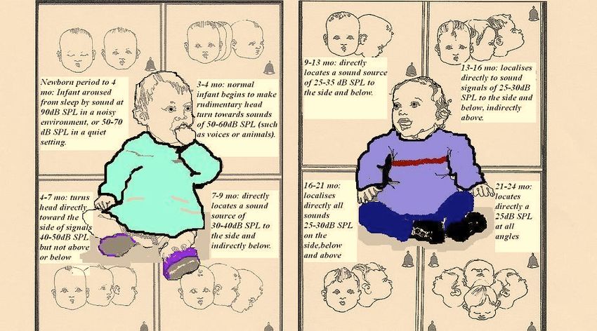
17DEVELOPMENT OF SOUND LOCALISATION
The distraction screen is based on the premise that for a 6-9 month old baby the normal response
to a quiet sound is to turn to locate the source of that sound, provided s/he is sufficiently free
from other distractions.
The development of localisation skills in young infants: the distraction test is only suitable for
children in a certain age range, when their ability to localise sound on a horizontal plane has
developed. This usually happens at about 6 months developmental age.
It is important to recognise the developmental stages below when screening infants at 7-9
months. The tester behind must be aware of positioning stimuli on the horizontal plane and
avoid holding the rattle/warbler too high up when the infant is not yet able to localise above ear
level. Stimuli position must be at ear level to ensure the infant has every chance to localise.
AGE WARBLE TONES SPEECH EXPECTED RESPONSE of INFANT
dB HL* DB HL
0-6 wks 75+ 40-60 Eye widening, eye blink, stirring or arousal from
sleep, startle response
6 wks -4 70 45-50 Similar to early responses although some head turning
m. starts around 4 mo.
4-7 m. 50 20 Head turn on lateral plane towards sound; listening
attitude; cannot find above or below
7-9 m. 45 15 Direct localisation of sounds on lateral plane;
indirectly below ear level
9-13 m. 35+HEARING SCREENING AND SURVEILLANCE
PROTOCOLS AND RECOMMENDATIONS:
Hearing Screening - Draft National Child Health Core Programme Review
Group Report 2005
Working Group Membership
Dr Theresa Pitt, (Chair) Senior Audiological Scientist, SEHB
Ms Majella Doherty, Child Health Development Officer, SHB
Ms Clare Farrell, Public Health Nurse, NAHB
Ms Maria Quaid, Audiological Scientist, OLHSC
Rationale
• Early intervention and habilitation of congenital hearing loss before 6 months of age is
best practice
Recommendations
• Early implementation of Universal Neonatal Hearing Screening (UNHS) programmes
• Retention of and staff training in Universal Distraction Hearing Test (UDHT) and School
Sweep Test as an interim measure
• Implementation of clear referral criteria as agreed by National Core Child Health
Programme Review Group
• Education of parents and professionals in using ‘Can your Baby Hear You’ surveillance
tool
Equipment
• “Can Your Baby Hear You?”
• Sound level meters
• Calibrated hand held warblers
• Free field audiometres
• Manchester rattle
• Distraction toys
• Low Table
• Audiometer
• Otoscopes
19Hearing Screening
Timing History Examination Equipment Health
Promotion
Birth Antenatal, birth UNHS is gold Otoscope Encourage
and family standard (two-stage parental
history, risk screen as per UNHS observation,
factors for report ‘Can your baby
hearing loss, recommendations). hear you?’
parental
concerns Inspection of ears,
facial morphology,
associated physical
findings or
syndromes
Postnatal visit As above As above As above
6-8 weeks As above Observation of Otoscope As above
auditory behaviour
3 months As above Observation of As above
auditory behaviour
7-9 months As above Distraction hearing Sound treated/Quiet room As above
test in the absence (ambient noise
of UNHSPARTNERSHIP WITH PARENTS
(Denyer et al, 1999; Hitching and Haggard, 1983)
Prior to 7–9 months of age parental observation should be encouraged to assess hearing. This
can be done using the following:
Tick if
EARLY DETECTION OF HEARING LOSS Response
Here is a checklist of some general signs you can look for in your baby’s first year: present
Shortly after birth:
Your baby should be startled by a sudden loud noise such as a hard clap or a door slamming and should
blink or open his/her eyes widely to such sounds.
By 1 month:
Your baby should be beginning to notice sudden prolonged sounds like the noise of a vacuum cleaner
and he/she should pause and listen to them when they begin.
By 4 months:
He/she should quieten or smile at the sound of your voice even when he/she cannot see you. He/she may also
turn his/her eyes towards you if you come up from behind or speak to him/her from the side.
By 7 months:
He/she should turn immediately to your voice across the room or to very quiet noises made on each side if not
too occupied with other things.
By 9 months:
He/she should listen attentively to familiar everyday sounds and search for very quiet sounds made out of
sight. He/she should also show pleasure in babbling loudly and tunefully.
By 12 months:
He/she should show some response to his/her own name and to other familiar words. He/she may also respond
when you say “no” and “bye bye” even when he/she cannot see any accompanying gesture.
PLEASE ATTEND YOUR LOCAL CLINIC WHEN YOUR BABY IS 7 MONTHS FOR A HEARING
SCREENING. IF YOU SUSPECT THAT YOUR BABY ISN’T HEARING NORMALLY EITHER BECAUSE
YOU CANNOT PLACE A DEFINITE TICK AGAINST THE ITEMS ABOVE OR FOR SOME OTHER REASON
THEN SEEK ADVICE FROM YOUR PUBLIC HEALTH NURSE OR G.P.
Based on original material produced by Professor Barry McCormick O.B.E. Children’s
Assessment Centre, Nottingham. Printed with permission.
A recent review by Clare O’Farrell, PHN (2002) recommends that guidelines be developed using
Guidance to Nurses and Midwives in the Development of Policies Guidelines and Protocols (An
Bord Altranais, 2000) as there is a danger that the leaflet may be distributed without systematic
questioning. Hitching and Haggard (1983) produced strong evidence suggesting that it was the
basic questioning procedure between the parent and the health visitor that reduced the age level
at which children with hearing loss were detected and produced a general re-orientation towards
hearing problems.
21A Standard Audit Tool and Action Plan (See Appendix 1) were developed to ensure that all
parents received the leaflet and had the contents discussed with them. In recognition of the
increasing diversity in our society, the leaflet has been translated into eleven different languages
to address the needs of our new citizens and clients. In collaboration with the NAD, a DVD is
currently being developed to address the needs of hearing impaired parents.
The leaflet should not be used in isolation. It complements Neonatal Hearing Screening and the
Distraction Hearing Test. (See Appendix 1)
ONGOING SURVEILLANCE IRRESPECTIVE OF ANY SCREEN OUTCOME
Parental concern about hearing should always be taken seriously. Latham and Haggard (1980)
state that up to 70% of childhood deafness, particularly severe hearing loss, can be detected by
parents. All professionals who may be in contact with a child should be able to refer to
Audiology if there is parental concern, or if they themselves are concerned.
Parental or professional concern about the infant’s hearing, development of auditory or vocal
behaviour should result in a hearing assessment carried out by appropriately trained audiology
staff. Checklists may also be useful in guiding the professionals and parents about expectations
for their child’s development of speech and language and listening behaviour
RISK FACTORS FOR ACQUIRED OR LATE ONSET HEARING IMPAIRMENT
The following groups require referral to audiology by whichever professional discovers or
becomes aware of the risk factor (may be screener, PHN, AMO, Paediatrician, GP or SLT)
• Confirmed or suspected bacterial meningitis or meningococcal disease within 4 weeks of
discharge from hospital (if assessment not already carried out in hospital).
• Those at high risk of chronic middle ear problems including children with Down syndrome
or cleft palate who require ongoing assessment by audiology.
• Other cranio-facial abnormalities (CFA) including chromosomal or syndromic conditions
such as Branchial Arch and Cervical Spine anomalies. CFA children are at risk of late onset
hearing loss although this may not develop until after the first year of life
(From www.nhsp.info website)
22OUTLINE CHECKLIST FOR SPEECH/LANGUAGE/AUDITORY MILESTONES
Time Speech Language / Social Auditory and Follow-up Health
Understanding if red flags Education
6-12 Cooing and Gurgling Uses own voice to attract Locates sounds Failure to develop Parents should ‘chat’
mths with inflection. attention; imitates sounds correctly in their mature babble & to their children often,
Babble, then like coughing /raspberries. immediate first word about books or toys,
‘Dada/Mama’ with Responds to ‘no’ & name environment by 12 progression, or lack play turntaking games,
meaning by 12m. being called. May wave months (localisation of response to name and reinforce baby’s
‘bye-bye’ and smile at skills well being called at home communication
familiar faces. developed). should be followed attempts.
Will recognise own up.
name readily.
12-24 Growing vocabulary Starts to join in with Knows some body If the child has no Parents: ensure that
mths of single words – up to simple conversations and parts; follows simple words and/or lacks your child
50 – then some 2-word understands commands e.g. ‘get interest/eye contact has a chance to learn
phrases should start to turntaking/holds eye your coat’, ‘pick up when ‘chatting’, words and feel
develop. contact when your bottle’- without they should be confident
communicating. visual cues. By 24m, followed up. when they try to
Often gives kisses/hugs. points to pictures if speak –
Plays alongside others. asked ‘where’s do not overcorrect
May copy older siblings. teddy’? mistakes in speech
at this age.
24-36 By 36 months, usually Able to maintain a Should be willing to If child is Parents: include your
mths has some 4-5 word conversation for a minute listen to stories, and unintelligible by 36 child in conversations
sentences with verbs, or two, and can tell little follow more months, says ‘uh?’ a and make sure they
(e.g. ‘The dog is stories about themselves. complex commands lot, or still depends can have friends of
running away’) even if Starting to play co- like: on gesture to make similar age to play
speech is not fully operatively with other ‘Get the milk and themselves with, not just older
intelligible to children; uses toys to you can give some understood, follow brothers and sisters.
strangers. May repeat pretend (cars or dolls). to the cat’. May them up.
part-words, or May talk to themselves enjoy looking at Focus on developing a
‘hesitate’ around 3 during play. books themselves. child’s understanding,
years, as if speaking rather than forcing
rate is slower their speech.
than thinking rate!
By 4 Fully understandable Holds normal Listens to stories If the child cannot Treat your child as a
years speech with almost no conversations and enjoys without pictures and make friends, or child and not a baby –
sound substitutions. make-believe games with can sing songs ‘forgets’ words or do not speak for them
Uses who, what, same-age friends. fluently. sentences, or in situations where
where, when – may Talks about their friends to Can learn and constantly asks for they can speak for
ask ‘why’ a lot too! others. May still make remember simple repetition, get themselves!
some grammatical errors ‘lines’ or poems for follow up before Encourage their
like school plays etc. e.g. they go to school. independence.
‘I runned to the shop’ or ‘Jesus was born in
‘The mans are coming’ the stable’.
(Also see: Cox and Cunningham, Am. Academy of Paediatrics, 2003).
23National Child Health Surveillance and Screening Core Programme Working Group:
Updated Recommendations for School Entry Hearing Screening
1. Timing: all children at school entry year or senior infants (4-5 years) to be screened ideally
by the Spring following school entry. Test younger children earlier in the day if possible.
Parents usually complete history & consent form in advance.
2. Equipment: quiet room, preferably carpeted with ambient noise =25dB HL if ‘average approach’ is adopted).
7. Referral to Audiology clinic or 2nd tier clinic as per local arrangements; send copies of both
screening audiograms dated with referral form.
8. History outline/form obtained should be sent with referrals, such that parental/teacher
concerns, ear infections/illnesses, risk factors, speech and language delay, may be taken into
account in follow-up.
Less than 10% of children should be referred by a School Screening Programme – many children
will have already attended other services and had hearing tests by this age, so the history form is
particularly important.
24DISTRACTION SCREENING TEST OF HEARING
(adapted from www.nhsp.info/workbook/shtml protocol by Prof. B Mc Cormick )
(cf Training Video:)
This is appropriate for children between 6 and 12 months developmental age
The recommended age of first screen is 7-8 months and second screen before 10 months.
The Essentials
A quiet room (see Appendix 2 and Appendix 3 for details.)
A low coffee table
A range of sound generators with known frequency content
A sound level meter
Two- people - well trained and working in partnership
Set-up for Screening Distraction Test
Desk with Sound Level
Meter available for use
Tester behind within 1 metre
of chair back
Noisemaker/warbler at 45-
50° angle, 50cm behind
baby’s right ear (to screen
right ear)
Chair
Parent with baby on front
of lap, both facing forward
LOW TABLE
Front tester/distractor on
working level for low table.
25PERFORMANCE OF THE DISTRACTION TEST: PARENT’S ROLE
Motto: support but do not join in
• The parent holds baby facing forward in midline, with knees together, holding baby just
slightly out from chest to allow freedom of movement.
• The parent places hands under the baby’s armpit with her thumbs at the baby’s back. This
steadies the baby and helps to prevent him/her from falling backwards. This distance
between parent and child also prevents the child getting too comfortable and sleepy and
promotes independent movement when searching for the sound.
• Avoid allowing the baby-sitting on the end of the parent’s knees. Babies need a firm-
sitting base, not a ‘wobbly’ one, to locate sound well.
• Parent should be told not to react to any sound or the tester behind.
PERFORMANCE OF THE DISTRACTION TEST: THE DISTRACTOR’S ROLE
Motto: control baby’s attention
• It is helpful to explain the screen to parents
• Arrange parent and baby’s sitting position i.e. baby upright, and not leaning back but
sitting securely on parent’s knees. If position alters during testing, direct parent to hold
baby back in the middle again. This is essential, as a baby will not respond on the correct
side if the sitting position is skewed. The baby must sit in centre of lap, not leaning to
either side. Break the test to gently inform parent to “straighten” child if necessary.
• Front tester sits or kneels at the low table and with various small toys, commands the
baby full attention. It is vital that the tester in front takes responsibility for control of the
baby’s attention.
• Person holding baby must be advised not to move or react to the sounds when they are
presented, as this can cue the baby.
Engage the attention of the baby but avoid over-stimulation.
To be successful here, you can use a soft voice to get the baby’s attention to
the table. A variety of 3-4 spinning tops/balls can be used on a low surface,
keeping the toys just below the baby’s eye level. It is important to keep the activity level quiet to
avoid the baby becoming less responsive to testing sounds.
Wait until the tester behind is in position before you phase or break the activity
The tester behind must be ready and in position to allow maximum opportunity for the baby to
turn, and minimum amount of movement/noise.
If baby becomes distracted before or during sound stimuli presentation, bring his/her attention
back to midline by tapping toy or table or clicking fingers
26Plateau child’s attention and let tester behind “enter” with sound.
Don’t alert baby to the tester behind by looking directly at the person. Minimise eye contact with
baby so that they do not fixate on tester in front.
Sound is presented after the stimulation level is dropped or phased by covering toy or
freezing activity.
Young babies will allow you to cover the toy with your hand, as they may not have yet
developed object permanence. It can help to move your fingers whilst covering the toy to help
keep the baby’s attention on the table.
Older babies may only allow a short break in activity i.e. freeze on the spot, as they may get
frustrated if the toy is constantly covered.
More mature infants will anticipate sound presentation if the routine becomes too predictable.
It helps to vary the routine.
Evaluate the responses of the baby
The majority of children will locate (correctly or incorrectly) the sound by turning 90 degrees. A
few will be too shy or unable physically to turn. In these cases, alert person behind that baby is
eye searching/pointing to sound sources i.e. nod gently.
Bring child’s attention back to the table
Bring child’s attention back to the table once he/she has located sound.
Don’t let child dwell on tester behind. Child’s attention should be brought forward before tester
behind moves away.
It is vital that the tester in front takes the responsibility for control of the baby’s attention.
PITFALLS FOR DISTRACTOR
If the distractor in front is uncertain, or not in control
This often happens with older or more active babies who are difficult to distract. To hold such a
child’s attention it may be necessary to engage in more sophisticated activities, such as symbolic
play with teddy/dolly and a sequence of activities with verbal commentary. “Look here’s teddy
and he’s hungry – Oh don’t cry teddy, let’s get your dinner”.
The activity of feeding teddy is often the signal for the child to watch and tester behind to enter
with sound presentation.
Other activities are building up a tower of blocks, man going in a car, man chasing a cow.
Once more remember, don’t over-stimulate and keep chat to a bare minimum. Be
prepared to change activities quickly for the very active ones! Use your voice and eyes to
engage the child’s attention.
Tester behind is not ready or not in the correct position
The tester behind is not ready to present the sound when toy is removed therefore there is a delay
and the child has either lost interest or picked up a clue. This can be avoided if the two testers
work as a team; there is an optimum number of seconds between the toy being removed and the
sound being presented, for the child to respond. The more active the child, the more necessary it
is to make sure that there is only a split second delay between cessation of the activity and
presentation of sound.
27You can also read