Groin Pain in Athletes - Chris Dolan, M.D - UNR Med
←
→
Page content transcription
If your browser does not render page correctly, please read the page content below
Groin Pain in Athletes

      Chris Dolan, M.D.
Differential Diagnosis
EXTRA ARTICULAR INTRA ARTICULAR
Muscle strains Femoroacetabular impingement
“Pulled Groin” Labral tears
Athletic Pubalgia / Sports Hip dysplasia
Hernia Osteoarthritis
Osteitis Pubis Inflammatory arthritis
Snapping Hip Hip dislocations – AVN
Nerve entrapment syndromes Osteonecrosis
Stress Fractures
Avulsion and apophyseal REFERRED / MEDICAL
injuries
Lumbar / Sacral pathology
Piriformis syndrome
Gynecologic
Bursitis
Urologic
Iliopsoas
Trochanteric
GI – Hernia, IBD
Hip and thigh contusions Neoplasm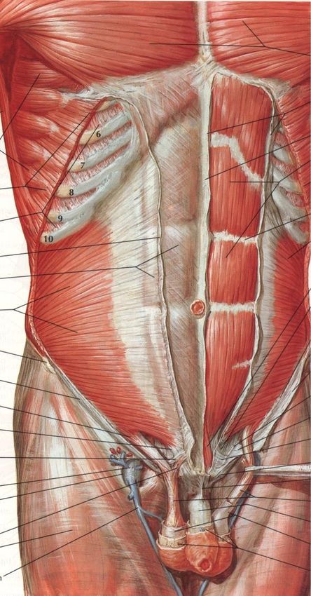
Groin Pain Epidemiology
Feeley et al. AJSM preview July 2008
NFL Injury Surveillance System 1997-2006
All injuries that caused athlete to miss >2
days; all hip & groin injuries recorded
23,806 total injuries; 738 hip (3.1%)
Avg 12.3 days lost
Muscle strain most common
“Sports hip triad” – labral tear, adductor
strain, rectus abdominus strain
Many players with labral tears demonstrated
persistent adductor strains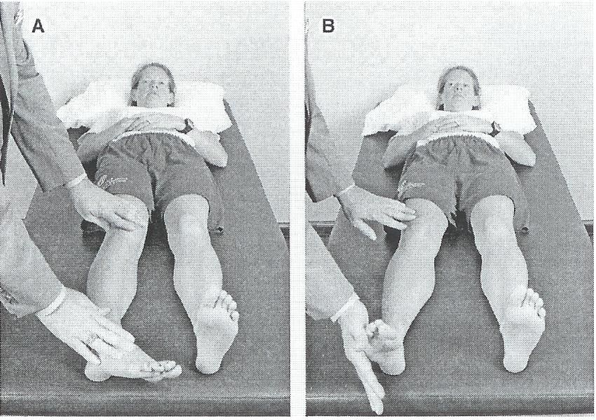
Groin Pain in the Athlete
Despite high prevalence the cause can be difficult
to elucidate
Complex local anatomy with large soft tissue sleeve
Complex biomechanics
Wide differential diagnosis
Often diffuse, insidious symptoms with nonspecific
presentation
Often multiple diagnoses
Ekberg Sports Med 1998 – Multi Disciplinary Approach
with G. Surg, Orthopaedics, Urology, Radiology
19/21 athletes 2 or more diagnosed causes
Inguinal hernia, neuralgia, adductor strain, osteitis pubis,
prostatitis
Difficult Dx, explanation for failed therapy
Patient History
Onset – acute or insidious (most common athletic groin injuries are hip
adductor and flexor strains which have an acute onset vs an athletic hernia
which is usually chronic in nature)
Location of pain
Duration
Severity
Mechanical symptoms – Intraarticular - chondral flaps and labral tears
Sporting activity
Recent increase in activity or vigorous new exercise
Long distance runners / Triathletes – stress fxs
Twisting type sports: Hockey, soccer, tennis, golf – adductor strains,
sports hernia, labral tears
Association with Trauma – history of hip dislocation; impact injury to greater
trochanter
History of ligamentous laxity
Hip dysplasia as a child
Previous hip problems – SCFE, Perthes
Safran. Op Tech Sports Med 2005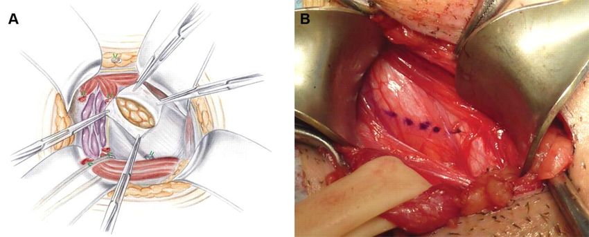
Patient History
Intra-articular Problems
Pts often have pain that is described as deep in the joint
and localized to anterior groin or inguinal region
May localize between a finger anteriorly in groin and
one at posterior aspect of trochanter or buttock
Discrete episodes of sharp pain with weight bearing
Pain with sitting with the hip flexed and pain or
catching on arising from a seated position
Catching, popping, locking
Safran. Op Tech Sports Med 2005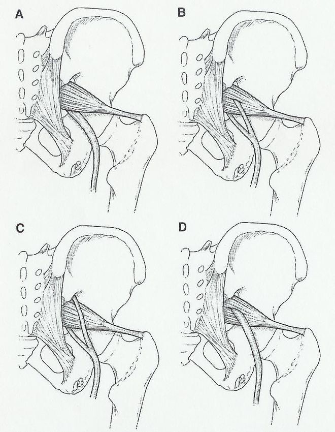
Patient History
Extra-articular Problems
Pain felt in the buttocks/posterior trochanteric
region, Lateral thigh
Low abdominal area
Pubic Symphysis and Adductors, Hip Flexors
Referred pain
Spine
Abdominal / Intrapelvic / Retroperitoneal
Remember hip problems may present as knee pain
Safran. Op Tech Sports Med 2005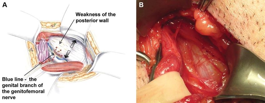
Physical Exam
Palpation
Muscles origins such as
sartorius, rectus femoris,
gluteus medius and adductors
Iliac crest – hip pointers
Hernias
Pubic symphysis – osteitis
pubis
Sciatic notch
With hip flexed 90deg palpate
half way between GT and
ischial tuberosity
Pts with sciatica, piriformis
syndrome
Greater trochanter
Bursitis
Snapping ITB
Safran. Op Tech Sports Med 2005
Adductor Muscle Strains
Adductor Muscle Strains
Most common injury about the groin
Injury occurs during eccentric contraction – hip hyperabduction
and hyperextension
Adductor muscles = pectineus, adductor longus, brevis and
magnus, gracilis and obturator externus
Adductor longus is the most frequently injured
More common in ice hockey and soccer, which require strong
eccentric contraction of the adductor musculature Anderson. AJSM 2001
Tyler et al. AJSM 2001 The association of hip strength and
flexibility on the incidence of groin strains in professional ice
hockey players.
The strength ratio of the adduction to abduction muscle groups (Adductor Muscle Strains
CLINICAL PRESENTATION / PHYSICAL EXAM
Usually sudden onset pain in groin region; insidious
Pts have pain on palpation of adductor tendons or insertion on
the pubic bone
Pain with resisted adduction or
passive abduction
Grade
First degree – pain, but minimal loss of strength and minimal restriction
of motion
Second degree – decreased strength
Third degree – complete disruption of muscle tendon unitAdductor Muscle Strains
Imaging
Plain films to rule out avulsions,
fractures, pathology
Bone scan to rule out osteitis
pubis
MRI muscle enhancement, ant
pubic bone
Robinson. Skeletal Rad 2004Adductor Muscle Strains
There is high incidence of recurrent strains
Likely due to incomplete rehab
Rehab program needs to emphasize eccentric resistive
exercise
Tyler et al showed that an 8-12wk active strengthening
program, consisting of progressive resistive adduction
and abduction exercises, balance training, abdominal
strengthening and skating movements on a slide board,
proved effective in treating chronic groin strains and
preventing new in season injuriesTyler et al. The effectiveness of a preseason exercise program to prevent adductor
muscle strains in professional ice hockey players. AJSM 2002Adductor Muscle Strains
TREATMENT
Rest, ice, NSAIDs, compressive shorts (skins)
Holmich. Lancet 1999. Effectiveness of Active Physical
Training as Treatment for Long-Standing Adductor-Related
Groin Pain in Athletes: randomized trail.
68 athletes >10 mos adductor related groin pain
8-12 weeks active vs passive therapy
Found that functional training with active exercise was far
superior to passive therapy with massage and modalities.
After RTP – only 40% athletes WITHOUT symptoms at end
of following seasonAdductor Muscle Strains
Surgical treatment = Adductor tenotomy
Akermark and Johansson. AJSM 1992
16athletes, all improved or were free of symptoms
All but one returned to the same sports at a mean of
6.6wks
10/16 (63%) were able to return to their previous sports
activities
5/16 returned but at a reduced level
All pts had decreased strengthSports Hernia
Sports Hernia
Athletic pubalgia, sportsman’s hernia,
Gilmore’s groin, hockey groin, slap shot gut,
Ashby’s inguinal ligament enthesopathy
Occult hernia caused by weakness or
deficiency of the posterior inguinal wall
without a clinically recognizable hernia,
leads to chronic groin pain
Almost always in men, most commonly in sports
such as hockey, soccer, Australian rules football,
and tennis
Often prolonged course before diagnosis
Cause of chronic groin pain in athletes
Primary dx in 39-50% of pts with chronic groin pain
(Lovell. Aust J Sci Med Sport. 1995; Kluin et al. AJSM
2004)Sports Hernia
Pathoanatomy Farber et al. JAAOS Aug 2007
Often from trunk hyperextension and thigh hyperabduction, leads to
shear across the pubic symphysis and stress on inguinal musculature
Shearing forces more prominent in athletes with an imbalance
between the strong adductors and relatively weak lower abdominal
musculature
Places stress on inguinal wall musculature leading to attenuation of the
soft tissuesSports Hernia
Pathologic findings have
included attenuation or
tearing of the transversalis
fascia, conjoined tendon,
the rectus abdominis
muscle insertion, or of the
Int O / Ext O muscles or
aponeurosesSports Hernia
“Syndrome of Muscle Imbalance of the Groin”
Additional stress on hemipelvis leads to
weakening or tearing of the transversalis fascia
and surrounding tissue
Results in tendonitis of adductor and/or
abdominal muscles
Hackney et al, Brit Jrnl Sports Med 1993Sports Hernia
Hallmark = asymptomatic with inactivity and pain returns with
activity
Usually an insidious onset of unilateral, deep groin pain that
often radiates to the perineum and inner thigh or across the
midline
Aggravated by sudden movements, valsalva, performing sit-ups,
sprinting and kicking
On exam findings may include local tenderness over the
conjoined tendon, pubic tubercle and midinguinal region;
tender, dilated superficial inguinal ring (up to 94%) and
tenderness of the post. wall of the inguinal canal; pain with
resisted hip adduction and resisted sit-up as well as valsalva or
coughing
Farber et al. JAAOS Aug 2007Sports Hernia
Imaging
Generally used to r/o other diagnoses
Plain films of hips, pelvis and LS spine
Bone scan may reveal increased uptake at superior pubis,
pubic symphysis or adductor origin but is nonspecific
Dynamic ultrasound – operator dependent
MRI – may show increased signal within the pubic bones or
within one or more groin muscles (rectus abdominus,
pectineus, adductors) but is also nonspecific
Albers et al. MR findings in athletes with pubalgia. Skel Radiol 2001.Sports Hernia
Conservative
NSAIDs
Deep massage
Prolonged rest
PT with emphasis on core strengthening
and resolving the imbalance of the hip and
pelvic muscle stabilizers
Pts with chronic groin pain do not get
better with conservative measures
Farber et al. JAAOS Aug 2007Sports Hernia
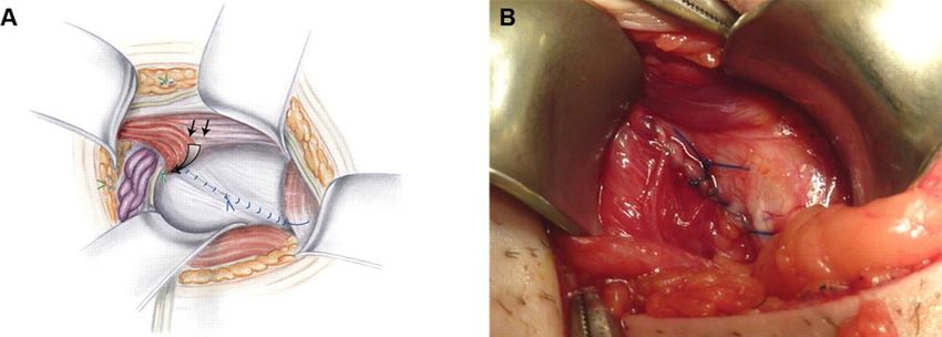
Surgical treatment
Consider after 8-12weeks of failed non-op Tx
Surgical repair of the weak posterior inguinal wall
with open or laparoscopic techniques leads to
success rates of 80-97%
Some also recommend adductor tenotomy in pts
who have symptomatic adductor abnormality that is
not corrected preoperativelySports Henria
Muschaweck argues if pain not improved in 4-6 weeks
athlete at high risk of chronic pain if not aggressively
treated
Tension free repair of the transversalis fasciaSports Hernia
Varied Surgical treatment
Meyers et al. AJSM 2000.
157 athletes, reattached the inferolateral edge of the rectus
abdominis to pubic bone and inguinal ligament: tightens
attachment around pubis and stabilizes pelvis
+/- adductor longus tenotomy;
f/u avg 3.9yrs, 95% success rate with 88% playing at or above
pre-injury level at 3mo and 96% at 6moSwan. CORR 2006 The Athletic Hernia: A Systematic Review
Sports Hernia
Only one prospective, randomized study
Ekstrand et al. Surgery versus conservative treatment in soccer
players with chronic groin pain: a prospective randomized study
in soccer players. Eur J Sports Traumatol Rel Res. 2001
66pts, four groups: surgery, individual training, NSAIDS and PT and
controls; only the surgical group showed substantial and statistically
significant improvement in symptoms and ability to return to sport
Caudill et al. BJSM July 2008. Sports Hernias: A Systematic
Literature Review
“Sports hernia anatomy, surgical procedures and rehabilitation strategies
are poorly described”
Majority Level IV
Call for better studiesSports Hernia
Should involve a multidisciplinary approach
Pts often have more than one diagnosis
Ekberg et al. Long-standing groin pain in athletes: a
multidisciplinary approach. Sports Med 1988.
Ortho surg, Gen. surg, Urologist evaluated 21pts
90% of pts had two or more positive findings, some >4
diagnoses
Most common diagnoses were osteitis pubis, inguinal hernia
and prostatitis(48%)Osteitis Pubis
Osteitis Pubis
Painful, inflammatory, non-infectious condition
of pubic symphysis and surrounding structures
Etiology considered to be associated with
muscle imbalance, pelvis instability and chronic
overuse injury
Abdominal and adductor muscle imbalance
(antagonists), prevalent in kicking sports
Abnormal vertical motion of the pubic
symphysis (>2mm) is a contributing factorOsteitis Pubis
Usually presents with
pubic symphysis or
adductor pain
Aggravated by activities
requiring sudden hip
flexion or rotation
Provocative maneuvers
Rocking cross-leg test –
pt sits with one knee
crossed over the other,
push down on knee and
hold down opposite iliac
crest
Lateral pelvic
compression testPhysical Exam
Lateral compression to evaluate
osteitis pubis
Lateral decubitus position with
downward force applied to iliac crest
Symptoms at the pubis
Gapping and
approximation/compression test
Supine (Transverse anterior stress test)
Downward and outward pressure on ASIS
Supine (Transverse posterior stress
test)
Pressure from iliac crests towards midlineOsteitis Pubis
Xrays findings –
marginal irregularity,
symmetrical bone
reabsorption, widening
of symphysis, reactive
sclerosis
Flamingo views – single
leg standing AP view of
PS, vertical motion
>2mm is abnormal
MRI – increased signal in
the PS because of bone
marrow edema
Radic et al. AJSM Jan 2008Osteitis Pubis
Paajanen et al. AJSM Jan 2008 Comparison of MRI findings for athletes with osteitis
pubis and asymptomatic athletes during heavy training
-65% of asymptomatic athletes demonstrated presence of bone marrow edema
-Decreases the value of MRI for surgical decision makingOsteitis Pubis
Nonoperative management
Rest from physical activity – average time to full
recovery was 9.6mo (Fricker et al. Sports Med 1991),
most studies indicate need for 3-6mo of rest
NSAIDS
Shock absorbing footwear
PT – Hip and adductor muscle
stretching/strengthening, core stability and
strengthening and muscle force balancing
Corticosteriod injection – some studies suggest a quicker
return to athletic activities if done early
Holt et al. AJSM 1995 – 8 athletes, after one injection 3/8
returned to sport within 3wks, four required a second injectionOsteitis Pubis
Surgical Management
Wedge Resection
Concern with late pelvic
instability (Grace et al. JBJS
1989)
Compression Plate arthrodesis
with bone graft
Williams et al. AJSM 2000 – 7
rugby players failed 13mo
non-op tx, at mean 52.4mo
f/u all were free of sxs and all
returned to sport
Currettage for resistant osteitis
pubis in athletes
Radic et al AJSM Jan 2008 –
23 athletes; 21 return to pain
free running at 3 mosOsteitis Pubis
Choi, et al. Br J Sports Med Sep 30 2008.
Treatment of Osteitis Pubis in Athletes: A
Systematic Review
25 articles (case series / reports) = all Level IV
No randomized controlled trials
195 athletes Dx with osteitis pubis
No comparisons of treatments, difficult to draw
accurate conclusions
Call for better studiesSnapping Hip
Snapping Hip / Coxa Saltans
Internal Type – Iliopsoas
External Type – TFL
Snapping of the hip is a normal occurrence
Many people experience benign, asymptomatic
snapping on an infrequent basis
It is the rare individual who experiences
symptomatic snapping
Allen. JAAOS 1995Snapping Hip / Coxa Saltans
Internal type – Iliopsoas tendon
Can mimic a mechanical intra-articular process
Is an asymptomatic incidental observation in 5% to
10% of the population
Commonly seen in ballet dancers
Occurs as the iliopsoas tendon subluxates from lateral
to medial while the hip is brought from a FABER
position into extension and IR
Debated whether the snapping is the tendon going back and
forth across the femoral head or across the pectineal
eminence
Allen. JAAOS 1995Snapping Hip
Snapping Hip

From FABER to extension
Snapping Hip
Conservative Treatment
Modify offending activities
Stretching and flexibility
Core stabilization program
NSAIDS
Corticosteroid injection in the iliopsoas bursa
Only a few case series reported
Vaccaro et al. Radiol 1995 – 8pts, 7 had between 2-8mo
relief however 4 went onto surgery
Wahl et al. AJSM 2004 – 2 pro football players, U/S
guided inj into bursa, both returned to sport in 4 weeks
with f/u 26mo and no return of snappingSnapping Hip
Surgical Treatment
Relaxation of the iliopsoas to eliminate the snapping
Different open approaches have been described depending
on the location of the snapping
Allen et al. AJSM 1990 – 20 hips, anterior approach, release of
posteromedial tendinous portion; 70% complete resolution, 25%
partial
Gruen et al. AJSM 2002 – 11pts, ilioinguinal approach with fractional
lengthening of the iliopsoas tendon within the psoas muscle; 100%
resolution of snapping and 83% pt satisfaction
Taylor et al. JBJS 1995 – 16hips, medial approach, tendinous portion
released from the lesser troch; all pts subjectively improved
Complications – reported to occur in 43-50% of patients; loss
of hip flexion strength, sensory disturbances, incisional
complications – hip arthroscopy results differSnapping Hip
Arthroscopic release of
tendinous portion of
iliopsoas at the lesser
troch
Also allows you to
address any intra-
articular problems
Avoids complications
due to incisionsSnapping Hip
Flanum et al. AJSM 2007 –
6pts, 5/6 also had intra-
articular pathology, none had
recurrence of snapping at
12mo
Ilizalitturi et al. Arthroscopy
2004 – 6pts, 4/6 had intra-
articular pathology, no
recurrence of snapping at 10-
27mo f/u
Hip flexor strength returned
by 8-12weeksSnapping Hip
External type – Iliotibial Band
Can often be seen from across the room
Pts describe a sense that the hip is subluxating or
dislocating
Classically described in the downside leg of runners
training on a sloped surface
With pt in lateral decubitus position flex and extend the
hip, snap palpated over greater troch which can be
blocked by applying pressure over GT
Often a dynamic process better demonstrated by the pt
then on passive exam
Ober test to evaluate for IT tightnessPhysical Exam
Ober Test
For hip abductor tightness or
IT band contracture
Patient in the lateral decub
position, with lower hip and
knee flexed
Flex hip to 90deg and fully
abduct then extend past
neutral with knee in 90deg
flexion. Now allow hip and
knee to adduct with hip
neutral
Hip should adduct such that
the knee is at or below the
midlineSnapping Hip
Snapping occurs from the IT
band flipping back and forth
across the greater troch
Often attributed to a
thickening of the post. part
of the IT tract or ant. border
of the glut. max.
Thickened portion lies post.
to the trochanter in extension
then flips forward over the
trochanter as the hip begins
to flexSnapping Hip
Xrays to r/o other pathology – coxa vara may
predispose
MRI may show evidence of trochanteric bursitis and
thickening of the tendon
Ultrasound
Treatment
Avoid offending activities
NSAIDS
PT – stretching program for the ITband
Corticosteroid injections into trochanteric bursaSnapping Hip
Surgical treatment
Goal is to eliminate the snapping by a relaxing procedure of
the ITband
Most use a Z-plasty technique with excision of the underlying
bursa (excision of an ellipsoid-shaped segment as well as a
cruciate incision of the IT band have also been described)
Proventure AJSM 2004 – 9hips, all had complete resolution of
snapping and all but one returned to unrestricted activitiesSnapping Hip
Arthroscopic releases
have also being described
Ilizalitturi et al.
Arthroscopy 2006 –
11pts, 2yr f/u 1 pt with
non-painful snapping,
rest had no sxs and
returned to previous
activityStress fractures
Generally felt to occur from a repetitive cycle overload
by submaximal forces – bone resorption > bone
formation
Muscle fatigue may lead to transmission of forces to the
underlying bone
Muscles may also contribute by concentrating forces
across a localized area of bone
Stress fxs of the pubic rami are particularly common
among long distance runners
Appears to be an association with anorexia and
amenorrhoea in female atheletes (female athlete triad –
eating disorder, amenorrhoea, osteoporosis)
Saha et al. AJSM 1988 – stress fxs occurred in 49% of
collegiate female distance runners that had less than 5
menses/yrStress fractures
Present with insidious onset of lower pelvic and groin
pain, worse after running and improves with rest
Often will have pain with axial loading or with standing
or hopping on the involved leg
Often the result of training errors
Imaging of choice is MRI or bone scan
Treatment of stress fxs about the pelvis
period of 4-6wks of rest and when pain free a graduated
program of return to activities
Address any dietary or hormonal issuesStress fractures
Femoral neck
Distance runners
F>M
Activity related anterior groin pain, have limitations of end
range of motion, active SLR and logrolling may cause pain
Xrays – may not show changes, if suspicious need to get MRI
or bone scanStress fractures
Femoral neck
Compression side =
inferior femoral neck
Good potential to heal
Treatment is generally
nonop. Crutches and non-
weight bearing until
asymptomatic. Weekly
xrays to ensure fx is not
progressing. Gradual
return to pain free
activities.
If fatigue line on MR is
>50% consider pinningStress fractures
Tension side = superior
femoral neck
Unstable
High rate of
complications if it
progresses to a
displaced fracture
Should be treated with
surgical fixationNerve Entrapments
Obturator nerve entrapment in skaters secondary to
adductor muscle development
Meralgia parasthetica
LFCN
Pudendal nerve cyclists
Treatment = removal of
the offending compression
Compression traumatic,
anatomic, nonanatomic
Sartorius and iliac fascia
Ilioinguinal neuralgia – nerve ablationApophyseal Avulsion Injuries
Operative fixation considered
for larger fragments and
displacement > 2cmThank You
Piriformis syndrome
Piriformis syndrome
Piriformis muscle
Origin from ant 2-4th sacral vertebrae,
sup margin of sciatic notch, exits
notch and inserts on superior aspect
of greater troch
In Extension it ER the hip and in
flexion it becomes an abductor
Sciatic nerve lies anterior to the
muscle and most commonly passes
underneath the muscle
Gluteal nerves and vessels, pudendal
n., PFCN also exit the notchPiriformis syndrome
Compression of the
sciatic nerve by the
piriformis muscle due to:
Overuse – muscle is
under strain during entire
gait cycle and may be
prone to hypertrophy
Acute trauma – blunt
blow to buttocks with
subsequent hematoma
and scarring
Anomalous anatomic
relationshipsPiriformis syndrome
Evaluation
Cardinal characteristic of the syndrome is sitting intolerance
Patients often will describe post. hip pain and variable pattern
of radicular sxs
Piriformis syndrome is a diagnosis of exclusion. Need to
evaluate for lumbar spine disease and SI joint as cause of pain
Imaging studies such as plain films and MRI and EMG/NCV
studies are used to r/o other causes
In pts with piriformis syndrome EMG may show involvement of the
peroneal division of the sciatic nerve or inferior gluteal nerve; NCV
may show delayed F wave and H reflex (which can be further delayed
with flexion adduction internal rotation)Piriformis syndrome
Exam
Gait – look for ER of
involved limb with
walking
Tenderness in the sciatic
notch
Freiberg sign – with hip
extended, passive IR
causes pain
Resisted ER of the leg
also reproduces pain
around the area of the
piriformisPiriformis syndrome
Exam
Pace sign – in flexion the
piriformis is an abductor,
so resisted ABD of the
flexed hip is a provocative
maneuver
Piriformis test – pt is in
lateral decub position
with hip flexed to 60deg
apply downward force to
knee
FAIR – flexion,
adduction and internal
rotationPiriformis syndrome
Treatment
PT – specific stretching of the piriformis, core strengthening
NSAIDS
Activity modification
Corticosteriod injection/Botox injection
Surgical release of the piriformis – tendon is released at
greater troch and followed back to the notch, in largest series
a sciatic neurolysis was also performed, good results in
carefully selected pts
Benson et al. JBJS 1999 – 15pts with h/o blunt trauma to buttocks,
all had immediate and long-lasting relief
Fishman et al. Arch Phys Med Rehab 2002 – 28/43(70%) showed
50% or greater improvementYou can also read