A new minimally invasive technique for the repair of diastasis recti: a pilot study
←
→
Page content transcription
If your browser does not render page correctly, please read the page content below
Surgical Endoscopy                                                                                                       and Other Interventional Techniques

https://doi.org/10.1007/s00464-021-08393-2

    DYNAMIC MANUSCRIPT

A new minimally invasive technique for the repair of diastasis recti:
a pilot study
Gabriele Manetti1 · Maria Giulia Lolli1 · Elena Belloni2 · Giuseppe Nigri2

Received: 2 November 2020 / Accepted: 9 February 2021
© The Author(s) 2021

Abstract
Background Diastasis recti is an abdominal wall defect that occurs frequently in women during pregnancy. Patients with
diastasis can experience lower back pain, uro-gynecological symptoms, and discomfort at the level of the defect. Diastasis
recti is diagnosed when the inter-rectus distance is > 2 cm. Several techniques, including both minimally invasive and open
access surgical treatment, are available. Abdominoplasty with plication of the anterior rectus sheath is the most commonly
used, with the major limitation of requiring a wide skin incision. The new technique we propose is a modification of Costa’s
technique that combines Rives–Stoppa principles and minimally invasive access using a surgical stapler to plicate the pos-
terior sheaths of the recti abdominis.
Methods It is a fully laparoscopic technique. The pneumoperitoneum is induced from a sovrapubic trocar, placed using an
open access technique. The posterior rectus sheath is dissected from the rectus muscle using a blunt dissector to create a
virtual cavity. The posterior sheets of the recti muscles are plicated using an endo-stapler. A mesh is then placed in the retro-
muscular space on top of the posterior sheet without any fixation. Using a clinical questionnaire, we analyzed the outcomes
in 74 patients who underwent minimally invasive repair for diastasis of the rectus abdominis sheath.
Results Seventy-four patients (9 men and 65 women) were treated using this technique. Follow-up was started two months
after surgery. All procedures were conducted successfully. There were no major complications or readmissions. No postopera-
tive infections were reported. There were two recurrences after six months. There was a significant reduction in symptoms.
Conclusions This new method is feasible and has achieved promising results, even though a longer follow-up is needed to
objectively assess this technique.

Keywords Diastasis recti · Surgical technique · Abdominal wall reconstruction

Diastasis recti is a very common acquired condition in which       after pregnancy, but obesity or previous abdominal surgeries
the rectus abdominis muscles are separated by an abnormal          can also be the cause [1, 2].
distance along their lengths. Unlike hernias, there is no fas-        Umbilical and epigastric hernias are often associated with
cial defect. The minimum inter-rectus distance to define a         diastasis due to the progressive laxity of the midline fas-
diastasis is 2 cm. The condition is due to an increase in intra-   cia. Surgery should correct diastasis and hernia at the same
abdominal pressure in which the forces applied to the linea        time, due to the high risk of recurrence if only the hernia is
alba cause it to stretch, resulting in a widening of the inter-    treated.
rectus distance. For these reasons, it occurs most frequently         Patients with diastasis can experience lower back pain,
                                                                   uro-gynecological symptoms, such as urinary incontinence,
                                                                   and pelvic organ prolapse. Discomfort at any level along
* Giuseppe Nigri                                                   the defect may be reported. Moreover, the appearance of
  giuseppe.nigri@uniroma1.it
                                                                   the abdominal wall is significantly altered, especially during
1
     Department of General Surgery, St. Giovanni Addolorata        contraction of the rectus abdominis muscles [2, 3]. There-
     Hospital, Rome, Italy                                         fore, diastasis recti has esthetic implications.
2
     Department of Medical and Surgical Sciences                      Several surgical procedures are available for the man-
     and Translational Medicine, St. Andrea University Hospital,   agement of recti diastasis, both open and laparoscopic.
     Sapienza University of Rome, Via di Grottarossa 1035/1039,    The choice of technique depends mainly on the inter-rectus
     00189 Rome, Italy

                                                                                                                      13
                                                                                                                 Vol.:(0123456789)
Surgical Endoscopy

distance and the laxity of the anterior abdominal wall, even       patients, and patients who had a previous abdominal wall
if there are no clear guidelines. For mild to moderate recti       surgery with the positioning of a mesh. The largest recti
diastasis, simple plication of the linea alba is usually consid-   diastasis observed was 12 cm.
ered. The plication-based techniques include open plication,          A polypropylene or composite mesh was used in the
laparoscopic plication, or hybrid plication of either the ante-    retrorectus pocket, and no fixation device was used for the
rior or posterior rectus fascia. Plication can be performed        mesh. The initial opening of the posterior sheath of the rec-
with single- or double-layer sutures, using an interrupted         tus abdominis muscles was closed with a barbed absorbable
or continuous, absorbable, slowly absorbable, or permanent         suture. Mesh was placed in all patients.
sutures according to the surgeon’s preferences [2–4]. In              All patients were requested to fill out a preoperative
the case of coexistence of extensive laxity of the abdomi-         questionnaire about symptoms associated with their diasta-
nal wall, onlay mesh reinforcement is generally used, even         sis before and at 2 and 6 months after surgery, simultane-
though there is a lack of evidence on which type of mesh           ously with clinical examination. Urinary incontinence, lower
should be used.                                                    back pain, shortness of breath, and abdominal swelling were
    In case of moderate to severe diastasis, retrorectus repair    evaluated.
reinforcement with sublay mesh, based on the Rives–Stoppa
principles, should be considered [5, 6]. The placement of a        Surgical technique (Video included)
mesh in a retrorectus plane allows greater improvement in
muscular strength and provides the most durable repair, with       The patient is placed in a supine/combined lithotomy posi-
no adherence to bowel loops [7]. Postoperative complica-           tion: thighs were extended (120°) (Fig. 1). Preoperative anti-
tions, include infection, seroma, mesh extrusion, recurrence,      biotics are administered, and general endotracheal anesthesia
nerve injury, postoperative pain, skin necrosis, and visceral      is induced. A Foley catheter is placed and then removed
injury.                                                            at the end of the procedure. The abdomen is prepped and
    The laparoscopic procedure we describe combines the            draped in the usual sterile fashion.
Rives–Stoppa principles with mechanical plication of the              Pneumoperitoneum is induced using an open technique,
rectus fascia using a stapler. Costa et al. described a similar    placing the first 12-mm trocar 2 cm above the pubic sym-
technique for incisional hernia repair in patients who previ-      physis. The abdomen is insufflated with carbon dioxide up
ously underwent laparoscopic gastric bypass [8–10].                to a pressure of 12 mmHg.
    We decided to apply a modified Costa’s technique as a             A 30° laparoscope is inserted, and the abdomen and
treatment option in patients affected by diastasis recti, and      abdominal wall are inspected.
we aimed to overcome some concerns raised by this tech-               Additional trocars are inserted under direct vision in the
nique when used in ventral hernias repairs, such as hernia         following locations: a 12-mm trocar in the left iliac fossa and
width, redundant hernia sac left in the subcutaneous tissue,       a 5-mm trocar in the right iliac fossa. If a 5-mm laparoscope
and the persistence of the pervious surgical scar [9].             is available, two 5-mm trocars and one 12-mm trocar can
    The aim of this study was to evaluate the effectiveness        be used (Fig. 2). When necessary, lysis of adhesions to the
and feasibility of using a laparoscopic approach to perform        abdominal wall is performed. Hernias possibly encountered
mechanical plication of the rectus fascia with a diastasis
recti > 2 cm using a linear endoscopic stapler.

Methods

Patients inclusion

Between April 2019 and July 2020, 74 patients (9 men and
65 women) were treated using this new technique. Informed
consent was obtained from all individual participants
included in this study. The procedure was in accordance with
the ethical standards of the institutional research committee
and with the 1964 Helsinki declaration.
   Inclusion criteria were as follows: recti abdominis dia-
stasis > 2 cm, symptomatic patients, and body mass index
(BMI) between 17 and 35. Exclusion criteria were as fol-
lows: recent pregnancy (at least 6 months), oncological            Fig. 1  Patient’s position

13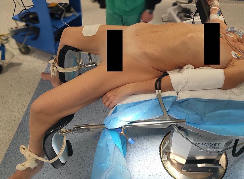
Surgical Endoscopy
Fig. 2 Ports’ position
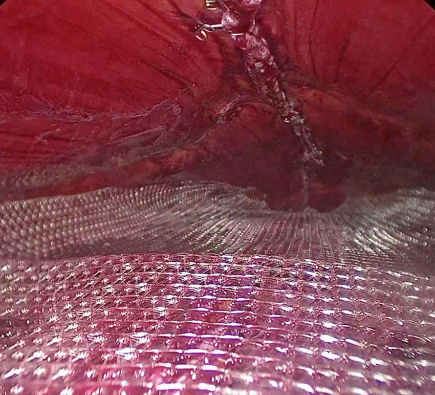
Fig. 4 Peritoneum and posterior sheath opening
Fig. 3 Dissection of the preperitoneal adipose tissue
along the median line are reduced into the abdominal cavity
and the sac is resected. Redundant adipose tissue is dissected
from the posterior layer of the anterior abdominal wall until Fig. 5 Dissection of the posterior sheath from the muscle
the defect is exposed (Fig. 3), and the falciform ligament is
dissected.
The peritoneum and the posterior rectus sheaths are
incised bilaterally where the diastasis begin, about 3 cm
below the umbilicus, using a monopolar energy device.
Then, the retromuscular space is exposed (Fig. 4).
At this point, the laparoscope is moved to the trocar in
the left iliac fossa. The rectus sheath is dissected from the
muscle using a blunt dissector introduced in the space previ-
ously created (Fig. 5). The abdominal pressure is lowered to
6–7 mmHg. A 60-mm endoscopic stapler (blue load), pos-
sibly reinforced, is introduced through the suprapubic trocar.
Its jaws are opened and then they are introduced into the two
retromuscular pockets previously created, 3 cm above the
umbilicus. The stapler is fired, joining together the rectus
sheaths. After this procedure, a cavity is created between
the rectum muscles anteriorly, and their sheaths posteriorly
(Fig. 6). The dissection of the muscle from the sheath is
carried out cranially and laterally to reduce lateral tension Fig. 6 Pocket between muscles and posterior sheath
13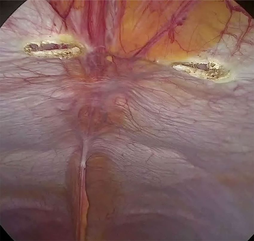
Surgical Endoscopy

and to reach the xiphoid process. During this procedure, dis-
section should reach the edge between the rectus muscle and
the transverse muscle laterally, and the lower margin of the
ribcage cranially. The firing process is repeated in a cranial
direction until the xiphoid process is reached (Fig. 7). A
polypropylene or dual mesh is placed in the space between
the rectus muscles and the posterior sheaths without using
any fixation device (Fig. 8).
   Subsequently, the laparoscope is moved back to the
suprapubic trocar. A barbered absorbable suture is used to
close the initial opening in the posterior sheath. A trans-
verse abdominis plane block is performed using ropivacaine
7.5 mg/mL in 20 mL of saline solution. The trocars are
removed under direct vision. Accesses for trocars greater
than 5 mm are closed with 2.0 polyglactin suture at the fas-
cial layer. At the end of this procedure, the medial margins of
the rectus muscles are approximated at the midline, thereby
repairing the diastasis and potential midline hernias. Very
large diastases can be treated with Botox infiltration before     Fig. 8  Mesh placement
surgery to ensure a greater laxity of the muscle structures.

                                                                      All procedures were performed using the same technique
Results                                                           and none needed open conversion.
                                                                      The average duration of surgery was 90 min. When only
The mean age was 46.3 (SD ± 11.3) years (Table 1). There          recti diastasis was corrected, patients were discharged on
were 9 men and 65 women. The mean BMI was 24.3                    postoperative day 2, while hospitalization was 4 days on
(SD ± 4.1). The mean diastasis width was 4.7 (SD ± 1.5),          average if an abdominoplasty was associated. One patient
as measured in the supra-umbilical region. Forty patients         had a subcutaneous hematoma treated conservatively, while
were affected by diastasis recti associated with umbilical        another patient presented with postoperative bleeding when
hernia. In 10 patients, it was associated with epigastric her-    abdominoplasty was performed at the same time. No post-
nia. Abdominoplasty was performed after laparoscopy in            operative infections or seromas were reported. There have
20 patients. Midline ventral hernia was repaired using the        been no readmissions, and only two recurrences have been
same technique in 4 patients. Characteristics of the patient      reported 6 months after surgery.
population are shown in Table 1.                                      On postoperative day one and two, 3 g of paracetamol
                                                                  was administered to all patients and 10 mg of ketorolac was
                                                                  administered to patients with persistent pain after surgery.
                                                                  A survey among patients showed a progressive reduction of
                                                                  postoperative pain until its disappearance on postoperative
                                                                  day 7. The median visual analog scale (VAS) score on the
                                                                  first postoperative day was 5.5 ± 2. The average follow-up
                                                                  period was 6 months (range 2–12 months).
                                                                      Of all patients, 57 (77%) completed the preoperative and
                                                                  postoperative questionnaires.

                                                                  Table 1  Patients characteristics

                                                                  Mean age                                       46.3 (SD 11.3)
                                                                  Men/women                                      9/65
                                                                  BMI                                            24.3 (SD 4)
                                                                  Diastasis size                                 4.7 (SD 1.5)
                                                                  Associated umbilical hernia                    40
                                                                  Associated epigastric hernia                   10
Fig. 7  Last stapler fired close to the xiphoid process

13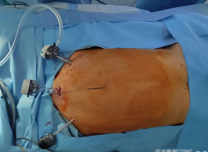
Surgical Endoscopy

   Of all the patients, 31 (42%) had urinary incontinence            technique repair has the lowest recurrences and major com-
before the surgery, while only 2 patients reported this symp-        plication rates [13].
tom after surgery. Lower back pain was reported before sur-             In fact, the placement of an intraperitoneal mesh, often
gery in 40 patients (54%), and 36 patients (90%) experienced         used in laparoscopic approaches, represents a risk for adhe-
improvement. 21 patients (28%) had respiratory symptoms,             sions with the abdominal viscera, which can lead to lesions
mostly shortness of breath, and 18 patients (86%) reported           of the small bowel loops enteric fistulae, infections, and
improvement with diastasis correction. Abdominal swelling            prosthesis displacement, despite the recent technological
and feeling of distress were also common symptoms in 43              advances in the introduction of biocompatible prosthetic
patients (60%), with a postoperative improvement of 84%.             materials [12].
In fact, only 7 patients reported this symptom 6 months after           The most commonly performed surgical procedure to
surgery. Even though these data are preliminary and based            treat diastasis recti is abdominoplasty with plication of the
on patients’ perception and personal experience, they seem           anterior rectus sheath, where a wide laparotomy and an
promising. Further follow-up studies are needed. The results         extended dissection of the subcutaneous tissue is required.
are shown in Table 2.                                                This technique is associated with high risks of infections,
                                                                     seromas, and significant postoperative pain, with an unclear
                                                                     durability of the plication over time, as shown by several
Discussion                                                           studies [15, 16].
                                                                        The present technique is a variation of Costa’s technique
The Rives–Stoppa technique was first described in the mid-           described in Hernia [10]. Our approach combines the advan-
1980s and since then, it has played a major role in abdominal        tages of laparoscopic surgery, with the consolidated results
wall surgery. To date, even though several other techniques          of the Rives–Stoppa repair. It is based on the use of a linear
have been described, it is still considered the gold standard.       stapler to join the posterior sheaths of rectus muscles to effi-
   The sublay position of the mesh proved to have the lowest         caciously repair the diastasis recti and a possible coexisting
risk of long-term recurrence and major complications due to          midline defect, as well as creating at the same time a space
the stability of the retromuscular mesh and absence of con-          for the placement of a retromuscular mesh.
tact with the abdominal content [11, 12]. According to the              The reconstruction of the midline using a stapler is a
La Place principle, a large overlap of underlay mesh allows          great advantage because it is easy and fast, thereby reducing
the distribution of forces over the entire mesh, decreasing the      operating time, distributing the tension evenly on the suture,
pressure on the fascia defect, leading to reduced recurrence         and preventing fascial tearing. In fact, it is crucial to have
rate [13]. For these reasons, a retromuscular proper-sized           an equally distributed tension on the suture. Therefore, it is
mesh is held in place by intra-abdominal pressure, making            very important to carefully dissect the preperitoneal adipose
fixation unnecessary, and thus reducing postoperative pain,          tissue posteriorly to the midline, to fully expose the defect
which is mainly caused by the transfacial mechanical fixa-           and to avoid any residue of adipose tissue between the jaws
tion devices [11, 14].                                               of the stapler.
   The major limitation of this technique is the wide inci-             An advantage of this technique is the concurrent closure
sion necessary to complete the subcutaneous dissection, long         of any midline defect with the stapler, thereby restoring the
operative time, long hospitalization, and increased postop-          anatomy and minimizing the risk of recurrence. Chelala
erative surgical site complications, such as infections, sero-       et al. in a paper published in Hernia in 2016 demonstrated
mas, skin necrosis, and hematomas.                                   the importance of closing the abdominal wall defect [17].
   For these reasons, since LeBlanc first described laparo-          They conducted a study on 1326 patients who underwent
scopic hernia repair in 1993, it has been widely used, and           laparoscopic hernia repair with routine suturing of the hernia
several techniques have been developed to benefit from the           gap and showed very good results in terms of recurrence and
advantages of minimally invasive surgery. However, when              complications. However, laparoscopic sutures are technically
compared with laparoscopic repairs, the Rives–Stoppa                 difficult to perform. They require advanced skills and a long
                                                                     learning curve, contrary to the use of a stapler [8, 17].
                                                                        The intraperitoneal approach allows to remove the pre-
Table 2  Symptoms assessment before and after surgery: UI uri-       peritoneal adipose tissue, that otherwise might be included
nary incontinence, LBP lower back pain, SB shortness of breath, AS   into the suture line, decreasing its strength; furthermore, this
abdominal swelling                                                   allows to discover the presence of epigastric hernias, that
                          UI (%)    LBP (%)     SB (%)    AS (%)     otherwise would be misdiagnosed. Also, the intraperitoneal
                                                                     technique allows to carefully monitor repetitive firing of
Preoperative symptoms     42        54          28        60
                                                                     the stapling device, avoiding damage to the intraperitoneal
Postoperative symptoms     3         5           4         9
                                                                     structures.

                                                                                                                          13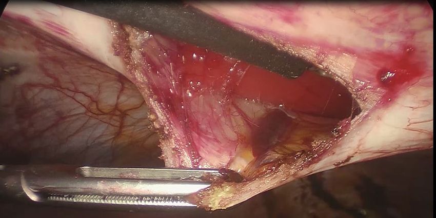
Surgical Endoscopy
In case of diastasis recti below the arcuate line, our Open Access This article is licensed under a Creative Commons Attri-
technique is not indicated, since the repair is based on bution 4.0 International License, which permits use, sharing, adapta-
tion, distribution and reproduction in any medium or format, as long
the plication of the anterior fascia only. This can be per- as you give appropriate credit to the original author(s) and the source,
formed by extending the sovrapubic port incision laterally provide a link to the Creative Commons licence, and indicate if changes
(2–3 cm) and plicating the anterior fascia of recti muscles were made. The images or other third party material in this article are
using a 2/0 barbed suture. Alternatively, THT technique included in the article’s Creative Commons licence, unless indicated
otherwise in a credit line to the material. If material is not included in
can be used [12]. However, this technique has the follow- the article’s Creative Commons licence and your intended use is not
ing limits: (a) it does not allow to carry out the abdomino- permitted by statutory regulation or exceeds the permitted use, you will
plasty since it would be necessary to detach the umbilicus need to obtain permission directly from the copyright holder. To view a
from its base; (b) it does not allow to remove the preperito- copy of this licence, visit http://creativecommons.org/licenses/by/4.0/.
neal adipose tissue, which would be included in the suture
line weakening it. References
An important advantage of our technique is the reduc-
1. Reinpold W, Köckerling F, Bittner R, Conze J, Fortelny R, Koch A,
tion of postoperative pain, mainly due to the absence of Kukleta J, Kuthe A, Lorenz R, Stechemesser B (2019) Classification
direct tacking of the mesh on the peritoneum, which is of rectus diastasis-a proposal by the German Hernia Society (DHG)
generally responsible for pain in laparoscopic repairs. and the International Endohernia Society (IEHS. Front Surg 28(6):1
As opposed to Costa’s article, we support not fixing the 2. Nahabedian MY (2018) Management strategies for diastasis recti.
Semin Plast Surg 32:147–154
mesh because of its retromuscular placement in a perfectly 3. Keshwani N, Mathur S, McLean L (2018) Relationship between
shaped pocket created by the junction of the posterior inter-rectus distance and symptom severity in women with diastasis
sheaths of the recti abdominis. Owing to this particular recti in the early postpartum period. Phys Ther 98(03):182–190
positioning, the mesh is held in place by intra-abdominal 4. Jessen ML, Öberg S, Rosenberg J (2019) Treatment options for
abdominal rectus diastasis. Front Surg 6:65
pressure, according to the La Place principle [18]. 5. Gama LJM, Barbosa MVJ, Czapkowski A, Ajzen S, Ferreira
To this date, we are not able to state whether the use LM, Nahas FX (2017) Single-layer plication for repair of diasta-
of a reinforced stapler provides better results in terms of sis recti: the most rapid and efficient technique. Aesthet Surg J
long-term recurrence. We used a reinforced recharge in 20 37(06):698–705
6. Batchvarova Z, Leymarie N, Lepage C, Leyder P (2008) Use of
patients, but in early follow-ups, there were no differences a submuscular resorbable mesh for correction of severe postpreg-
between the two groups. nancy musculoaponeurotic laxity: an 11-year retrospective study.
Plast Reconstr Surg 121(04):1240–1248
7. Cheesborough JE, Dumanian GA (2015) Simultaneous prosthetic
mesh abdominal wall reconstruction with abdominoplasty for ven-
Conclusions tral hernia and severe rectus diastasis repairs. Plast Reconstr Surg
135(01):268–276
This procedure was performed on a small number of 8. Emanuelsson P, Gunnarsson U, Strigard Stark KB (2014) Early
patients and longer follow-up is needed. The short-term complications, pain, and quality of life after reconstructive surgery
for abdominal rectus muscle diastasis: a 3-months follow up. J Plast
outcomes showed good results in terms of postopera- Reconstr Aesthet Surg 67(08):1082–1088
tive pain and decrease of preoperative symptoms. This 9. Montgomery A (2016) The best of two words: a new innovative
technique is feasible and requires a short operating time. laparoscopic Rives-Stoppa technique for ventral/incisional hernias—
Associated ventral hernias can be repaired easily. Since “The Brazilian Technique.” Hernia 20:267–270
10. Abdalla RZ, Garcia RB, Costa RID, Abdalla BMZ (2013) Treatment
this procedure avoids disconnection of the umbilicus, in of mid-line abdominal wall hernias with the use of endo stapler for
contrast to other similar laparoscopic procedures, it allows mid-line closure. Arq Bras Cir Dig 26(4):335–337
synchronous abdominoplasty. 11. Helgstrand F, Rosenberg J, Kehlet H, Jorgensen LN, Bisgaard T
(2013) Nationwide prospective study of outcomes after elective
incisional hernia repair. J Am Coll Surg 216(2):217–228
12. Carrara A, Lauro E, Fabris L, Frisini M, Rizzo S (2018) Endo-lap-
aroscopic reconstruction of the abdominal wall midline with linear
Supplementary Information The online version contains supplemen- stapler, the THT technique. Early results of the first case series. Ann
tary material available at https: //doi.org/10.1007/s00464 -021-08393- 2. Med Surg 38:1–7
13. Williams RF, Martin DF, Mulrooney MT, Voeller GR (2008) Intra-
Funding Open access funding provided by Università degli Studi di peritoneal modification of the Rives-Stoppa repair for large inci-
Roma La Sapienza within the CRUI-CARE Agreement. sional hernias. Hernia 12(2):141–145
14. Jensen KK, Henriksen NA, Jorgensen LN (2014) Endoscopic com-
ponent separation for ventral hernia causes fewer wound complica-
Compliance with ethical standards tions compared to open components separation: a systematic review
and meta-analysis. Surg Endosc 28(11):3046–3052
Disclosure Drs. Gabriele Manetti, Maria Giulia Lolli, Elena Belloni, 15. Van Uchelen JH, Kon M, Werker PM (2001) The long-term durabil-
and Giuseppe Nigri have no conflicts of interest or financial ties to ity of plication of the anterior rectus sheath assessed by ultrasonog-
disclose. raphy. Plast Reconstr Surg 107(6):1578–1584
13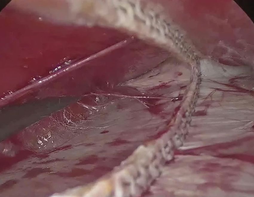
Surgical Endoscopy

16. Tadiparthi S, Shokrollahi K, Doyle GS (2012) Rectus sheath plica-          Publisher’s Note Springer Nature remains neutral with regard to
    tion in abdominoplasty: assessment of its longevity and a review of        jurisdictional claims in published maps and institutional affiliations.
    the literature. J Plast Reconstr Aesthetic Surg 65:328–332
17. Chelala E, Baraké H, Estievenart J, Dessily M, Charara F, Allé JL
    (2016) Long-term outcomes of 1326 laparoscopic incisional and
    ventral hernia repair with the routine suturing concept: a single insti-
    tution experience. Hernia 20(1):101–110
18. Awaiz A, Rahman F, Hossain MB et al (2015) Meta-analysis and
    systematic review of laparoscopic versus open mesh repair for elec-
    tive incisional hernia. Hernia 19(3):449–463

                                                                                                                                         13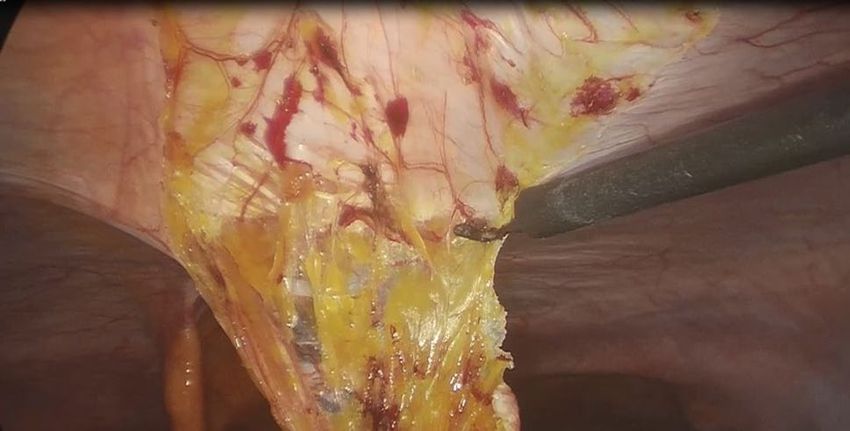
You can also read