Infected transcartilaginous ear piercings. A case report and review of the literature
←
→
Page content transcription
If your browser does not render page correctly, please read the page content below
Caso clínico
                                                                                                                  DOI: http://dx.doi.org/10.11565/arsmed.v44i2.1550

   Infected transcartilaginous ear piercings. A case report and review of the literature
   Pericondritis del pabellón auricular por piercings. Reporte de un caso clínico y revisión de la literatura
                                         Javier Rodríguez, 1,2,*, Natalie Thone, 2,3, Josefina Duque4, Rocío Brañes4

Abstract
Background: transcartilaginous perforations have become a prominent practice among adolescents and young adults in recent years,
which are associated with an increased risk of complications since it is frequently performed without sterile technique and by unqualified
individuals. The transgression of the integrity of the skin and cartilage of the ear favors infections such as cellulitis, chondritis, perichon-
dritis or abscesses that can cause serious deformities. Methods: we present a clinical case compatible with a perichondritis secondary
to ear perforations with three abscesses. Results: the three abscesses were drained with sterile technique and successfully managed
with outpatient antibiotic treatment. In relation to the pathophysiology, the trauma in the auditory pavilion produces the extraction
of the adjacent perichondrium, causing devascularization of the cartilage and microfractures, which together with the transgression
of the skin, increase the susceptibility to infection. In addition, subpericardial bleeding and inflammatory reaction decrease the blood
supply, which limits the immune response and the effectiveness of antibiotics. In some cases, incision and drainage are required. The
signs of perichondritis include pain, swelling, and erythema of the skin. Clinically, perichondritis can be differentiated from cellulitis of
the pinna, in that the first usually does not involve the earlobe. The fluctuating swelling leads us to an abscess. Conclusions: the admi-
nistration of broad-spectrum antibiotics should be immediately administered and include coverage for Pseudomonas aeruginosa since
it is responsible for the majority of post-perforation cartilage infections (up to 95% of cases). Due to the increase of post-perforation
infectious complications, all physicians should be familiar with its diagnosis and treatment.

Keywords: ear piercing; perichondritis; transcartilaginous ear perforations; pseudomonas infections.

Fecha de envío: 4 de diciembre de 2018 - Fecha de aprobación: 30 de mayo de 2019

Introduction                                                                       Case presentation

Transcartilaginous ear piercings (TEPs) have become a prominent                    A 21-year-old female patient with no medical or surgical history,
practice among adolescents and young adults in the last years,                     with 3 TEPs on scapha performed four weeks ago in a piercing
and unlike the earlobe piercings, TEPs are associated with an                      studio (for the procedure, we wiped the ear with 70% alcohol
increased risk for the patients (Keene et al., 2004; Perry & Sosin                 and the cartilage was penetrated with a sharp, hollow ste-
2014; Sosin et al., 2015). It is frequently performed without sterile              rile needle). The patient arrived at our department with a
technique by unqualified individuals, and inadequate aftercare                     4-day history of upper right ear pain and erythema of the
instructions (without an explanation of potential complications)                   scaphoid fossa associated with localized swellings in the
(Keene et al., 2004; Lee & Gold 2011). Transgression of skin and                   piercing sites. There was no history of fever or any other
cartilage integrity favors potential infections like ear cellulitis,               symptoms. Vital signs were stable. Upon ear examination,
chondritis, perichondritis or abscesses; such complications may                    we noted three small swellings on the ear cartilage with mild
cause severe ear deformities (Sosin et al., 2015). It is important                 pus discharge about the piercings, in addition to extensive
to determine the best approach to deal with infections resulting                   redness on the upper ear (Figure 1). The ear was exquisitely
from TEPs. We present a case report of a young adult with a pinna                  tender to palpation, without cervical lymphadenopathies.
perichondritis secondary to ear piercings managed with outpatient                  Due to these signs, we suspected an auricular perichondritis and
antibiotic treatment.                                                              three little abscesses.

(1) Department of Surgery, Facultad de Medicina, Pontificia Universidad Católica de Chile, Santiago, Chile.
(2) Department of Medical Education, Facultad de Medicina, Pontificia Universidad Católica de Chile, Santiago, Chile.
(3) Department of Otorhinolaryngology, Facultad de Medicina, Pontificia Universidad Católica de Chile, Santiago, Chile.
(4) Medical School, Facultad de Medicina, Pontificia Universidad Católica de Chile, Santiago, Chile.
* Corresponding author: jirodri4@uc.cl

ARS MEDICA Revista de Ciencias Médicas Volumen 44 número 2 año 2019
ISSN: 0719-1855 © Dirección de Extensión y Educación Continua, Escuela de Medicina, Pontificia Universidad Católica de Chile. http://arsmedica.cl               23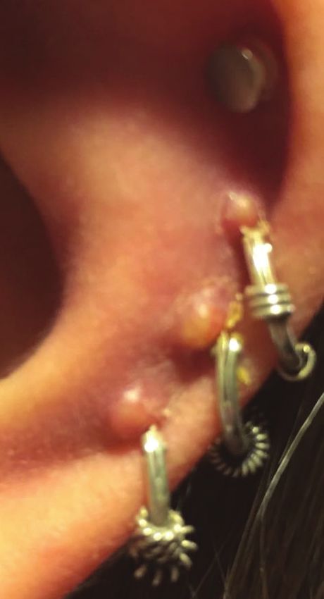
Rodríguez et al.
The three hoops were removed using sterile technique, drained Discussion
the abscesses and a 10-day course of levofloxacin (750 mg
once daily) was prescribed to cover for empiric Pseudomonas The practice of body piercing (BP) in adolescents and young adults,
aeruginosa and Staphylococcus aureus. The patient was mo- principally high TEPs (in the upper third of the pinna), have become
nitored daily and educated in alarm symptoms to consult in increasingly popular and prevalent in the last 20 years (Hanif et
emergency service. Four days after the first visit, and having al., 2001; Bone et al., 2008; Liu & Chokkalingam, 2013; Sosin et al.,
received levofloxacin, the erythema and swellings in the right 2015). TEPs have been reported as the third most common body
ear had decreased. There was no cosmetic deformity of the piercing other than the earlobe (Bone et al., 2008). Unfortunately,
pinna at follow up (Figure 2). cartilage ear piercing is frequently performed without sterile
technique and by unqualified individuals that are unaware of the
severity of potential complications after the procedure (Keene
et al.,2004; Perry & Sosin, 2014). Before the procedure, the ear
commonly is disinfected with isopropyl alcohol (like in the case
reported) or benzalkonium chloride, which are ineffective against
the Pseudomonas species and spore-forming bacteria ( Stewart et
al., 2006; Lee & Gold 2011; Perry & Sosin 2014).
Regardless of the technique, the localized trauma on the pinna
produces extraction of the adjacent perichondrium (which causes
cartilage devascularization) and microfractures, which together
with the transgression of the skin, increase the susceptibility to
infection (Sosin et al., 2015). Also, the subperichondrial bleeding
and inflammatory reaction decrease an already limited blood
supply of the cartilage (Perry & Sosin, 2014; Sosin et al., 2015).
The avascular environment limits the immune response and the
effectiveness of antibiotics, so in some cases, incision and drainage
are required (ideally obtaining cultures and sensitivities to guide
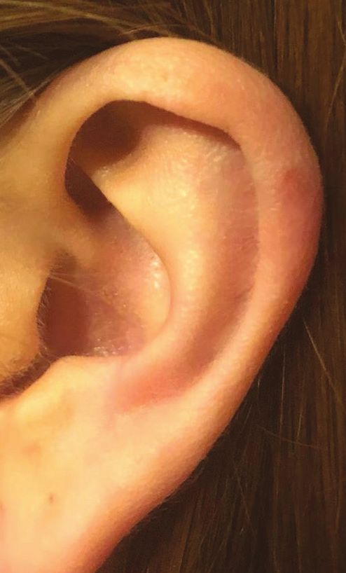
Figure 1: Three small swellings on the helix of the left pinna with mild pus
discharge in relation to the piercings. Extensive redness on cartilaginous pinna. the optimal antibiotic therapy) (Lee & Gold, 2011; Perry & Sosin,
2014; Sosin et al., 2015).
A recent systematic review confirms that post piercing infectious
complications are increasing, and identifies that adolescent and
young adult females most frequently develop post piercing pe-
richondritis (Sosin et al., 2015). Furthermore, the use of piercing
guns and exposure of the wound to fresh water, water in swimming
pools or hot tubs after the procedure, may additionally increase
the risk of infection ( Keene et al., 2004; Fisher et al., 2005; Lee &
Gold, 2011). The lag time between the onset of symptoms and
medical attention is about one week, but a delay greater than
five days since the beginning of the symptoms is more likely to
result in hospitalization (Sosin et al., 2015). Signs of perichondritis
or chondritis in patients with an embedded earring are similar
(these include pain, swelling and erythema of the overlying skin)
and fluctuant swelling indicate an abscess that we should drain
(typically associated with chondritis) (Fisher et al., 2005; Sosin et al.,
Figure 2: Left pinna after treatment, without sign of cosmetic deformity. 2015). Clinically, perichondritis can be differentiated from cellulitis
of the pinna, in that the first usually does not involve the earlobe
(Kullar & Yates, 2015).
ARS MEDICA Revista de Ciencias Médicas Volumen 44 número 2 año 2019
ISSN: 0719-1855 © Dirección de Extensión y Educación Continua, Escuela de Medicina, Pontificia Universidad Católica de Chile. http://arsmedica.cl 24
Rodríguez et al.

Once the clinical diagnosis of a post piercing infection has been                 With this report, we want to raise awareness about the risk of in-
made, antibiotic therapy should be administered immediately,                      fection and deformity of the ear after ear piercing. We also want to
since perichondritis can result in permanent ear deformity (cau-                  report a case of infected TEPs managed with outpatient antibiotic
liflower ear) (Hanif et al., 2001; Stewart et al., 2006; Lee & Gold,              treatment with excellent response.
2011; Liu & Chokkalingam, 2013; Perry & Sosin, 2014). It is relevant
to select a broad spectrum antibiotic that covers Pseudomonas                     Patient consent
and Staphylococcus species and includes oral fluoroquinolones                     Patient consent was obtained for the publication of this article
(e.g., levofloxacin) in adolescents and adults (Lee & Gold, 2011;                 and associated images
Liu & Chokkalingam, 2013; Perry & Sosin, 2014; Sosin et al., 2015).
It is also necessary to remove the foreign body from the pinna.                   References:
Pseudomonas aeruginosa is responsible for most post piercing
                                                                                  Bone A, Ncube F, Nichols T & Norman D.N. (2008). Body Piercing in
cartilage infections (in 87 up to 95% percent of cases) (Fisher et
                                                                                  England: A Survey of Piercing at Sites Other than Earlobe. British
al., 2005; Liu & Chokkalingam, 2013; Sosin et al., 2015). Infections
                                                                                  Medical Journal: BMJ 336, 1426–28.
may require prolonged antimicrobial therapy, plus incision and
drainage (due to the poor blood supply of the ear cartilage). When
                                                                                  Fisher C, Kacica M.A & Bennett N.M. (2005). Risk Factors for Cartilage
abscesses are present, they should be drained promptly and cultured
                                                                                  Infections of the Ear. American journal of preventive medicine 29, 204–9.
to reduce the risk of cartilage damage induced by pressure and
provides a microbiological diagnosis and sensitivity results (Sosin               Hanif J, Frosh A, Marnane C, Ghufoor K, Rivron R & Sandhu G.(2001).
et al., 2015). However, even with timely and adequate treatment,                  Lesson of the Week:‘High’ Ear Piercing and the Rising Incidence of
these infections may result in cartilage necrosis and deformity of                Perichondritis of the Pinna. British Medical Journal: BMJ 323, 400.
the pinna (Lee & Gold, 2011).
                                                                                  Keene W, Markum A & Samadpour M. (2004). Outbreak of Pseudomonas
In patients without fluctuant swelling, outpatient antibiotic treat-              Aeruginosa Infections Caused by Commercial Piercing of Upper Ear
ment may be attempted, with daily observation (and drainage or                    Cartilage. Journal of the American Medical Association 291, 981–85.
debridement if necessary) (Perry & Sosin, 2014). Nonetheless, if the
patient has no response to oral antibiotics within 24 hours, they                 Kullar P & Yates P. (2015). Infections and Foreign Bodies in ENT.
must be hospitalized for intravenous antibiotics and eventually                   Surgery 33, 593–99.
surgical drainage (Perry & Sosin, 2014; Sosin et al., 2015).
                                                                                  Todd L. C. & Gold W. (2011). Necrotizing Pseudomonas Chondritis
Conclusion                                                                        after Piercing of the Upper Ear. Canadian Medical Association journal
                                                                                  = journal de l’Associationmedicale Canadienne: CMAJ 183, 819–21.
Post-piercing infectious complications are increasing. All physicians
should be familiar with its diagnosis and treatment. Signs of pe-                 Liu, Z. W. & Chokkalingam P. (2013). Piercing Associated Perichondritis
richondritis or chondritis include pain, erythema of the overlying                of the Pinna: Are We Treating It Correctly?. Journal of Laryngology
skin and swelling. A fluctuant swelling indicates an abscess that                 and Otology 127, 505–8.
we should drain.
                                                                                  Perry, Arthur W & Sosin M. (2014). Reconstruction of Ear Deformity
Pseudomonas and Staphylococcus species are the most frequently                    from Post Piercing Perichondritis. Archives of Plastic Surgery 41, 609–12.
isolated pathogens on post piercing infections. Treatment should
be administered immediately after diagnosis to avoid devastating                  Sosin M, Weissler J, Pulcrano M & Rodriguez E. (2015).
complications. It is important to select a broad spectrum antibiotic              Transcartilaginous Ear Piercing and Infectious Complications: A
that covers these bacteria, and that includes oral fluoroquinolones               Systematic Review and Critical Analysis of Outcomes. The Laryn-
(eg, Levofloxacin) in adolescents and adults.                                     goscope 125, 1827–34.

In the absence of fluctuant swelling, outpatient antibiotic treatment             Stewart, Gail M, Thorp A & Brown L. (2006). Perichondritis -- A Com-
may be attempted, with daily observation. If the patient has no res-              plication of High Ear Piercing. Pediatric Emergency Care 22, 804–6.
ponse to oral antibiotics within 24 hours, they must be hospitalized.

ARS MEDICA Revista de Ciencias Médicas Volumen 44 número 2 año 2019
ISSN: 0719-1855 © Dirección de Extensión y Educación Continua, Escuela de Medicina, Pontificia Universidad Católica de Chile. http://arsmedica.cl        25
You can also read