International Journal of Gastrointestinal Intervention
←
→
Page content transcription
If your browser does not render page correctly, please read the page content below
Int J Gastrointest Interv 2021;10:42–48

                      International Journal of Gastrointestinal Intervention
                                                             journal homepage: www.ijgii.org

Review Article
Percutaneous endoscopic gastrostomy
Xudong Liu1, Zhengqiang Yang2, Shun He1, and Guiqi Wang1,*

A B S T R A C T

Percutaneous endoscopic gastrostomy (PEG) is a feasible and safe technique for patients who need long-term feeding and cannot eat orally. With the
increasing maturity of PEG technique, a large number of patients receive PEG tube placement every year in the world. However, PEG tube placement
is not necessary in some patients, and some other patients are not evaluated strictly, leading to serious complications. In a broad sense, the mainly
two indications for PEG include long-term enteral nutrition and gastric decompression. On the other hand, the main contraindications of PEG are
distal intestinal obstruction, severe coagulation abnormalities, and severe infection at the PEG site. In the first section of this review, the indications
and contraindications of PEG are introduced. Although PEG tube placement is a relatively safe technique, it can still cause a number of complica-
tions, including minor and major complications. Through standard management and treatment, the outcome of most patients is very good. In the
second section of this review, we describe a variety of minor and major tube-related complications, and the treatment and prevention of these com-
plications. In addition, the preparation and post-insertion care are also very important for PEG, which can reduce the incidence of complications. In
the last section of this review, we describe related issues about the preparation and post-insertion care of PEG. In conclusion, PEG tube placement is
a widely accepted technique that can bring benefits to the right patients.
                                                                                                      Copyright © 2021, Society of Gastrointestinal Intervention.

Keywords: Complications; Indication; Management; Percutaneous endoscopic gastrostomy

Introduction                                                                            technique, outcome, and complications of PEG.

     A number of cancers of larynx and gastrointestinal tract, and                      Indications and Contraindications
various swallowing difficulties, can block the passage of food
along the digestive tract. There are several approaches available to                        There are so many studies already proved that enteral
provide nutritional support;1 nasogastric tube (NGT) and gastros-                       nutrition have many positive effects compared with parenteral
tomy. NGT is hardly accepted as it stimulates the nasopharynx,                          nutrition. These effects include preservation of the intestinal
increase the risk of aspiration pneumonia caused by reflux, and                         mucosal barrier, reduction of intestinal and other infections and
reduce the quality of life.2,3 If enteral nutrition is required for more                improvement of the overall prognosis of patients with long-
than 4 weeks, percutaneous access should be considered, depend-                         term artificial nutrition.8 European Society for Clinical Nutrition
ing on the clinical setting. Access to insert the gastrostomy tube                      and Metabolism (ESPEN) guidelines also recommends enteral
can be achieved by the use of endoscopy,4 radiological guidance                         over parenteral nutrition in order “to support intestinal functions
or surgical techniques.5 Surgical gastrostomy needs anesthesia or                       to the greatest possible extent.”9 Enteral nutrition via PEG is
open placement of gastric fistula, which is painful and risky for                       technically and functionally feasible.10 However, PEG feeding
patients.6 A meta-analysis demonstrated that percutaneous endo-                         has an uncertain benefit for some patients, such as those with
scopic gastrostomy (PEG) is associated with a lower probability                         diabetes or advanced dementia and in elderly patients aged over
of 30-day mortality compared to percutaneous radiologic gas-                            80 years.11 So, the patient’s needs, diagnosis, long-term survival
trostomy (PRG), suggesting that PEG should be considered as the                         rate, general condition should to be considered when deciding
first choice for most patients.7 This article reviews the indications,                  on PEG placement. The decision for tube placement is not only
1
Department of Endoscopy, National Cancer Center/Cancer Hospital, Chinese Academy of Medical Sciences and Peking Union Medical College, Beijing, China
2
Department of Radiology Intervention, National Cancer Center/Cancer Hospital, Chinese Academy of Medical Sciences and Peking Union Medical College, Beijing, China
Received March 2, 2021; Revised March 9, 2021; Accepted March 9, 2021
  * Corresponding author. Department of Endoscopy, National Cancer Center/Cancer Hospital, Chinese Academy of Medical Sciences and Peking Union Medical College, 17
    Panjiayuan, Chaoyang District, Beijing 100021, China.
    E-mail address: wangguiq@126.com (G. Wang).

pISSN 2636-0004 eISSN 2636-0012 https://doi.org/10.18528/ijgii210015
            This is an open-access article distributed under the terms of the Creative Commons Attribution Non-Commercial License (http://creativecommons.org/licenses/by-
            nc/4.0) which permits unrestricted noncommercial use, distribution, and reproduction in any medium, provided the original work is properly cited.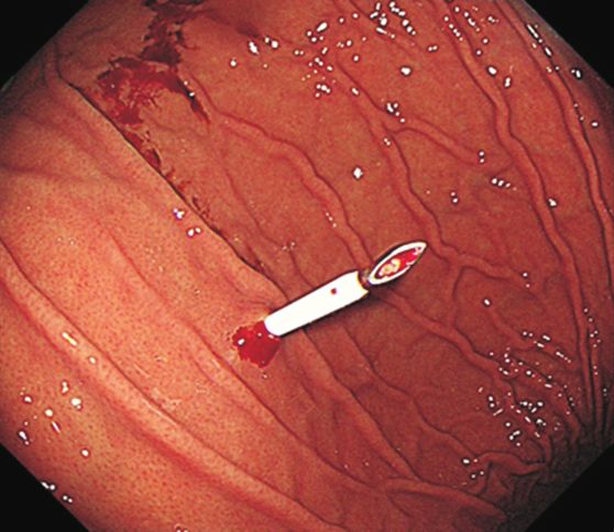
Xudong Liu et al. / Percutaneous endoscopic gastrostomy                                                      43

to improve the patient’s survival and nutritional status, but also                    Neurodegenerative disease: The feeding and nutritional
to improve their quality of life. So the patients must be strictly               challenges of neurodegenerative diseases are significantly
evaluated before PEG tube placement.                                             different from those of stroke, PEG is a standard method of
    Of course, many diseases are accepted and data-supported as                  feeding in those patients. But in a recent study, short-term
indications for PEG (Table 1).                                                   mortality and morbidity associated with PEG were significant
                                                                                 in patients with neurological disease, Age older than 75 years
Indications                                                                      was associated with poor outcome.19 For these patients, careful
                                                                                 patient selection, optimal timing of PEG tube insertion, and
Cancer                                                                           periprocedural care, comprehensive education of patients and
    Head and neck cancer, pharyngeal cancer, esophageal cancer:                  carers to reduce morbidity, mortality, and cost-effectiveness are
More than 40% of patients with head and neck cancer have some                    very important.19
degree of dysphagia,12 because of the obstruction of the tumor,                       Dementia: The 2015 worldwide prevalence of dementia was
local edema due to high dose radiotherapy and/or chemotherapy                    estimated to be 47.5 million, and it is expected to increase to 75.6
and reduced appetite. The PEG tube can be inserted either pro-                   million by 2030.20 Dysphagia also becomes more prevalent in
phylactically or therapeutically in these patients.13 Prophylactic               elderly dementia. Even in its early stages, Alzheimer’s dementia
PEG was widely placed. However, a predictive model is needed to                  impairs the ability to focus on mastication and impacts the sen-
identify patients at high risk of malnutrition, and to prevent un-               sory aspects of swallowing.21 Similar to stroke, vascular dementia
necessary PEG-placement.14                                                       can affect the motor aspect of swallowing, resulting in difficulty
    Cancer with mechanical or functional bowel obstruction: Gas-                 in mastication and swallowing.22 There is much controversy about
trointestinal decompression is one of the most useful therapeutic                the use and timing of enteral feeding support in these patients
method for patients with mechanical or functional bowel obstruc-                 with dysphagia. There is no evidence to suggest long-term sur-
tion caused by cancer. Compared with NGT, PEG is more com-                       vival rates improved in patients with advanced dementia who
fortable and can be used to drain gastric secretions and resolve                 underwent PEG placement.23 PEG may not provide any clinical
persistent nausea and vomiting in long term.15                                   benefit to these patients. So European Society of Gastrointestinal
                                                                                 Endoscopy (ESGE) recommends refraining from PEG placement in
Neurological disorders                                                           patients with advanced dementia.
    Cerebrovascular disease/stroke : Stroke is the most common                        Severe brain damage: It is very difficult to start enteral nu-
cause of acute dysphagia which can lead to malnutrition and af-                  trition in the patients with severe brain damage, because their
fect quality of life. The prevalence of malnutrition following an                recovery time is uncertain. Some authors suggest that PEG tube
acute stroke ranges from 8% to 34%.16 Most acute stroke patients                 should be placed in severe brain damage patients if they do not
recover from dysphagia within the first four weeks, although 15%                 recover in 14 days.24
of patients may develop long-term dysphagia.17 Some experts
recommend that NGT feeding alone may be enough in patients                       Contraindications
who need nutritional support for less than four weeks, but PEG
tube placement needs to be considered for longer periods.18 How-                     Absolute and relative contraindications of PEG tube place-
ever, for patients with temporary dysphagia or with a shorter life               ment are summarized in Table 2.
expectancy due to an underlying disease, this decision must be                       Absolute contraindications : Mechanical obstruction of the
weighed. At least a two week wait time for PEG insertion is clini-               digestive tract (unless the procedure itself is indicated for de-
cally appropriate to evaluate its medical necessity. After insertion             compression), active peritonitis, uncorrectable coagulopathy, or
of the PEG tube, routine follow-up of patients should be per-                    ongoing bowel ischemia, sepsis, abdominal wall infection at the
formed to evaluate the recovery of their swallowing ability. PEG                 selected site of placement, history of total gastrectomy.
tubes can be removed at any time if patients regain spontaneous
swallowing.5
                                                                                 Table 2 Contraindications
                                                                                         Variable                            Contraindications
Table 1 Accepted and Data-Supported Indications for Percutaneous Endo-            Absolute                  Mechanical obstruction of the digestive tract
scopic Gastrostomy (PEG)
                                                                                   contraindications        Active peritonitis
    Variable                            Indications
                                                                                                            Uncorrectable coagulopathy (INR > 1.5, platelets
 Cancer          Head and neck cancer                                                                        < 50,000/mm3)
                 Pharyngeal cancer                                                                          Ongoing bowel ischemia
                 Esophageal cancer                                                                          Sepsis
                 Cancer with mechanical or functional bowel obstrction                                      Abdominal wall infection at the selected site of
                  (PEG used as a decompression measure)                                                      placement
 Neurological    Cerebrovascular disease                                                                    History of total gastrectomy
  disorders      Amyotrophic lateral sclerosis                                    Relative                  Recent gastrointestinal bleeding due to peptic ulcer
                 Parkinson’s disease                                               contraindications         disease
                 Cerebral tumor                                                                             Hemodynamic or respiratory instability
                 Cerebral palsy                                                                             Ascites
                 Dementia                                                                                   Severe obesity
                 Multiple sclerosis                                                                         Abdominal wall defects
                 Severe brain damage from various reasons (trauma, per-                                     Peritoneal adhesions
                  sistent vegetative state, psychomental retardation, etc.)      INR, international normalized ratio.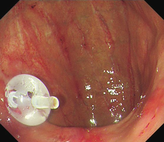
44 International Journal of Gastrointestinal Intervention 2021 10(2), 42–48
A B C
D E F
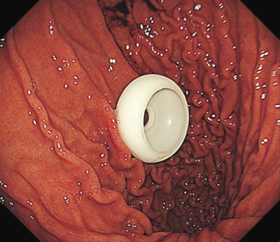
Fig. 1. Percutaneous endoscopic gastrostomy (PEG) using the pull (A–C) or the push (D–F) techniques. (A) A needle is inserted into the stomach. (B) The guidewire is
grasped by the endoscopist using a snare. (C) The tube is pulled from the puncture site until the internal fixation bumper apposes the anterior wall of the stomach. (D)
Double-lumen gastropexy needle is inserted into the stomach. (E) Penetration of the gastric wall using trocar and a peel-away sheath. (F) The tube balloon has been
filled with sterile water. Reused from the article of Cui et al (Med Sci Monit. 2019;25:9651-7).26
Relative contraindications : Recent gastrointestinal bleeding placement in clinical practice.
due to peptic ulcer disease with high risk of rebleeding, as well The “push” technique, or namely the “introducer” technique
as hemodynamic and respiratory instability.25 Generally, ascites was first introduced by Russell et al.28 In this technique, double-
is considered a relative contraindication for PEG tube placement lumen gastropexy needle is used (Fig. 1D), then operator uses the
due to concerns of ascitic fluid leakage. In these cases, Gastropexy dedicated trocar and overlying peel-away sheath for puncture
devices could be used to secure the stomach to the anterior of the abdominal wall and anterior gastric wall through the skin
abdominal wall, reducing the risk of ascitic fluid re-accumulation incision (Fig. 1E). A balloon-type tube is then introduced through
and leakage.25 Severe obesity, abdominal wall defects and the the sheath and once the tube balloon has been filled with sterile
presence of peritoneal adhesions are also relative contraindications water under endoscopic visualization, the sheath is peeled away
for PEG tube placement, in such cases, more careful planning of (Fig. 1F), The “push” technique for PEG placement in cases where
the potential target location for PEG placement should be given. the “pull” technique is contraindicated, for example in severe
esophageal stenosis or in patients with head and neck cancer or
Insertion Techniques esophageal cancer. In order to prevent deflection of the stomach
and tube misplacement, T-fasteners or a dedicated suturing device
There are two major techniques for PEG tube placement: the should be used in the “pull” technique.29
“pull” technique and the “push” technique (Fig. 1).26
As introduced before, the “pull” technique introduced by Complications
Gauderer et al4 in 1980. In this technique the dedicated wire is
inserted through a needle in the abdominal wall into the stomach Since the PEG tube was introduced in 1980,4 it is considered
(Fig. 1A), grasped by the endoscopist using a endoscopic biopsy as a feasible and safe procedure. PEG has no inferiority to surgical
forceps or snare (Fig. 1B), and then taken out through the esopha- gastrostomy in terms of morbidity or mortality,30,31 with success
gus and mouth. Subsequently the string is fixed to the external rates of 95% to 100%.25 However, major and minor complications
end of the feeding tube and the tube is pulled from the mouth to may occur depending on a variable reason. These complications
the stomach and then out through the abdominal wall and the are summarized in Table 3.
puncture site until the internal fixation bumper apposes the ante-
rior wall of the stomach (Fig. 1C). PEG placement using the “pull”
technique has replaced surgical gastrostomy.27 In nowadays, the
“pull” technique is the most widely accepted technique for PEG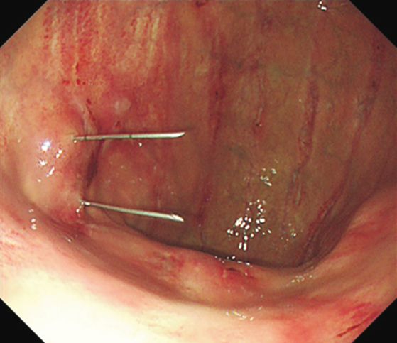
Xudong Liu et al. / Percutaneous endoscopic gastrostomy                                         45
Table 3 Complications of Percutaneous Endoscopic Gastrostomy (PEG)                  Subcutaneous emphysema and pneumoperitoneum
       Variable                             Complications                               Subcutaneous emphysema is a very rare complication of
 Minor complications    Wound infection/wound bleeding                              PEG.40 When an emphysema is suspected, a ultrasonography or
                                                                                    CT scan will confirm the diagnosis. If there are no accompanying
                        Peristomal leakage
                                                                                    symptoms, most subcutaneous emphysema is self-limited and be-
                        Tube blockage
                                                                                    nign. It may be treated by repositioning the tube or replacement
                        PEG site herniation                                         of the tube with one of a large enough.
                        Subcutaneous emphysema and pneumoperitoneum                     Pneumoperitoneum is common after PEG procedure and its
                        Self-extraction                                             prevalence is reported to be as high as 50%. 41 It is thought that
                        Gastric outlet obstruction                                  air escapes through the small opening in the stomach during the
                        Granuloma formation
                                                                                    interval between the initial needle puncture and the PEG tube
                                                                                    passage through the abdominal wall.42 Pneumoperitoneum is usu-
 Major complications    Bleeding
                                                                                    ally self-limiting, it does not cause any unfavorable consequences.
                        Internal organ injury
                        Buried bumper syndrome                                      Self-extraction
                        Aspiration pneumonia                                            Inadvertent PEG tube removal occurs in 1.6% to 4.4% of pa-
                        Metastatic seeding                                          tients.43 If a PEG tube is displaced less than one month after place-
                        Necrotizing fasciitis                                       ment, the stomach may have separated from the abdominal wall,
                                                                                    resulting in a free perforation. If recognized early, the replacement
                        Fistula formation
                                                                                    PEG tube can be placed close to or even through the same PEG
                                                                                    tube site.44 If recognized too late, a NGT should be placed, and
                                                                                    antibiotic should be started, a new PEG should be placed within 7
Minor complications                                                                 to 10 days.45 In patients with a mature abdominal tract, the PEG
                                                                                    tube can be replaced without endoscopy.
Wound infection/bleeding
    PEG site infection or bleeding is the most common minor                         Gastric outlet obstruction
complication. Bleeding from the PEG tract can usually be                               Gastric outlet obstruction is a rare complication of PEG tubes,
controlled with simple pressure over the wound or tightening                        because of part of the PEG tube migrates to the pyloric area.46 It
the external bolster against the skin.32 The prevalence of wound                    can be avoided by properly using external bolster to anchor the
infection in PEG varies between 5%–65%.33 Minor wound                               PEG tube.
infections usually resolve with the application of local antiseptics
and daily dressing changes, but in cases of persistent infection                    Granuloma formation
further investigation is warranted. Wound swabs can be cultured                         The granuloma formation around the gastrostomy tube is a
to tailor the systemic or local antibiotic treatments. Besides,                     common complication in patients with a PEG tube.47 The reason
antibiotic prophylaxis can reduce the infection rate to 3%.33,34 The                for granulation tissue develops around the tube site may be the
current gold standard for antibiotic prophylaxis is the intravenous                 leakage from the edge of the tube or insufficient care in a long
administration of a single dose of a beta-lactam antibiotic (or                     time.48 Leakage and bleeding may be seen at the edge of the tube.
appropriate alternative antibiotic, in the case of allergy). The                    The granulating tissue can be treated with surgical or chemical
optima timing is 30 minutes before the procedure.35 Besides                         cauterization and wound care.
prophylactic antibiotic administration, the adherence to a full
sterile, aseptic technique and avoidance of excessive pressure                      Peristomal leakage
between the skin and the external bumper have also been shown                           The incidence of peristomal leakage is 1% to 2%, it especially
to decrease the risk of wound infection.36                                          occurs within the first few days after PEG tube placement.42 Sev-
                                                                                    eral factors increased the risk of peristomal leakage, such as ex-
Tube blockage                                                                       cessive cleansing with hydroperoxide, infections, gastric hyperse-
    Tube blockage is a common problem in patients with long-                        cretion, buried bumper syndrome (BBS) and excessive side torsion
term enteral nutrition by PEG. During 18 months of follow-up,                       along the PEG tube, and lack of external bolster to stabilize the
16% to 31% of PEG tubes had at least 1 time of significant block-                   tube.48 Patient-specific factors inhibit wound healing (malnutrition,
age, of which 7% needed to be removed.37 To avoid tube block-                       immunodeficiency, diabetes) and can lead to peristomal leakage.
age, there are several tips, such as frequent flushing with water                   Prevention of peristomal leakage must focus on the reduction of
(before and after feeding), administering liquid medication or                      risk factors, while barrier creams containing zinc and skin protec-
well-ground pills.                                                                  tants are also recommended.48

Percutaneous endoscopic gastrostomy site herniation                                 Major complications
    Herniation through PEG site is an extremely rare complica-
tion with only six other cases reported in the literature.38,39 When                Bleeding
a hernia is suspected, a computed tomography (CT) scan will                             Acute bleeding is not common after PEG tube placement,
confirm the diagnosis, as this is a surgically correctable entity,                  with a reported incidence of 2.5%.43 The most common causes of
and can be safely managed via laparoscopic or open techniques.                      acute bleeding are vessel injury. Bleeding from the gastric artery,
To prevent this complication, operator should avoid inserting the                   superior mesenteric artery, splenic or mesenteric vein injuries
PEG tube through linea alba, as this is an area of potential weak-                  have been reported.49,50 Risk factors include anatomic aberration,
ness; and use cut and push technique rather than traction which                     anticoagulation, and antiplatelet therapy.25 To prevent bleeding,
can create a more permanent passage.38                                              with consideration of abnormal anatomy and correcting coagula-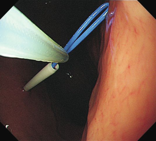
46                                       International Journal of Gastrointestinal Intervention 2021 10(2), 42–48

tion disorders before PEG tube placement can be useful. In cases                size. In order to reduce this complication, it is reasonable to place
of hemodynamic instability, supporting therapy should be the                    the PEG use the pull-string or direct-introducer technique.30
critical therapy, then angiographic embolization or surgery can be
operated.                                                                       Fistula formation
                                                                                     Actually, this complication includes two; gastrocolocutaneous
Internal organ injury                                                           fistula and gastrocutaneous fistula. Gastrocolocutaneous fistula
    Any organ around the stomach can be injured during the                      occur rarely after PEG placement, and resulting from the displace-
PEG tube placement. The injury of colon, small bowel, liver, and                ment of the colon over the anterior gastric wall. It is usually dis-
spleen have been reported.49,51–53 The displacement of the trans-               covered months after the PEG placement when the original PEG
verse colon can result in colonic injury during PEG placement.                  tube is removed. The treatment is PEG tube removal to allow the
The most common causes are inadequate gastric insufflation. As                  fistulous tract to heal. If it failed, an endoscopic approach, using
we all know, colonic injuries usually result in peritonitis and the             endo clips, or surgery is necessary.
treatment is surgery in most cases. The injury of small bowel af-                    Gastrocutaneous fistula occurs after PEG removal. Generally,
ter PEG placement are rare. It can cause intraabdominal spillage                the gastrocutaneous tract usually starts to heal in 24 hours, and
acutely or result in an entero-cutaneous fistula. To diagnose early,            complete in a few days. However, in some cases the tract fails to
a watchful follow-up is important after the PEG tube placement.                 heal and a gastrocutaneous fistula persists. Studies showed that
Liver and spleen injury during PEG placement is not common,                     longer PEG duration has a more possibility of fistula formation.62
but is a potential life-threatening complication.54 If liver or spleen          Once diagnosed, endoscopic approach, using endo clips to close
injury is suspected, transabdominal ultrasound or a CT scan must                the fistula is first choice, if it fails, surgery is necessary.63
be performed. A surgery is the best treatment for liver or spleen
injury. To avoid the injuries, the tube insertion site should be cho-           Preparation
sen accurately.
                                                                                    Firstly, ensure that sufficient informed consent of patients or
Buried bumper syndrome                                                          their relatives. The purpose of informed consent is not only to
    BBS is a rare but serious complication of PEG, in which the                 declare the risks and benefits of PEG, but also to guide the patient
internal bolster migrates from the gastric and lodges anywhere                  and their carers how to use and care the tube.
between the gastric wall and the skin along the PEG tract.55 It has                 Then, before the procedure, patient should fast overnight (6
a reported prevalence of in 1% to 4% of cases.55,56 BBS occurs                  hours for solids and 2 hours for clear liquids, longer if there is
as result of excessive tension between the internal and external                gastric motility disorder).
bumpers leading to ischemic necrosis of the gastric wall and sub-                   At last, patient should receive prophylactic antibiotic (single
sequent migration. Other possible contributing factors include                  intravenous dose of a beta-lactam antibiotic, or suitable alterna-
obesity, weight gain, and chronic cough. Common symptoms                        tive in case of allergy) 30 minutes before PEG tube placement.
include feeding difficulties, peristomal leakage, the occurrence of
abdominal pain.55 BBS is diagnosed by endoscopic or CT dem-                     Post-Insertion Care
onstration of the migrated internal bumper. The tube should be
removed as soon as diagnosed. To prevent BBS, The 2020 ESGE                          Peristomal pain is common after PEG tube placement, the pre-
guideline recommends that daily tube mobilization (pushing in-                  vention of peristomal pain after PEG placement should be admin-
ward) along with a loose position of the external PEG bumper (1–2               istered when ensure the procedure is achieved. To avoid peritoneal
cm from the abdominal wall).                                                    leakage, feeding was delayed to the next day, even a longer time
                                                                                in some centers. But two meta-analyses showed no differences in
Aspiration pneumonia                                                            complications and early mortality (< 72 hours) when feeding was
    Aspiration pneumonia is a potentially life-threatening com-                 started within 3 to 4 hours after PEG placement as compared with
plication of PEG tube feeding. It is more common when patients                  feeding was delayed (> 24 hours).64,65 Moreover, another study
are fed with NGT. It has a reported prevalence of in 0.3% to 1.0%               showed that early feeding after PEG tube placement could also
PEG cases.57 Risk factors for aspiration include supine position,               help reduce inpatient stay.66 So feeding was started within 3 to 4
sedation, neurological dysphagia, and advanced age.58 To prevent                hours after PEG placement is safe and effective.
aspiration pneumonia, a jejunal extension can be considered.                         After the PEG tube placement, the skin and the PEG site
                                                                                should be cleaned using sterile saline every day. The external
Necrotizing fasciitis                                                           bolster should be placed tightly, about 0.5 cm above the skin, to
    Necrotizing fasciitis is a very rare, but serious complication              prevent leakage during the first 3 to 5 days. A sterile “Y” shaped
of PEG placement.59 Risk factors for necrotizing fasciitis are dia-             dressing should be applied under the external site for the first
betes, wound infections, malnutrition, and impaired immunity.                   week. After 7 to 10 days, the wound has completely healed, the
To prevent necrotizing fasciitis, keep the external bumper 1 to                 tube can be slightly moved up and down about 2 to 5 cm in order
2 cm away from the abdominal wall can relieve the pressure on                   to prevent infection and BBS.67
the PEG wound. Once necrotizing fasciitis is diagnosed, the stan-                    The tube should be flushed before and after every time of
dard treatment is immediate wide surgical debridement, broad-                   feeding. Medication administration through PEG tube requires
spectrum empiric antibiotics and intensive care support.5                       careful evaluation. The use of medication in liquid form is pre-
                                                                                ferred. If crushed solid forms are administered through PEG tube,
Metastatic seeding                                                              these should be optimally flushed through, in order to avoid tube
    This is a late complication at the PEG site; it is seen mainly              blockage. If the tube is blocked, it can be cleared by a 20/50 mL
with head and neck cancer and esophagus cancer. It has been                     syringe filled with warm water attaching to the tube and carrying
reported with an incidence of < 1%.60,61 Risk factors for it include            out a pull and push technique. Pancreatic enzymes mixed with
primary head and neck cancer, less differentiated, large tumor                  bicarbonate is useful in some studies.68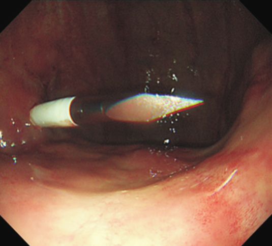
Xudong Liu et al. / Percutaneous endoscopic gastrostomy                                                                47

Removal of Percutaneous Endoscopic Gastrostomy                                                      35:545-56.
                                                                                                10. Masaki S, Kawamoto T. Comparison of long-term outcomes between enteral nutri-
                                                                                                    tion via gastrostomy and total parenteral nutrition in older persons with dyspha-
    When a PEG tube is no longer needed, it should be removed.                                      gia: a propensity-matched cohort study. PLoS One. 2019;14:e0217120.
In some cases with complications such as peristomal leakage                                     11. Blomberg J, Lagergren J, Martin L, Mattsson F, Lagergren P. Complications after
                                                                                                    percutaneous endoscopic gastrostomy in a prospective study. Scand J Gastroen-
and BBS, the tube should also be removed. Before removing the                                       terol. 2012;47:737-42.
tube, a strict evaluation must be done. Make sure the patient can                               12. Bassett MR, Dobie RA. Patterns of nutritional deficiency in head and neck cancer.
keep weight stable without using the PEG tube. Besides, in order                                    Otolaryngol Head Neck Surg. 1983;91:119-25.
                                                                                                13. Pulkkinen J, Rekola J, Asanti M, Grénman R. Prophylactic percutaneous endo-
to avoid the risk of internal leakage and peritonitis, a PEG tube                                   scopic gastrostomy in head and neck cancer patients: results of tertiary institute.
should not be removed within 4 weeks after the PEG tube place-                                      Eur Arch Otorhinolaryngol. 2014;271:1755-8.
ment.69 In patients with previous bowel surgery, the use of en-                                 14. Lang K, ElShafie RA, Akbaba S, Koschny R, Bougatf N, Bernhardt D, et al. Percu-
                                                                                                    taneous endoscopic gastrostomy tube placement in patients with head and neck
doscopic removal of PEG tubes is recommended.70 Generally, the                                      cancer treated with radiotherapy. Cancer Manag Res. 2020;12:127-36.
PEG tract usually starts to heal in 24 hours, and complete in a few                             15. Kawata N, Kakushima N, Tanaka M, Sawai H, Imai K, Hagiwara T, et al. Percuta-
days. However, in some cases the tract fails to heal and a gastro-                                  neous endoscopic gastrostomy for decompression of malignant bowel obstruction.
cutaneous fistula persists. Once diagnosed, endoscopic approach,                                    Dig Endosc. 2014;26:208-13.
                                                                                                16. FOOD Trial Collaboration. Poor nutritional status on admission predicts poor out-
using endo clips to close the fistula is first choice, if it fails, sur-                            comes after stroke: observational data from the FOOD trial. Stroke. 2003;34:1450-
gery is necessary.63                                                                                6.
                                                                                                17. Rowat A. Enteral tube feeding for dysphagic stroke patients. Br J Nurs. 2015;24:
                                                                                                    138, 140, 142-5.
Conclusions                                                                                     18. Corrigan ML, Escuro AA, Celestin J, Kirby DF. Nutrition in the stroke patient. Nutr
                                                                                                    Clin Pract. 2011;26:242-52.
    PEG had proven to be a very safe technique. PEG tube place-                                 19. Sarkar P, Cole A, Scolding NJ, Rice CM. Percutaneous endoscopic gastrostomy
                                                                                                    tube insertion in neurodegenerative disease: a retrospective study and literature
ment has many indications and contraindications, the patient                                        review. Clin Endosc. 2017;50:270-8.
must be carefully evaluated. PEG may result in many compli-                                     20. WHO. 10 facts on dementia. WHO Website. Available from: http://www.who.int/
cations, mainly including minor and major. Through standard                                         features/factfiles/dementia/en/. Published 2016. Accessed January 16, 2016.
                                                                                                21. Humbert IA, McLaren DG, Kosmatka K, Fitzgerald M, Johnson S, Porcaro E, et al.
management and treatment, the outcome of most patients is very                                      Early deficits in cortical control of swallowing in Alzheimer's disease. J Alzheimers
good. Therefore, it is very important to know how to perform a                                      Dis. 2010;19:1185-97.
PEG, choose the right patients and operation timing, and how to                                 22. Roden DF, Altman KW. Causes of dysphagia among different age groups: a sys-
                                                                                                    tematic review of the literature. Otolaryngol Clin North Am. 2013;46:965-87.
manage after the operation, how to reduce the incidence of com-                                 23. Goldberg LS, Altman KW. The role of gastrostomy tube placement in advanced
plications, and how to deal with complications after the occur-                                     dementia with dysphagia: a critical review. Clin Interv Aging. 2014;9:1733-9.
rence.                                                                                          24. Akkersdijk WL, Roukema JA, van der Werken C. Percutaneous endoscopic gas-
                                                                                                    trostomy for patients with severe cerebral injury. Injury. 1998;29:11-4.
                                                                                                25. Itkin M, DeLegge MH, Fang JC, McClave SA, Kundu S, d'Othee BJ, et al. Multidis-
Conflicts of Interest                                                                               ciplinary practical guidelines for gastrointestinal access for enteral nutrition and
                                                                                                    decompression from the Society of Interventional Radiology and American Gas-
                                                                                                    troenterological Association (AGA) Institute, with endorsement by Canadian Inter-
    No potential conflict of interest relevant to this article was re-                              ventional Radiological Association (CIRA) and Cardiovascular and Interventional
ported.                                                                                             Radiological Society of Europe (CIRSE). Gastroenterology. 2011;141:742-65.
                                                                                                26. Cui N, Zhao Y, Cao J. Clinical features and advantages of a novel percutaneous
                                                                                                    endoscopic gastrostomy method. Med Sci Monit. 2019;25:9651-7.
ORCID                                                                                           27. Stiegmann GV, Goff JS, Silas D, Pearlman N, Sun J, Norton L. Endoscopic versus
                                                                                                    operative gastrostomy: final results of a prospective randomized trial. Gastrointest
    Xudong Liu, https://orcid.org/0000-0003-0080-7878                                               Endosc. 1990;36:1-5.
                                                                                                28. Russell TR, Brotman M, Norris F. Percutaneous gastrostomy. A new simplified and
    Zhengqiang Yang, https://orcid.org/0000-0001-9103-3534                                          cost-effective technique. Am J Surg. 1984;148:132-7.
    Shun He, https://orcid.org/0000-0002-3472-9532                                              29. Chadha KS, Thatikonda C, Schiff M, Nava H, Sitrin MD. Outcomes of percutaneous
    Guiqi Wang, https://orcid.org/0000-0003-3226-9285                                               endoscopic gastrostomy tube placement using a T-fastener gastropexy device in
                                                                                                    head and neck and esophageal cancer patients. Nutr Clin Pract. 2010;25:658-62.
                                                                                                30. Shastri YM, Hoepffner N, Tessmer A, Ackermann H, Schroeder O, Stein J. New
References                                                                                          introducer PEG gastropexy does not require prophylactic antibiotics: multicenter
                                                                                                    prospective randomized double-blind placebo-controlled study. Gastrointest En-
 1. Nugent B, Lewis S, O'Sullivan JM. Enteral feeding methods for nutritional man-                  dosc. 2008;67:620-8.
    agement in patients with head and neck cancers being treated with radiotherapy              31. Bravo JG, Ide E, Kondo A, de Moura DT, de Moura ET, Sakai P, et al. Percutaneous
    and/or chemotherapy. Cochrane Database Syst Rev. 2013;2013:CD007904. doi: 10                    endoscopic versus surgical gastrostomy in patients with benign and malignant
    .2174/1389450120666190307095720.                                                                diseases: a systematic review and meta-analysis. Clinics (Sao Paulo). 2016;71:169-
 2. Wang J, Liu M, Liu C, Ye Y, Huang G. Percutaneous endoscopic gastrostomy ver-                   78.
    sus nasogastric tube feeding for patients with head and neck cancer: a systematic           32. Schurink CA, Tuynman H, Scholten P, Arjaans W, Klinkenberg-Knol EC, Meuwis-
    review. J Radiat Res. 2014;55:559-67.                                                           sen SG, et al. Percutaneous endoscopic gastrostomy: complications and sugges-
 3. Gundogan K, Yurci A, Coskun R, Baskol M, Gursoy S, Hebbar G, et al. Outcomes                    tions to avoid them. Eur J Gastroenterol Hepatol. 2001;13:819-23.
    of percutaneous endoscopic gastrostomy in hospitalized patients at a tertiary care          33. Preclik G, Grüne S, Leser HG, Lebherz J, Heldwein W, Machka K, et al. Prospective,
    center in Turkey. Eur J Clin Nutr. 2014;68:437-40.                                              randomised, double blind trial of prophylaxis with single dose of co-amoxiclav
 4. Gauderer MW, Ponsky JL, Izant RJ Jr. Gastrostomy without laparotomy: a percu-                   before percutaneous endoscopic gastrostomy. BMJ. 1999;319:881-4.
    taneous endoscopic technique. J Pediatr Surg. 1980;15:872-5.                                34. Dormann AJ, Wigginghaus B, Risius H, Kleimann F, Kloppenborg A, Rosemann J,
 5. Rahnemai-Azar AA, Rahnemaiazar AA, Naghshizadian R, Kurtz A, Farkas DT.                         et al. Antibiotic prophylaxis in percutaneous endoscopic gastrostomy (PEG)--results
    Percutaneous endoscopic gastrostomy: indications, technique, complications and                  from a prospective randomized multicenter trial. Z Gastroenterol. 2000;38:229-34.
    management. World J Gastroenterol. 2014;20:7739-51.                                         35. ASGE Standards of Practice Committee, Khashab MA, Chithadi KV, Acosta RD,
 6. Kim J, Koh H, Chang EY, Park SY, Kim S. Single center experience with gastrosto-                Bruining DH, Chandrasekhara V, et al. Antibiotic prophylaxis for GI endoscopy.
    my insertion in pediatric patients: a 10-year review. Pediatr Gastroenterol Hepatol             Gastrointest Endosc. 2015;81:81-9.
    Nutr. 2017;20:34-40.                                                                        36. DeLegge M. Gastrostomy tubes: complications and their management. UpToDate
 7. Lim JH, Choi SH, Lee C, Seo JY, Kang HY, Yang JI, et al. Thirty-day mortality after             Website. Available from: https://www.uptodate.com/contents/gastrostomy-tubes-
    percutaneous gastrostomy by endoscopic versus radiologic placement: a system-                   complications-and-their-management/print#!. 2019. Accessed February 21, 2021.
    atic review and meta-analysis. Intest Res. 2016;14:333-42.                                  37. Blacka J, Donoghue J, Sutherland M, Martincich I, Mitten-Lewis S, Morris P, et al.
 8. Ralls MW, Demehri FR, Feng Y, Woods Ignatoski KM, Teitelbaum DH. Enteral nu-                    Dwell time and functional failure in percutaneous endoscopic gastrostomy tubes:
    trient deprivation in patients leads to a loss of intestinal epithelial barrier function.       a prospective randomized-controlled comparison between silicon polymer and
    Surgery. 2015;157:732-42.                                                                       polyurethane percutaneous endoscopic gastrostomy tubes. Aliment Pharmacol
 9. Druml C, Ballmer PE, Druml W, Oehmichen F, Shenkin A, Singer P, et al. ESPEN                    Ther. 2004;20:875-82.
    guideline on ethical aspects of artificial nutrition and hydration. Clin Nutr. 2016;        38. Boldo-Roda E, Peris-Trias A, de Lucia-Peñalver GP, Martinez-Ramos D, Miralles-
48 International Journal of Gastrointestinal Intervention 2021 10(2), 42–48
Tena JM. Reflections in front of a case of ventral hernia after PEG tube removal. Gastroenterol Belg. 2011;74:312-6.
Gastrointest Endosc. 2005;62:323-4. 57. Grant JP. Percutaneous endoscopic gastrostomy. Initial placement by single
39. Navarro F, Loflin C, Diegidio P, Atwez A, Reeves J. Herniation through endoscopic technique and long-term follow-up. Ann Surg. 1993;217:168-74.
gastrostomy site: case report. Int J Surg Case Rep. 2016;25:165-6. 58. Finucane TE, Bynum JP. Use of tube feeding to prevent aspiration pneumonia.
40. Stathopoulos G, Rudberg MA, Harig JM. Subcutaneous emphysema following Lancet. 1996;348:1421-4.
PEG. Gastrointest Endosc. 1991;37:374-6. 59. MacLean AA, Miller G, Bamboat ZM, Hiotis K. Abdominal wall necrotizing
41. Gottfried EB, Plumser AB, Clair MR. Pneumoperitoneum following percutaneous fasciitis from dislodged percutaneous endoscopic gastrostomy tubes: a case series.
endoscopic gastrostomy. A prospective study. Gastrointest Endosc. 1986;32:397-9. Am Surg. 2004;70:827-31.
42. Schrag SP, Sharma R, Jaik NP, Seamon MJ, Lukaszczyk JJ, Martin ND, et al. 60. Cruz I, Mamel JJ, Brady PG, Cass-Garcia M. Incidence of abdominal wall
Complications related to percutaneous endoscopic gastrostomy (PEG) tubes. A metastasis complicating PEG tube placement in untreated head and neck cancer.
comprehensive clinical review. J Gastrointestin Liver Dis. 2007;16:407-18. Gastrointest Endosc. 2005;62:708-11.
43. Larson DE, Burton DD, Schroeder KW, DiMagno EP. Percutaneous endoscopic 61. Volkmer K, Meyer T, Sailer M, Fein M. [Metastasis of an esophageal carcinoma at
gastrostomy. Indications, success, complications, and mortality in 314 consecutive a PEG site--case report and review of the literature]. Zentralbl Chir. 2009;134:481-
patients. Gastroenterology. 1987;93:48-52. 5. German.
44. Galat SA, Gerig KD, Porter JA, Slezak FA. Management of premature removal of 62. Janik TA, Hendrickson RJ, Janik JS, Landholm AE. Analysis of factors affecting
the percutaneous gastrostomy. Am Surg. 1990;56:733-6. the spontaneous closure of a gastrocutaneous fistula. J Pediatr Surg. 2004;39:
45. Lynch CR, Fang JC. Prevention and management of complications of percutaneous 1197-9.
endoscopic gastrostomy (PEG) tubes. Pract Gastroenterol. 2004;28:66-76. 63. Gay-Chevallier S, Lupu A, Rivory J, Rostain F, Ponchon T, Saurin JC, et al. Closure
46. Date RS, Das N, Bateson PG. Unusual complications of ballooned feeding tubes. Ir of non-healing gastrocutaneous fistula after percutaneous endoscopic gastrostomy
Med J. 2002;95:181-2. by endoscopic submucosal dissection and over-the-scope clip. Endoscopy. 2019;
47. Warriner L, Spruce P. Managing overgranulation tissue around gastrostomy sites. 51:E125-6.
Br J Nurs. 2012;21:S14-6, S18, S20. 64. Bechtold ML, Matteson ML, Choudhary A, Puli SR, Jiang PP, Roy PK. Early versus
48. McClave SA, Chang WK. Complications of enteral access. Gastrointest Endosc. delayed feeding after placement of a percutaneous endoscopic gastrostomy: a
2003;58:739-51. meta-analysis. Am J Gastroenterol. 2008;103:2919-24.
49. Lau G, Lai SH. Fatal retroperitoneal haemorrhage: an unusual complication of 65. Szary NM, Arif M, Matteson ML, Choudhary A, Puli SR, Bechtold ML. Enteral
percutaneous endoscopic gastrostomy. Forensic Sci Int. 2001;116:69-75. feeding within three hours after percutaneous endoscopic gastrostomy placement:
50. Lee SH, Moon HS, Park JH, Kim JS, Kang SH, Lee ES, et al. Percutaneous a meta-analysis. J Clin Gastroenterol. 2011;45:e34-8.
endoscopic gastrostomy tube insertion-induced superior mesenteric artery injury 66. Vyawahare MA, Shirodkar M, Gharat A, Patil P, Mehta S, Mohandas KM.
treated with angiography. Korean J Gastroenterol. 2018;72:308-12. A comparative observational study of early versus delayed feeding after
51. Guloglu R, Taviloglu K, Alimoglu O. Colon injury following percutaneous percutaneous endoscopic gastrostomy. Indian J Gastroenterol. 2013;32:366-8.
endoscopic gastrostomy tube insertion. J Laparoendosc Adv Surg Tech A. 67. Roveron G, Antonini M, Barbierato M, Calandrino V, Canese G, Chiurazzi LF,
2003;13:69-72. et al. Clinical practice guidelines for the nursing management of percutaneous
52. Karhadkar AS, Schwartz HJ, Dutta SK. Jejunocutaneous fistula manifesting as endoscopic gastrostomy and jejunostomy (PEG/PEJ) in adult patients: an executive
chronic diarrhea after PEG tube replacement. J Clin Gastroenterol. 2006;40:560-1. summary. J Wound Ostomy Continence Nurs. 2018;45:326-34.
53. Chaer RA, Rekkas D, Trevino J, Brown R, Espat J. Intrahepatic placement of a PEG 68. Sriram K, Jayanthi V, Lakshmi RG, George VS. Prophylactic locking of enteral
tube. Gastrointest Endosc. 2003;57:763-5. feeding tubes with pancreatic enzymes. JPEN J Parenter Enteral Nutr. 1997;21:
54. Gubler C, Wildi SM, Bauerfeind P. Liver injury during PEG tube placement: report 353-6.
of two cases. Gastrointest Endosc. 2005;61:346-8. 69. Bischoff SC, Austin P, Boeykens K, Chourdakis M, Cuerda C, Jonkers-Schuitema C,
55. Klein S, Heare BR, Soloway RD. The “buried bumper syndrome": a complication of et al. ESPEN guideline on home enteral nutrition. Clin Nutr. 2020;39:5-22.
percutaneous endoscopic gastrostomy. Am J Gastroenterol. 1990;85:448-51. 70. Waxman I, al-Kawas FH, Bass B, Glouderman M. PEG ileus. A new cause of small
56. El AZ, Arvanitakis M, Ballarin A, Devière J, Le Moine O, Van Gossum A. Buried bowel obstruction. Dig Dis Sci. 1991;36:251-4.
bumper syndrome: low incidence and safe endoscopic management. ActaYou can also read