IVERMECTIN AS A PROMISING RNA-DEPENDENT RNA POLYMERASE INHIBITOR AND A THERAPEUTIC DRUG AGAINST SARS-COV2: EVIDENCE FROM IN SILICO STUDIES
←
→
Page content transcription
If your browser does not render page correctly, please read the page content below
Ivermectin as a promising RNA-dependent RNA
polymerase inhibitor and a therapeutic drug against
SARS-CoV2: Evidence from in silico studies
Ananta Swargiary ( ananbuzoo101@gmail.com )
Department of Zoology, Bodoland University https://orcid.org/0000-0001-9594-3666
Research Article
Keywords: COVID-19, SARS-CoV2, ivermectin, RdRp, Docking
DOI: https://doi.org/10.21203/rs.3.rs-73308/v1
License: This work is licensed under a Creative Commons Attribution 4.0 International License.
Read Full License
Page 1/12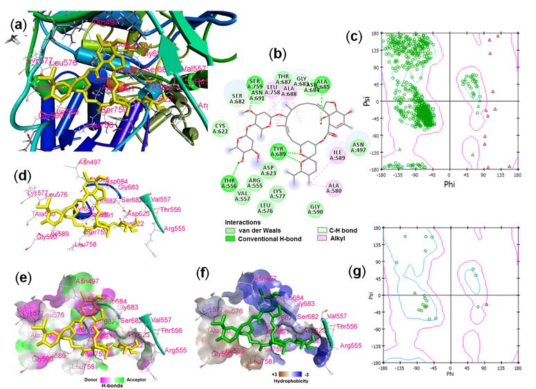
Abstract
Purpose: COVID-19, caused by SARS-CoV2 virus is a contagious disease affecting millions of lives
throughout the globe. Currently, there are no clinically approved drugs for SARS-CoV2 although some
drugs are undergoing clinical trials. The present study investigates the binding property of ivermectin on
four important drug targets, spike protein, RNA-dependent RNA polymerase, 3-chymotrypsin- and papain-
like proteases of SARS-CoV2.

Methods: The 3D structure of ivermectin along with known antiviral drug lopinavir, simeprevir and four
nucleotides ATP, GTP, CTP, and UTP were downloaded from PubChem database. Crystal structures of
proteins were downloaded from PDB database. PDB les were converted into pdbqt le using AutoDock
tools. After proper processing and grid formation, docking was carried out in AutoDock vina. Furthermore,
the co-crystallized RNA and its binding interactions with RdRp were studied using various visualization
tools including Discovery studio.

Results: Docking study showed that ivermectin is the best binding drug compared to lopinavir and
simeprevir. The best binding interaction was found to be -9.7kcal/mol with RdRp suggesting potential
inhibitor of the protein. Twenty-one amino acid residues of RdRp were found to interact with ivermectin
including the catalytic residue Asp760. Furthermore, RNA-RdRp complex revealed that the catalytic active
residues Ser759 and Asp760 of RdRp formed strong interactions with RNA chain. Binding of ivermectin in
the active site of RdRp make clash with the nucleotides of RNA chain suggesting the possible inhibition
of replication.

Conclusions: The present study suggests ivermectin as a potential inhibitor of RdRp which may be crucial
to combat the SARS-CoV2.

Introduction
The pandemic of COVID-19 has disastrously affected human health and wealth since its emergence in
2019 from the Wuhan city of China. Severe Acute Respiratory Syndrome corona virus -2 (SARS-CoV2), a
single-stranded positive-sense RNA virus belonging the family Coronaviridae is the causative organism of
COVID-19 disease (Benvenuto et al. 2020). According to the latest WHO update (2:09 pm CEST, 22 August
2020), there have been 22,812,491 con rmed cases of COVID-19, including 795,132 deaths globally.
Today, there are seven different species of human coronaviruse (HCoV) - 229E, NL63, OC43, HKU1, SARS-
CoV, MERS-CoV, and SARS-CoV2 (Benvenuto et al. 2020). Most of the HCoVs causes mild infections such
as common cold while SARS, MERS, and SARS-CoV2 are highly pathogenic, and contagious causing
severe respiratory problems and even death (Cui et al. 2019). Genomic and sequence homology studies
emphasize on the common origin of all the HCoVs (from bats?) except HKU1 and HCoV-OC43 which are
linked to rodents (Forni et al. 2017). The attack and pathogenicity of SARS-CoV2 progress with four
stages – asymptomatic, moderate, extreme, and clinical which starts at the lower respiratory tract
                                                 Page 2/12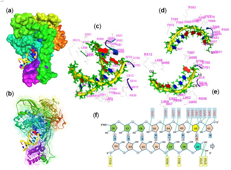
followed by an invasion of pulmonary epithelial cells and hijacking the entire host cell machinery. The
most common symptoms of COVID-19 include cough, fever, malaise, gastrointestinal symptoms, loss of
smell, sore throat, heart failure, and acute kidney injury (Gandhi et al. 2020).

Currently, there is no clinically approved drug for SARS-CoV2. However, recent publications reveal that
several medications such as ivermectin, hydroxychloroquine, and remdesivir are being used to reduce the
virus load and improve disease symptoms (Caly et al. 2020; McKee et al. 2020; Wang et al. 2020).
Researchers around the world are working diligently to develop effective drugs based on the
pathogenicity and molecular details of the SARS CoV-2. Four key targets proteins such as spike protein
(S-protein), virus mail protease (3Clpro), papain-like protease (Plpro), and RNA-dependent RNA
polymerases (RdRp) are being explored by researchers around the world to develop medicines against
COVID-19. The SARS-CoV S-protein is an important drug target which is a quaternary protein composed
of two subunits - S1 subunit that contains a receptor-binding domain (RBD) which engages with the host
cell receptor angiotensin-converting enzyme-2 and the S2 subunit that mediates fusion between the viral
and host cell membranes (Du et al. 2009; Zhou et al. 2019). 3Clpro and Plpro are two important virus
proteases that catalyse the processing of polyproteins pp1a and pp1ab leading to the formation of RdRp
and other important non-structural proteins (nsp) (Anand et al. 2003; Chen et al., 2020). The RNA-
dependent RNA polymerase, also known as nsp12 is the central component of CoV replication and
transcription machinery, and therefore, it appears to be a primary target for the antiviral drug (Xu et al.
2003). Given the pivotal roles of S-protein, 3Clpro, PL-pro, and RdRp in the infection, replication, and
generation of viral particle, these proteins are widely regarded as an important and attractive target for
the rational design of anti-SARS-CoV2 drugs.

Ivermectin is a widely used FDA approved broad-spectrum antihelmintic drug. It causes stimulation of
gamma amino-butyric acid (GABA)-gated-Cl-channels, leading to hyperpolarization, and resulting in
paralysis of helminth parasites (Canga et al. 2008). Furthermore, ivermectin is also known to act as
inhibitor of importin-α/β-mediated nuclear import and suppress the replication of several RNA viruses,
including HIV, chikungunya virus, and yellow fever virus (Lv et al. 2018). Recent studies have shown that
ivermectin can inhibit the replication of SARS-CoV-2 in vitro (Sharun et al. 2020). The present study
investigates the in silico binding a nity of ivermectin drug with S-protein, 3Clpro, PL-pro, and RdRp
proteins of SARS-CoV2.

Materials And Methods
Ligand Selection and Preparation

The 3D structure of ivermectin (Drugbank ID: DB00602) along with two antiviral drugs simeprevir
(DB06290), and lopinavir (DB01601) from Drugbank database (https://www.drugbank.ca/). Four
nucleotides ATP (PubChem CID: 5957), GTP (CID: 135398633), CTP (CID: 6176), and UTP (CID: 6133)
were downoloaded from PubChem database. SDF les were converted into pdb le using PyMol

                                                   Page 3/12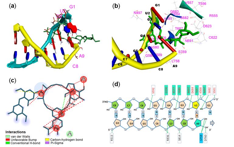
software. All the PDB les of the ligands were processed and nally converted into .pdbqt le using
AutoDock tool (Trott and Olson 2010).

Collection and Preparation of Proteins

Three-dimensional structures of S-protein (RBD) (PDB ID: 7BZ5), 3Clpro (PDB ID: 6M2N), Plpro (PDB ID:
7JN2), and RdRp (PDB ID: 6XQB) were downloaded from PDB database. The protein structures were
cleaned by removing the water and other hetatms. Polar hydrogen atoms and Kollman charges were
added to the structure and nally converted into .pdbqt le format for docking using AutoDock Tools.

Molecular Docking

After the ligand drugs and the target enzymes were prepared docking was carried out in AutoDock Vina
(Trott and Olson 2010). The grid box parameters for docking of all the four proteins are presented in table
1. The docking algorithm was carried out by keeping the default exhaustiveness at 8. After docking, the
pose scoring the lowest binding energy (kcal/mol) was selected and visualize in Discovery Studio.

Table 1. Docking grid box parameters for AutoDock Vina docking.
  Proteins   Active site amino acid residues                          Center co-ordinates           Size co-
                                                                                                  ordinates
                                                                       x         y         z      x     y    z
  S-         Leu455, Phe486, Gln493, Ser 494, Asn501, Tyr505        -57.021   -34.024   14.358   42 74 58
  protein
  3Clpro     His41, Thr45, Met49, Phe140, Asn142, Cys145, Asp187,   -13.137   14.854    69.809   46   46   66
             Arg188, Gln189, Met165, His172, Glu166
  Plpro      Cys112, Leu163, Gly164, Asp165, Pro248, Pro249,        -37.547   22.916    -9.733   58   52   64
             Tyr269, Gln270, His273, Tyr274, Asp-287, Thr302
  RdRp       Gly616, Trp617, Asp618, Tyr619, Leu758, Ser759,        93.080    81.814    97.063   58   48   48
             Asp760, Asp761, Ala762, Lys798, Cys799, Trp800,
             Glu811, Phe812, Cys813, Ser814.

Results
The present study investigates the binding a nity of ivermectin, a broad-spectrum antiparasitic FDA
approved drug against four key enzymes of SARS-CoV2, the spike protein, 3Clpro, Plpro, and RdRp
enzymes. Figure 1 showed the binding energies of ivermectin with all the four protein. Ivermectin showed
the best and strongest binding a nity with RdRp (-9.7kcal/mol) followed by S-protein, Plpro, and 3Clpro.
The reference antiviral drug simeprevir also showed strong a nity to all the proteins. Highest binding
a nity was observed in Plpro. Lopinavir showed the weakest a nity among all the three drugs. To
compare the a nity of nucleotides to the active site of RdRp enzymes, all the four nucleotides ATP, GTP,
CTP, and UTP were also docked with the protein. All the four nucleotides showed weaker a nity to the
enzyme compared to ivermectin. ATP showed the highest binding energy -8.1 kcal/mol followed by UTP
(-8.0 kcal/mol), CTP (-7.8 kcal/mol), and lowest in GTP (-7.6 kcal/mol).

                                                 Page 4/12
Fig. 2 showed the binding interactions of the ivermectin with the RdRp of SARS-CoV2 and Ramachandran
plot of the amino acid residues. A total of 21 amino acid residues (Asn497, Arg555, Thr556, Val557,
Leu576, Lys577, Ala580, Ile589, Gly590, Cys622, Asp623, Ser682, Gly683, Asp684, Ala685, Thr687,
Ala688, Tyr689, Asn691, Leu758, and Ser759) were found to be interacting with the ivermectin. The
ligand-binding site in the RdRp protein and the schematic view of the complex is shown in Fig. 2a. Five
conventional H-bonds were seen between the ligand-protein complexes while majority of the binding
interactions were found to be van der Waal’s interaction (Fig. 2b,d). The H-bond donor and acceptor and
the hydrophobicity property of the ligand surrounding amino acid residues were shown in Fig. 2e and 2f.
Most of the surrounding amino acid residues showed hydrophilicity while eight residues showed
hydrophobic property. Ramachandran plot study revealed that 11 amino acid residues (all glycine) of
RdRp apoprotein were found to be distributed outside the allowed region of the plot.
The various amino acid residues interacting with the replicating RNA in the active site of RdRp enzyme is
presented in Fig. 3. The co-crystallized structure of RNA-RdRp complex (Pdb ID: 6xqb) has been dissected
to visualize the amino acids attaching to the dsRNA chain. A total of 41 amino acid residues were found
to form the surrounding surface of the dsRNA (Fig. 3c). Out of 41 amino acid residues, 23 amino acids
form interactions with the template RNA strand (3’ – 5’ strand, Fig. 3d) while 20 residues interact with the
newly replicating strand (5’- 3’ strand, Fig. e) of RNA. Two amino acids, Thr687 and Glu857 formed
interactions with both the stands of dsRNA. Fig. 3f showed the H-bonding amino acid residues with the
dsRNA strand. Out of 41 interacting residues, only 15 residues formed hydrogen bonding with the RNA
chain. Ten amino acid residues formed eleven H-bonds with the template strand (3’-5’ strand) while ve
amino acids form seven H-bonds with the complementary new strand of RNA. Asn497 of template strand
form 2-H-bonds with the uracil (U2) nucleotide while Arg836 and Ser759 formed 2H-bonds each with
cytosine (C7) and adenine (A9) nucleotides of new RNA strand (Fig. 3f). It is also observed that out of 21
amino acid residues of RdRp that are interacting with the ivermectin, 16 residues also made interactions
with the dsRNA.
The interaction of ivermectin with active site amino acid and RNA strands is shown in Fig. 4. The RNA-
ligand interactions and surrounding amino acids were shown in Fig. 4a and 4b. The co-crystallized
dsRNA structure of RdRp and the best docking pose of ivermectin when tted into the binding site ve
nucleotides – G1, U2, G3, and G4 from template strand (3’ – 5’ strand) and adenine-9 from newly
synthesized RNA strand made unfavourable interactions with the ivermectin (Fig. 4c). Fig. 4d showed
that 6 active site amino acid residues (out of 15) that were making H-bonds with dsRNA were also found
to make interactions with the ligand.
Discussion
SARS-CoV2 is a positive-sense single-stranded RNA virus. The entry of viral genome into the host body
and its generation and invasion to the other cells is mediated by several proteins including spike proteins,
3chymotrypsin- and papain-like proteases. A polyprotein complex, RNA-dependent RNA-polymerase
(RdRp) is a major protein in SARS-CoV2 that regulates viral replication (Yin et al. 2020). Inhibition of
Page 5/12replication may be an important strategy to combat SARS-CoV2 and COVID-19. RdRp protein, therefore, is
an important and potential therapeutic target of antiviral drugs. Compounds that bind effectively with the
active site of the RdRp and inhibit the enzyme’s catalytic activity shall be an important aspect of
controlling virus replication and generation. Several molecules and antiviral drugs have been screened by
many researchers to nd suitable molecule with high e cacy (El ky 2020; Babadaei et al. 2020; Touret et
al. 2020). Ivermectin, a broad-spectrum antihelmintic drug has been recently established to be a potential
inhibitor of SARS-CoV2 RdRp protein in in vitro study (Caly et al. 2020). It has also been opined that
hydroxychloroquine and ivermectin may show effective result if administered simultaneously (Patri and
Fabbrocini, 2020). Ivermectin is also known to inhibit DNA polymerase of pseudorabies virus (Lv et al.
2018). The present study revealed strong binding a nity of ivermectin to the RdRp protein. In association
to surrounding key residues, Ser759, Aspertate760 and -761 forms the core catalytic site amino acid
residues of RdRp in SARS-CoV2 (Yin et al. 2020). The present study observed that Ser759 form strong
interaction with ivermectin and adenine-9 nucleotide as well. The study, however, did not nd any
interaction with catalytically active Asp760 and -761 residues of RdRp. Ornipressin, an FDA approved
drug for vasoconstriction and liver cirrhosis, is reported to have strong interactions with eight amino acid
residues of RdRp including the catalytic site residue Asp760 (Ahmad et al. 2020). In an in silico study, Yin
et al. (2020) reported that remdesivir, a nucleoside analog used to inhibit the action of RNA polymerase in
Ebola virus make phosphodiester bond with the 3’-OH group of the newly synthesized RNA strand as well
as strong interaction with the Asp760 residue of SARS-CoV2 leading to the inhibition of replication.
Ivermectin is not a nucleotide analog and therefore did not bind with the 3’OH- end of the leading strand.
However, the study showed unfavourable interactions with the parental (template) RNA strand up to four
nucleotides upstream of the replicating site. Binding of ivermectin to the active pocket of RdRp may
prevent the catalytically favourable accommodation of the template RNA chain in the enzyme and hence
inhibition of replication. The ndings of the present study along with in vitro validation by earlier studies
suggest the potentiality of ivermectin to be an effective inhibitor of SARS-CoV2 RNA-dependent RNA-
polymerase.
Conclusion
COVID-19 pandemic has tremendously affected the health and wealth of global community. Speedy trial
and approval of new drug is the only strategy to combat SARS-CoV2 along with social distancing. The
present study revealed the structural inhibition of RdRp protein by ivermectin drug and therefore
ivermectin may be a potential drug to combat COVID-19 and SARS-CoV2.
Declarations
Acknowledgment
AS would like to acknowledge the necessary facility provided by Head, Department of Zoology, Bodoland
University, Kokrajhar, Assam, India.
Page 6/12Funding: There is no speci c funding for the current study.
Con icts of interest/Competing interests: There is no con ict of interest.
Ethics approval: NA
Consent to participate: NA
Consent for publication: Author gives the consent to publish the manuscript
Availability of data and material: No
Authors' contributions: NA
References
Ahmad J, Ikram S, Ahmad F, Rehman IU, Mushtaq M (2020) SARS-CoV-2 RNA Dependent RNA
polymerase (RdRp) – A drug repurposing study. Heliyon 6(7):e04502.
https://doi.org/10.1016/j.heliyon.2020.e04502.
Anand K, Ziebuhr J, Wadhwani P, Mesters JR, Hilgenfeld R (2003) Coronavirus main proteinase (Mpro)
structure: basis for design of anti-SARS drugs. Science 300:1763e1767. Doi: 10.1126/science.1085658.
Babadaei MMN, Hasan A, Vahdani Y, Bloukh SH, Shari M, Kachooei E, et al. (2020) Development of
remdesivir repositioning as a nucleotide analog against COVID-19 RNA dependent RNA polymerase. J
Biomol Struct Dyn. Doi: 10.1080/07391102.2020.1767210.
Benvenuto D, Giovanetti M, Ciccozzi A, Spoto S, Angeletti S, Ciccozzi M (2020) The 2019-new Coronavirus
epidemic: evidence for virus evolution. J Med Virol 92:455–459. doi: 10.1002/jmv.25688.
Caly L, Druce JD, Catton MG, Jans DA, Wagstaff KM (2020) The FDA-approved drug ivermectin inhibits
the replication of SARS-CoV-2 in vitro. Antiviral Res 178:104787.
Doi.org/10.1016/j.antiviral.2020.104787.
Canga AG, Prieto AMS, Liebana MJD, Martinez NF, Vega MS, Vieitez JJG (2008) The Pharmacokinetics
and Interactions of Ivermectin in Humans - A Mini-review. AAPS J 10:42–46.
https://doi.org/10.1208/s12248-007-9000-9.
Cui J, Li F, Shi Z (2019) Origin and evolution of pathogenic coronaviruses. Nat Rev Microbiol 17:181-192.
https://doi.org/10.1038/s41579-018-0118-9.
Chen YW, Yiu CPB, Wong KY (2020) Prediction of the SARSCoV-2 (2019-nCoV) 3C-like protease (3CL (pro)
structure: Virtual screening reveals velpatasvir, ledipasvir, and other drug repurposing candidates.
F1000Research 9:129. https://doi.org/10.12688/f1000research.22457.1
Page 7/12Du L, He Y, Zhou Y, Liu S, Zheng BJ, Jiang S (2009) The spike protein of SARS-CoV—a target for vaccine
and therapeutic development. Nat Rev Microbiol 7(3):226-236. doi.org/10.1038/nrmicro2090.
El ky AA (2020) Anti-HCV, nucleotide inhibitors, repurposing against COVID-19. Life Sci 248:117477.
https://doi.org/10.1016/j.lfs.2020.117477.
Forni D, Cagliani R, Clerici M, Sironi M (2017) Molecular evolution of human coronavirus genomes. Trends
Microbiol 25:35–48. https://doi.org/10.1016/j.tim.2016.09.001.
Gandhi RT, Lynch JB, Rio C (2020) Mild or Moderate Covid-19. New Engl J Med.
https://doi.org/10.1056/NEJMcp2009249.
Lv C, Liu W, Wang B, Dang R, Qiu L, Ren J, et al. (2018) Ivermectin inhibits DNA polymerase UL42 of
pseudorabies virus entrance into the nucleus and proliferation of the virus in vitro and vivo. Antiviral Res
159:55-62. Doi.org/10.1016/j.antiviral.2018.09.010.
McKee DL, Sternberg A, Stange U, Laufer S, Naujokat C (2020) Candidate drugs against SARS-CoV-2 and
COVID-19. Pharmacol Res 157:104859. doi.org/10.1016/j.phrs.2020.104859
Patrì A, Fabbrocini G (2020) Hydroxychloroquine and ivermectin: A synergistic combination for COVID-19
chemoprophylaxis and treatment? J Am Acad Dermatol 82(6):e221. Doi: 10.1016/j.jaad.2020.04.017.
Sharun K, Dhama K, Patel SK, Pathak M, Tiwari R, Singh BR, et al. (2020) Ivermectin, a new candidate
therapeutic against SARS-CoV-2/COVID-19. Ann Clin Microbiol Antimicrob 19:23.
https://doi.org/10.1186/s12941-020-00368-w
Touret F, Gilles M, Barral K, Nougairede A, Helden J, Decroly E, et al. (2020) In vitro screening of a FDA
approved chemical library reveals potential inhibitors of SARS-CoV-2 replication. Sci Rep 10:13093.
https://doi.org/10.1038/s41598-020-70143-6.
Trott O, Olson AJ (2010) AutoDock Vina: improving the speed and accuracy of docking with a new
scoring function, e cient optimization and multithreading. J Comput Chem 31:455–461. http://doi:
10.1002/jcc.21334
Wang M, Cao R, Zhang L, Yang X, Liu J, Xu M, et al. Remdesivir and chloroquine effectively inhibit the
recently emerged novel coronavirus (2019-nCoV) in vitro. Cell Res 30:269–271.
https://doi.org/10.1038/s41422-020-0282-0.
Xu X, Liu Y, Weiss S, Arnold E, Sara anos SG, Ding J. (2003) Molecular model of SARS coronavirus
polymerase: implications for biochemical functions and drug design. Nucleic Acids Res 31(24):7117-
7130. Doi: 10.1093/nar/gkg916.
Yin W, Mao C, Luan X, Shen D, Shen Q, Su H, et al. (2020) Structural basis for inhibition of the RNA-
dependent RNA polymerase from SARS-CoV-2 by remdesivir. Science 368, 1499–1504. DOI:
Page 8/1210.1126/science.abc1560.
Zhou J, Fang L, Yang Z, Xu S, Lv M, Sun Z, et al. (2019) Identi cation of novel proteolytically inactive
mutations in coronavirus 3C-like protease using a combined approach. The FASEB Journal 33(12):14575-
14587. https://doi.org/10.1096/fj.201901624RR.
Figures
Figure 1
Docking energy and binding a nities of drug wit spike protein, 3chymotrypsin-like protease (3Clpro),
papain-like protease (Plpro), and RNA-dependent RNA polymerase (RdRp).
Page 9/12Figure 2
Binding interactions of SARS-CoV2 RNA-dependent RNA polymerase and ivermectin. (a) RdRp-ligand
complex schematic, (b) 2D display of RdRp-ligand interactions, (c) Ramachandran plot of PLpro, (d)
amino acid residues surrounding the ligand, (e) H-bond property of binding pocket, (f) Hydrophobicity
pro le of binding pocket, and (g) Ramachandran plot of ligand-interacting amino acid residues.
Page 10/12Figure 3
Binding interactions of RNA and RNA-dependent RNA polymerase. (a & b) surface view of RNA-RdRp
complex, (c) RNA and surrounding amino acid residues, (d) 3’-5’ RNA strand with surrounding amino
acids, (e) 5’-3’-strand with surrounding amino acids and (f) amino acid residues forming H-bond with both
the strand of dsRNA.
Page 11/12Figure 4
Interactions between RNA, RdRp, and ivermectin. (a) rode and ring view of dsRNA with attached
ivermectin, (b) rode and ladder view of dsRNA with attached ivermectin and interacting amino acid
residues of RdRp, (c) 2D display of ivermectin and nucleotide interactions, and (d) amino acid residues
forming H-bond with RNA chain and ivermectin (cyan/blue coloured boxes).
Page 12/12You can also read