Journal of Clinical Images and Medical Case Reports
←
→
Page content transcription
If your browser does not render page correctly, please read the page content below
www.jcimcr.org

                                   Journal of
                                   Clinical Images and Medical Case Reports
                                                                                                                  ISSN 2766-7820

Case Report
Open Access, Volume 2

Tuberculosis of bilateral ankles: Misdiagnosed
as rheumatoid arthritis
Haiyao Wang1; Ping He2; Gailian Zhang3,*; Jinfang Gao1; Liyun Zhang1
1
 Department of Rheumatology, Shanxi Bethune Hospital, Shanxi Academy of Medical Sciences, Shanxi Bethune Hospital Affiliated
to Shanxi Medical University, Taiyuan 030032, Shanxi, China.
2
 Department of Nephrology, Changzhi People’s Hospital, Changzhi 046000, Shanxi, China.
3
 Department of Rheumatology, Shanxi Provincial People’s Hospital, Taiyuan 030012, Shanxi, China.

                                                              Abstract
  *Corresponding Authors: Gailian Zhang
 Department of Rheumatology, Shanxi Provincial                   The osteoarticular tuberculosis is a uncommon diease, which is ac-
                                                              cout for 0.1%-0.3% of Tuberculosis (TB). The involoment of foot and
 People’s Hospital, Taiyuan 030012, Shanxi, China.
                                                              ankle is only 0.01%-0.03% of TB, particularly the bilateral feet and
 Tel: 13007016221; Email: 13754820091@sina.com                ankles are affected is extremely rare. Futhermore, its clinical manifes-
                                                              tation is atypical and similar with autoimmune disorder, subacute or
                                                              chronic pyogenic arthritis, osteochondrosis, benign bone tumors and
Received: May 01, 2021                                        so on, which conduce to the delay of its diagnose and therapy along
                                                              with unfavourable prognosis. Here we present a case of a 52-year-old
Accepted: Jun 15, 2021                                        man with bilateral feet and ankles tuberculous arthritis, misdiagnosed
Published: Jun 18, 2021                                       successively Rheumatoid Arthritis (RA) and Poncet’s diease. It is hope
Archived: www.jcimcr.org                                      that can enhance the alertness about osteoarticular tuberculosis.
Copyright: © Zhang G (2021).                                  Keywords: Osteoarticular tuberculosis; ankle; foot; misdignose.

   Introduction                                                          Case report
    TB is a chronic infection caused by mycobacterium tubercu-           A 52-year-old workman was admitted to our hospital com-
losis. Osteoarticular Tuberculosis (Osteoarticular TB) is scarce,    plaining of a painful swelling of his both feet which had lasted
accounting for about 0.1% - 0.3% of TB. Among the osteoar-           for over a year. In April 2015, the patient suffered from swell-
ticular tuberculosis, the foot and ankle suffered only 8%-10%        ing and pain on his both ankles and the dorsum of both feet
[1]. Extremely rare, patients with osteoarticular TB develop a       without any cause. There was no limitation of movement. No
form of bilateral ankles and feet arthritis. Additionally, because   other symptoms were reported. He had a history of hyperten-
of its insidious onset and the atypical clinical symptoms that can   sion and appendectomy, but no history of trauma. The tradi-
mimic other diseases like subacute or chronic pyogenic arthri-       tional Chinese drug and acupuncture treatment were showed
tis, traumatic arthritis, rheumatoid arthritis, tuberculous rheu-    poor efficacy and thus he was admitted to local hospital at the
matism, osteochondrosis and benign bone tumors, the misdiag-         first time. Physical examination revealed his bilateral ankles and
nosis rate is high [2-4]. Here we present a case of a 52-year-old    the dorsum of feet were swelling and tender. Haematological
man with bilateral feet and ankles tuberculous arthritis, misdi-     parameters (complete blood count, hepatic function and kid-
agnosed successively Rheumatoid Arthritis (RA) and Poncet’s          ney function, erythrocyte sedimentation rate and C-reactive
diease.                                                              protien) were normal. Autoantibody test revealed Antinuclear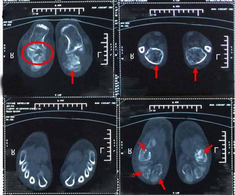
Citation: Wang H, He P, Zhang G, Gao J, Zhang L. Tuberculosis of bilateral ankles: Misdiagnosed as rheumatoid arthritis. J
Clin Images Med Case Rep. 2021; 2(3): 1192.
Antibody (ANA) posive 1:100S and The feet radiographs were mostly without bone destruction and anti-tuberculosis treat-
no obvious abnormity. And then he was diagnosed as RA, ment is effective [6]. Although the clinical manifestations of
treated with methylprednisolone (intravenous, 160mg twice a this patient are similar to that of this disease and effective in
day for 3 days), leflunomide (10 mg daily) and hydroxychloro- anti-tuberculosis treatment, the imaging findings of both feet
quine sulfate (200 mg twice daily). After 2 months of treatment, showed obvious bone destruction and tuberculous bacilli can
the swelling improved, but relapsed when drug withdrawal. In be found in joint fluid, which could be clearly diagnosed as bi-
October 2016, he was hospitalized again beacuse of his more lateral ankles tuberculosis.
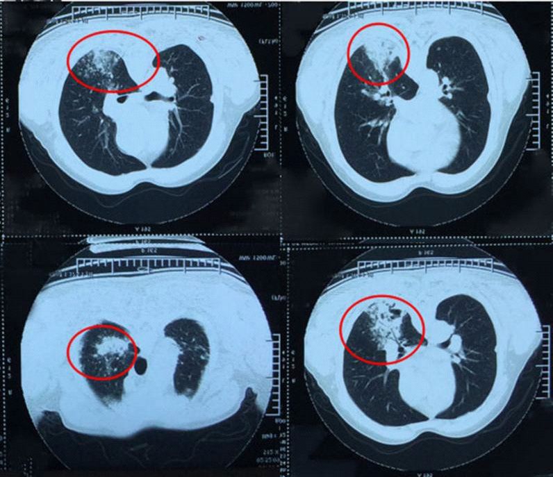
swollen feet and ankles and underwent chest CT, suggesting the
possibility of tuberculosis (Figure 1). Then he was suspected of
“Tuberculous Rheumatism” and transferred to a tuberculosis
hospital for treatment. Pertinent laboratory tests revealed the
Erythrocyte Sedimentation Rate (ESR) was 81 mm/hr, C-Reac-
tive Protein (CRP) was 42.51 mg/L and tuberculosis antibody
(IgG, 38 KDa) was positive. Empiric Anti-Tuberculous (anti-TB)
treatment was instituted, using a regimen of isoniazid, rifam-
picin, pyrazinamide for half a month. However his bilateral feet
and ankles got more swelling and difficult to walk, coming along
with fever. Consequently, he was admitted to our hospital. Phys-
ical examination was same as before. The leukocyte cout was
8.0×10⁹/L with a predominance of neutrophil (N 83.3%), ESR 50
mm/hr, CRP 46.79 mg /L and Rheumatoid Factor (RF) was nega-
tive. Immune indicator (IgG, IgA, IgM, C3, C4) were normal and
immune-related antibodies and tumor markers were negative.
Computed Tomography scan (CT) of the foot showed multi-focal Figure 1: CT image of chest: Showing multiple irregular lesions in
destruction, sclerosis and sequestrum formation of tarsal bone, both lungs, mostly in the right lung, of which upper middle lobe
the bony destruction in calcaneus, talus and navicular and os- can find inflammatory lesions and old TB of pulmonary apex.
teoporsis in the lower tibia (Figure 2). Ultrasound of the ankles
indicated multiple well-defined ellipse shadows, of which the
internal was poor-medium echo and hyper-echo on edge, sur-
rounded by the PD signal, existed on the bone surface. In addi-
tion, the bipedal Magnetic Resonance Imaging (MRI) revealed
joint effusion, multiple bony destruction, bone marrow edema
and subcutaneous soft tissue edema, with abnormal signals
around ankles (Figure 3). The acid-fast bacillus was postive in
the fifth examination of sputum smears. The ultrasound-guided
ankle arthroparacentesis was performed for three times and
mycobacterium tuberculosis was detected for 3.44×10³/L. The
tuberculosis of bilateral ankles was established and multidrug
anti-tuberculosis treatment was administered daily with iso-
niazid 300 mg, rifampin 450 mg, ethambutol 750 mg and pyra-
zinamide 500 mg. After two weeks, his swollen feet and ankles
were improved without pain and fever. Then he was discharged
and continued anti-tuberculosis treatment. At follow-up, the
patient’s general health condition is satisfactory. Figure 2: CT scan of foot: Showing multi-focal destruction, sclerosis
and sequestrum formation of tarsal bone, the bony destruction in
Nowadays, TB is still a serious threat to human health. Pul- calcaneus, talus and navicular and osteoporsis in the lower tibia.
monary TB is the most common pattern of manifestaion, but
Extrapulmonary TB (EPTB) contributes significantly to disability
and mortality [3]. Timely diagnosis and correct treatment are
critical to avoid functional disability. In this paper, The patient
failed to be present the classical systemic symptoms and un-
derwent two misdiagnosis successively. The patient character-
ized by swelling and pain of both feet, without typical systemic
symptoms and the ANA is postive, which is easily misdiagnosed
as RA. But he was failing to respond to the treatment using glu-
cocorticoid and immunosuppressive agents, thus excluding the
diagnosis of RA. And Jae Hyoung Im et al’s research shows that
there is a certain association between tuberculosis and ANA,
and most frequently in the patients with extrapulmonary tuber- Figure 3: MRI of the bipedal: Showing joint effusion, multiple bony
culosis [5]. Tuberculous rheumatism, also known as ‘Poncet’s destruction, bone marrow edema and subcutaneous soft tissue
disease’, is a sterile reactive arthritis caused by tuberculotoxin, edema, with abnormal signals around ankles.a T2; b T2 STIR; c T1.
www.jcimcr.org Page 2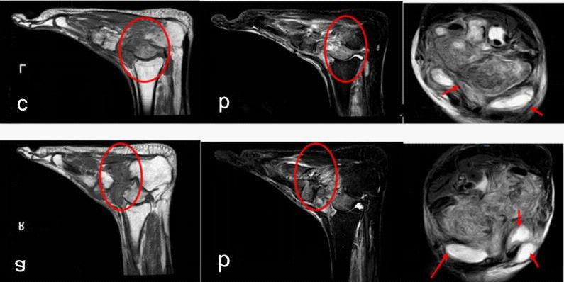
Conclusion                                                              References
   In the clinicial, the patient characterized by swelling and pain   1.     Dhillon MS, Aggarwal S, Prabhakar S, Bachhal V. Tuberculosis of
of both feet, without typical systemic symptoms and immune-                  the foot: An osteolytic variety. INDIAN J ORTHOP. 2012; 46: 206-
related antibodies should carry out imaging examination of pul-              211.
monary actively to determine whether there are tuberculose            2.     Faroug R, Psyllakis P, Gulati A, Makvana S, Pareek M, et al. Di-
focus in the lung and to be vigilant against the occurrence of os-           agnosis and treatment of tuberculosis of the foot and ankle-A
teoarthrosis in rare sites. Beause of paucibacillary character of            literature review. Foot (Edinb). 2018; 37: 105-112.
the disease, the risk of TB infection cannot be ruled out even if
                                                                      3.     Rando MM, De Matteis G, Gessi M, Bartoli M, Galli M, et al.
the acid-fast bacilli staining and culture are negative. We should
                                                                             Tuberculous Arthritis of the Ankle. Eur J Case Rep Intern Med.
promote acid-fast bacilli staining and culture repeatedly. Fur-
                                                                             2018; 5: 870.
thermore, we should pay close attention to the imaging process
of the lesions. If necessary, the diagnosis should be confirmed       4.     Grubisic F, Boric I, Segota A, Kruslin B, Grazio S. An unusual mani-
by surgical intervention.                                                    festation of osteoarticular tuberculosis: case report. ACTA CLIN
                                                                             CROAT. 2014; 53: 237-241.
   Declarations
                                                                      5.     Im JH, Chung MH, Park YK, Kwon HY, Baek JH, Lee SY, et al. An-
   Funding: Not applicable.                                                  tinuclear antibodies in infectious diseases. Infect Dis (Lond).
                                                                             2020; 52: 177-185.
  Conflicts of interest: The authors declare that they have no
competing interest.                                                   6.     Sait S, Mubashir M, Anwar R, Khan N. Poncet’s disease (tubercu-
                                                                             lar rheumatism) with primary involvement of the foot - A case
   Availability of data and material: The dataset supporting                 report. FOOT ANKLE SURG. 2016; 22: e17-20.
the conclusions of this manuscript is included with in the manu-
script.
   Authors' contributions: All authors participated in the com-
pleting the manuscript. All authors read and approval the final
manuscript.
   Acknowledgements: Not applicable.

www.jcimcr.org			                       									                                                                                        Page 3
You can also read