Limbic Encephalitis as a Presenting Complication for Small Cell Lung Cancer - Cureus
←
→
Page content transcription
If your browser does not render page correctly, please read the page content below
Open Access Case
                                          Report                                               DOI: 10.7759/cureus.9623

                                          Limbic Encephalitis as a Presenting
                                          Complication for Small Cell Lung Cancer
                                          Ahmad Abu-Hashyeh 1 , Abdulrahman Katabi 1 , Fuad Zeid 2

                                          1. Internal Medicine, Joan C. Edwards School of Medicine, Marshall University, Huntington, USA 2.
                                          Pulmonary Medicine, Joan C. Edwards School of Medicine, Marshall University, Huntington, USA

                                          Corresponding author: Abdulrahman Katabi, katabi@marshall.edu

                                          Abstract
                                          Limbic encephalitis (LE) is a rare neurological paraneoplastic complication that occurs
                                          secondary to malignant tumors. It is commonly presented as refractory seizures that are
                                          resistant to most anti-epileptics. We are presenting a unique case of small cell lung cancer
                                          complicated with LE. The challenging part of our case is that the patient had a history of
                                          seizure disorder in the past, and she was treated initially as an anti-epileptic treatment failure.
                                          A 68-year-old patient with a history of epilepsy was admitted to the ICU with resistant status
                                          epilepticus (SE), and respiratory failure secondary to pneumonia. Further workup revealed that
                                          the patient has small cell lung carcinoma. An extensive workup done to investigate resistant
                                          seizures revealed that she had a rare type of paraneoplastic autoantibodies (Anti-Hu) in the
                                          cerebrospinal fluid, which supported the diagnosis of the paraneoplastic autoimmune LE. High
                                          dose steroids helped to decrease the seizures episodes, but the family decided to proceed with
                                          palliative measures only at the end. Diagnosing LE requires ruling out other common causes of
                                          SE. Treatment options include treating underlying cancer as well as means of
                                          immunosuppression or antibody removal by tacrolimus and cyclophosphamide and even
                                          intravenous immunoglobulin (IVIG) or plasma exchange. It is important to consider LE in
                                          the differential diagnosis when managing patients with resistant SE in the ICU, even if the brain
                                          imaging and cerebrospinal fluid (CSF) analysis were within normal limits.

                                          Categories: Neurology, Oncology, Pulmonology
                                          Keywords: paraneoplastic neurological syndromes, small-cell lung carcinoma, status epilepticus,
                                          limbic encephalitis, autoimmune encephalitis

                                          Introduction
                                          Paraneoplastic limbic encephalitis (LE) is an autoimmune-mediated disease that happens in
                                          association with different cancers. It is a rare complication that occurs in 1 per 10,000
                                          patients [1]. Common cancers include lung cancer (50%), testicular cancer (20%), and breast
Received 06/04/2020
                                          cancer (8%) [2]. It is challenging to manage these cases, especially when the underlying cancer
Review began 07/14/2020
Review ended 07/26/2020                   remains occult at the time of presentation. The main signs and symptoms of LE are loss of
Published 08/09/2020                      short-term memory, confusion, seizures, or psychiatric symptoms.
© Copyright 2020
Abu-Hashyeh et al. This is an open
access article distributed under the
                                          Case Presentation
terms of the Creative Commons             A 68-year-old female patient with past medical history important for seizure disorder
Attribution License CC-BY 4.0., which     secondary to traumatic brain injury in the past, chronic obstructive pulmonary disease (COPD),
permits unrestricted use, distribution,
                                          cerebral aneurysm, type-2 diabetes mellitus, and hypertension, presented to the ER for acute
and reproduction in any medium,
provided the original author and          respiratory distress and several days’ history of progressive altered mental status, repetitive
source are credited.                      epileptic movements, and recurrent falls without significant head injuries. She was not on any
                                          seizure-predisposing medications at home. The patient was intubated in the ER emergently for

                                          How to cite this article
                                          Abu-Hashyeh A, Katabi A, Zeid F (August 09, 2020) Limbic Encephalitis as a Presenting Complication for
                                          Small Cell Lung Cancer. Cureus 12(8): e9623. DOI 10.7759/cureus.9623
progressive severe hypoxic respiratory failure and for controlling status epilepticus (SE). Initial
arterial blood gases on room air were pH 7.30, pCO2 56, and pO2 89. Other labs showed
leukocytosis white blood cell (WBCs) 18.6 k/cmm with neutrophils predominance, elevated
lactic acid at 4.3 mmol/L (normal range 0.7-2.1), hypokalemia at 2.5 mEq/L, and
hypomagnesemia at 1 mg/dL. Troponin-I trended from 516 to 1615 pg/mL. Initial
electrocardiogram (EKG) showed diffuse ST-segment depression (Figure 1). Chest X-ray showed
right lower lobe infiltrate. Head CT scan without contrast showed chronic small vessel ischemic
disease, without acute pathological changes (Figure 2). Brain MRI showed chronic small vessel
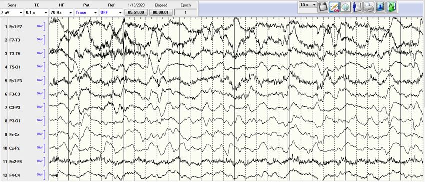
cerebrovascular disease as well. Electroencephalography (EEG) demonstrated left rhythmic
hemispheric spikes without evolving into seizures (Figures 3-4). Cardiac ECHO showed basal
inferior hypokinesis with normal ejection fraction. She was admitted to the ICU for sepsis and
acute respiratory failure secondary to pneumonia, SE, non-ST elevation myocardial infarction
(NSTEMI), and multiple electrolyte abnormalities. Initial management included IV fluids,
broad-spectrum antibiotics, ipratropium bromide/albuterol nebulizer, aspirin, atorvastatin,
metoprolol, heparin drip, and levetiracetam. Electrolytes replaced as well. Sodium valproate
was added later on due to repeated epileptic movements.
FIGURE 1: Initial EKG showing non-specific ST changes.
EKG, electrocardiogram
2020 Abu-Hashyeh et al. Cureus 12(8): e9623. DOI 10.7759/cureus.9623 2 of 6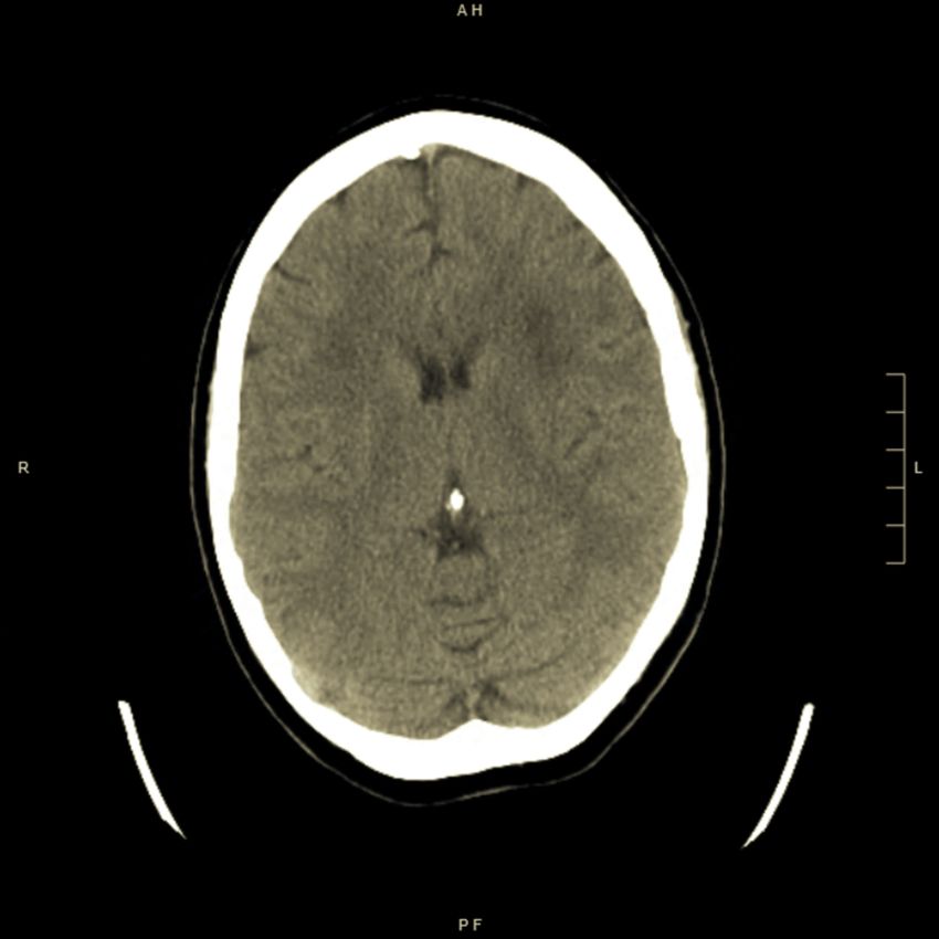
FIGURE 2: Head CT showing chronic ischemic changes.

                                     FIGURE 3: EEG showing rhythmic spikes.
                                     EEG, electroencephalography

2020 Abu-Hashyeh et al. Cureus 12(8): e9623. DOI 10.7759/cureus.9623                       3 of 6
FIGURE 4: EEG showing rhythmic spikes.
EEG, electroencephalography
The respiratory status initially improved, and the patient failed extubation trial and she
required reintubation for severe stridor. Video EEG showed a picture of diffuse cerebral
dysfunction with epileptogenicity in the temporal regions and interictal activity in the left
hemisphere. The patient was difficult to wean from the ventilator after the second intubation.
CT chest with IV contrast was done to investigate extubation failure and it ruled out pulmonary
embolism but it showed right paratracheal lymph node measuring 3.6 cm, subcarinal lymph
node measuring 5.9 cm, and left hilar lymph node measuring 2.8 cm highly suspicious for
malignancy. Endobronchial ultrasound (EBUS) was performed to biopsy the lymph nodes, and
it showed no endobronchial abnormalities. The lymph nodes biopsy pathology result was
positive for small cell carcinoma with positive for CD56 and synaptophysin, and negative for
CD20. Brain MRI showed no acute abnormalities. Lumbar puncture was performed,
cerebrospinal fluid (CSF) studies showed: opening pressure 10 cm H2O, clear CSF, Protein 44,
Glucose 110, negative meningitis panel, and negative for malignant cells. Paraneoplastic
evaluation of the CSF identified anti-neuronal nuclear antibody type-1 (Anti-Hu) 1:512
(referenceLack of these antibodies does not rule out the disease given that a minority of cases (10%-20%)
can have a seronegative disease, which appears to carry a slightly worse prognosis.
MRI can be helpful in diagnosis by showing amygdala-hippocampal signals especially in fluid-
attenuated inversion recovery (FLAIR), and some temporal atrophy may be seen later in the
course of the disease. These changes can be found in around 50%-60% of the cases and
sometimes MRIs need to be repeated as they may be normal in the early stages of the
disease [4].
Of note, the seizures accompanying LE can be refractory to a wide variety of anti-epileptic drugs
as in our case due to the origin of the seizures being in the medio-temporal lobe, which was the
trigger for the extensive evaluation that was pursued in our case after having resistant severe
seizures and an EEG concerning for temporal lobe seizures [5].
Paraneoplastic autoimmune encephalitis can be treated with an approach that focuses on
treating underlying cancer as well as means of immunosuppression or antibody removal.
Medications include steroids tacrolimus and cyclophosphamide and even intravenous
immunoglobulin (IVIG) or plasma exchange in some cases. The choice of therapy may be
influenced by the location of the antibody (intra-cellular vs extra-cellular) [6].
Conclusions
Paraneoplastic LE is a rare condition that presents with common symptoms or signs like
seizures or altered mental status. Although LE is a rare paraneoplastic complication, it is
important to consider it in the differential diagnosis list when managing patients with resistant
SE in the ICU even if the head imaging and CSF analysis were within normal limits.
Additional Information
Disclosures
Human subjects: Consent was obtained by all participants in this study. Conflicts of interest:
In compliance with the ICMJE uniform disclosure form, all authors declare the following:
Payment/services info: All authors have declared that no financial support was received from
any organization for the submitted work. Financial relationships: All authors have declared
that they have no financial relationships at present or within the previous three years with any
organizations that might have an interest in the submitted work. Other relationships: All
authors have declared that there are no other relationships or activities that could appear to
have influenced the submitted work.
References
1. Paraneoplastic Limbic Encephalitis. (2020). Accessed: 26 July 2020:
https://europepmc.org/books/n/statpearls/article-26632/?extid=29083631&src=med.
2. Gultekin SH, Rosenfeld MR, Voltz R, et al.: Paraneoplastic limbic encephalitis: neurological
symptoms, immunological findings and tumour association in 50 patients. Brain. 2000,
123:1481-1494. 10.1093/brain/123.7.1481
3. Pastuszak Ż, Stępień A, Tomczykiewicz K, et al.: Limbic encephalitis - a report of four cases .
Cent Eur J Immunol. 2017, 42:213-217.
4. Kacem M, Belloumi N, Bachouche I, et al.: Paraneoplastic limbic encephalitis revealing a small
cell carcinoma of the lung. Respir Med Case Rep. 2018, 26:157-160.
5. Munawar M, Iftikhar PM, Khan JA, et al.: A case report of seronegative limbic encephalitis .
Cureus. 2019, 11:e4681.
6. Nagafuji H, Yokoi H, Fujiwara M, Sato D, Saito K: Paraneoplastic limbic encephalitis
associated with mixed olfactory neuroblastoma and craniopharyngioma: a case report and
2020 Abu-Hashyeh et al. Cureus 12(8): e9623. DOI 10.7759/cureus.9623 5 of 6literature review. Medicine. 2018, 97:e10932. 2020 Abu-Hashyeh et al. Cureus 12(8): e9623. DOI 10.7759/cureus.9623 6 of 6
You can also read