PANCYTOPENIA SECONDARY TO SARS-COV 2 INFECTION-A CASE REPRT
←
→
Page content transcription
If your browser does not render page correctly, please read the page content below
Pancytopenia Secondary to SARS-CoV 2 Infection-A
Case Reprt
Neeraj Sharma ( dmt18sharma@gmail.com )
Military Hospital Namkum https://orcid.org/0000-0002-6676-6100
Rajat Shukla
Military Hospital Namkum
Rachna Warrier
Military Hospital Namkum
Kunal Kumar
Military Hospital Namkum
Nalin Singh
Military Hospital Namkum
Sourav Ghose
MH Namkum
Vivek Kumar
Military Hospital Namkum
Research Article
Keywords: Pancytopenia, SARS CoV-2 infection, COVID 19 disease
DOI: https://doi.org/10.21203/rs.3.rs-657352/v1
License: This work is licensed under a Creative Commons Attribution 4.0 International License.
Read Full License
Page 1/10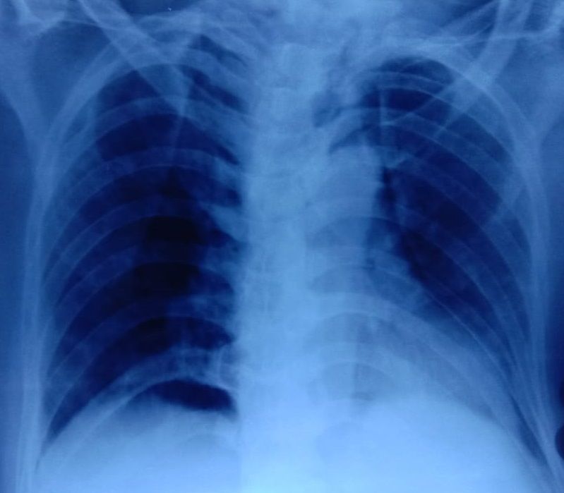
Abstract
Pancytopenia is a condition when person has low count of all three types of blood cells causing a triage
of anemia, leukopenia and thrombocytopenia. It should not be considered as a disease in itself but rather
the sign of a disease that needs to be further evaluated. Among the various causes, viral infections like
Human Immunodeficiency Virus, Cytomegalovirus, Epstein-Barr virus and Parvovirus B19 have been
implicated. Pancytopenia is a rare complication and not commonly seen in patients with COVID 19
disease. Here, we report a case of pancytopenia in previously immunocompetent elderly male patient with
SARS-CoV2 infection.

Introduction
The most common haematological and immunological findings of coronavirus disease 2019 (COVID-19)
caused by Severe Acute Respiratory Syndrome Coronavirus 2 (SARS-CoV2) are elevated inflammatory
markers, hypercoagulable state and lymphopenia. Pancytopenia is a rare complication and not
commonly seen in immunocompetent patients with COVID 19 disease. The common causes of
pancytopenia in clinical practice are megaloblastic anemia, hypersplenism, drug induced bone marrow
toxicity, leukaemia, radiation therapy, chemotherapy, immunosuppressive medications, connective tissue
diseases and infections1. Pancytopenia as a result of direct bone marrow suppression has been
previously reported in viral infections like Human Immunodeficiency Virus, Cytomegalovirus, Epstein-Barr
virus and Parvovirus B19. Here, we report a case of pancytopenia in previously immunocompetent elderly
male patient with SARS-CoV2 infection.

Case Presentation
A 69-year‐old male presented to the hospital emergency department with complaints of fever, dry cough,
fatigue and progressive breathlessness for 15 days. He gave history of hypertension but was not on any
regular treatment and follow up. On examination he had high blood pressure recording (150/90mm Hg
right arm supine) with tachycardia, tachypnoea and was not maintaining oxygen saturation at room air
(SpO2 80% on room air). He was febrile (101-degree fahrenheit) and had pallor. Initial laboratory workup
on admission showed pancytopenia and deranged serum creatinine of 2.4 mg/dl (findings are
summarized in Table 1). It showed total white blood cell count (WBC) 2,100/µL, haemoglobin 2.2gm/dl,
platelet count 65,000/µL, urea 46mg/dl, creatinine 2.4mg/dl, total bilirubin 0.8mg/dl, lactate
dehydrogenases (LDH) 353IU/L, aspartate aminotransferase (AST) 32U/L and alanine aminotransferase
(ALT) 84U/L. His nasopharyngeal swab test for COVID-19 RT-PCR was positive. The chest radiograph
showed non homogenous opacities involving left mid and lower zones (Fig. 1). He was diagnosed as a
case of severe COVID 19 disease with pancytopenia and was transferred to COVID intensive care unit. He
was managed with oxygen therapy, parenteral broad-spectrum antibiotics, packed red blood cell
transfusion, injection dexamethasone (6mg IV once a day), antihypertensives, anticoagulant therapy
(renal modified dose of injection low molecular weight heparin) and other supportive measures. His
inflammatory markers were raised (C‐reaction protein 20.5mg/L, D‐dimer 0.80µg/L, ferritin 900 ng/mL,
                                                Page 2/10
lactate dehydrogenase 353 IU/L). His iron studies, serum folate and vitamin B12 were within normal
range. His coagulation profile was normal. He was evaluated for the possible causes of pancytopenia
which included leukaemia, myelodysplastic syndrome, malignancy, bone marrow failure, infections, drug
induced bone marrow toxicity, connective tissue diseases and immunosuppressive medications. His
serum antinuclear antibody test was negative. Ultrasound of abdomen showed features of bilateral
medical renal disease with no organomegaly. Peripheral blood microscopic examination showed
pancytopenia and normocytic normochromic anaemia with moderate anisocytosis. No evidence of
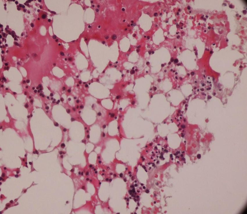
haemolysis, sepsis or atypia seen (Fig. 2). Bone marrow aspiration and biopsy was done to determine the
cause of pancytopenia which showed hypocellular marrow with cellularity less than 20% and increase in
fat spaces (Fig. 3). There were focal areas of marrow elements showing trilineage haematopoiesis with
markedly diminished myeloid, erythroid and megakaryocytic series. There was no evidence of any
leukaemia, myelodysplastic syndrome (MDS), infections or metastatic deposits. Peripheral blood smear
and bone marrow microscopic examination was suggestive of pancytopenia with hypocellular bone
marrow (Figs. 2 and 3). During the hospital course, three units of packed red blood cells were transfused
to the patient. The patient's clinical condition and haematology parameters improved with therapy
(Fig. 4). He was discharged on Day 15 with WBC 7,200/ µL, haemoglobin 7gm/dl and platelet count of
1,42,000/ µL. His throat swab for COVID 19 PCR was negative at the time of discharge.
Discussion
The novel SARS-CoV 2 virus is the etiological agent for COVID 19 disease and the respiratory system is
involved in majority of patients. The clinical presentation of the disease is variable, including
asymptomatic infection, mild upper respiratory infection and severe pneumonia with respiratory failure2.
The involvement of haematological system in COVID-19 disease has been reported in an increasing
number of patients. The most common and well reported haematological complications in patients of
severe COVID-19 disease are hyperinflammatory state, coagulopathic complications and lymphopenia3.
Though platelets and haemoglobin levels are normal infrequently thrombocytopenia is also observed4.
Pancytopenia is rarely reported in immunocompetent patients with severe COVID 19 disease.
Pancytopenia is a condition when person has low count of all three types of blood cells causing a triage
of anemia, leukopenia and thrombocytopenia. It should not be considered as a disease in itself but rather
the sign of a disease that needs to be further evaluated. The possible causes are nutritional deficiencies,
megaloblastic anemia, hypersplenism, malignancies, radiation therapy, chemotherapy drug induced bone
marrow toxicity, connective tissue diseases and immunosuppressive medications1. All these conditions
were ruled out in our patient during hospital stay. Pancytopenia as a result of bone marrow suppression
has been reported in viral infections and commonly implicated viruses are Human Immunodeficiency
Virus, Parvovirus B19 Epstein-Barr virus and Cytomegalovirus5. The decreased myeloid, erythroid and
megakaryocytic series was observed in bone marrow biopsy of our patient indicating bone marrow
suppression. There was no evidence of lymphoma, fibrosis and myelodysplasia in bone marrow biopsy.
There are very few case reports on SARS-CoV2 induced pancytopenia6. Issa N et al reported the first case
Page 3/10of persistent pancytopenia associated with SARS-CoV2 bone marrow infiltration in an
immunocompromised patient7. However, our patient was immunocompetent. He was found to have
chronic kidney disease during evaluation which alone could not explain the severe anemia and
associated pancytopenia. Ufuk F et al reported a case of COVID-19 associated pancytopenia which was
complicated by neutropenic enterocolitis8. Our patient responded well to the supportive care and there
were no complications during hospital stay. Once the patient’s infection resolved, his blood counts
improved and at the time of hospital discharge his leucocyte and platelet count had normalized and only
anemia remained. Hersby DS et al also reported a similar self-limiting clinical course of patient with
COVID 19 induced pancytopenia9.
The possible pathophysiology of pancytopenia secondary to SARS CoV2 infection could be linked to the
angiotensin converting enzyme 2 receptor(ACE 2 receptor), which is present in bone marrow in lower
levels 10 .It is possible that direct infection of myelocytes by SARS CoV-2 virus could lead to bone marrow
suppression as seen in other viral infections like HIV,Parvovirus B19,Epstein-Barr virus and
Cytomegalovirus. Other possibility is that after viral infection an antigenic epitope on myelocytes could
be exposed which can lead to the production of autoantibody and destruction of blood cells. Also,
hyperinflammatory state is a key feature of severe COVID-19 disease and It is well known that certain
cytokines, such as the interferons and tumor‐necrosis factor‐α can affect hematopoietic stem cells and
thus impair hematopoiesis11. Lung is a site for platelet biogenesis and a reservoir for hematopoietic
progenitors and with SARS‐CoV 2 infection leading to lung injury, it is possible that the destruction of
lung hematopoietic progenitors could also contribute to the pancytopenia 12.
Conclusion
SARS CoV 2 infection leading to pancytopenia is rare. We reported a case of pancytopenia associated
with COVID-19 disease likely caused by bone marrow suppression.
Page 4/10Table 01
Test Name Result
Haemoglobin 2.2 g/dl
Total leucocyte count 2,100/µL
Differential leucocyte count N-68%,L-29%,M-2%,E-1%
Platelet count 65,000/µL
INR 1.2
Urea 46 mg/dL
Creatinine 2.4 mg/dL
Uric Acid 8.7 mg/dL
Albumin 2.1 gm/dI
Bilirubin Total 0.3 mg/dL
Protiens 6.0 gm/dL
SGOT/AST 32 U/L
SGPT/ALT 84 U/L
Albumin 2.6 g/dL
Globulin 3.4 g/dL
Sodium 134 meq/L
Potassium 5.1 meq/L
LDH 353 U/L
D-dimer 0.8
Ferritin 900 ng/ml
CRP 20.3mg/dL
Procalcitonin < 0.5ng/ml
Declarations
Funding-Not applicable.
Conflicts of interest - The authors declare no competing interests.
Ethics approval- Not applicable
Page 5/10Consent to participate-Not applicable
Consent for publication-Taken from patient
Availability of data and material-Not applicable
Code availability-Not applicable
Authors contribution- All the authors have been involved in the review of the case report.
References
1. Jain A, Naniwadekar M. An etiological reappraisal of pancytopenia - largest series reported to date
from a single tertiary care teaching hospital. BMC Hematol. 2013;13(1):10.
https://doi.org/10.1186/2052-1839-13-10.
2. Huang C, Wang Y, Li X, Ren L, Zhao J, Hu Y, et al. Clinical features of patients infected with 2019
novel coronavirus in Wuhan, China. Lancet. 2020;395:497–506.
3. Al-Samkari H, Karp Leaf RS, DzikWH, et al. COVID-19 and coagulation: bleeding and thrombotic
manifestations of SARS-CoV-2 infection. Blood. 2020;136(4):489–500.
https://doi.org/10.1182/BLOOD.2020006520.
4. Huang I, Pranata R. Lymphopenia in severe coronavirus disease- 2019 (COVID‐19): systematic review
and meta‐analysis. J Intensive Care. 2020;8:36.
5. Pascutti MF, Erkelens MN, Nolte MA. Impact of viral infections on hematopoiesis: from beneficial to
detrimental effects on bone marrow output. Front Immunol. 2016;7:364.
6. Hersby DS, Do TH, Gang AO, Nielsen TH. COVID-19-associated pancytopenia can be self-limiting and
does not necessarily warrant bonemarrow biopsy for the purposes of SARS-CoV-2 diagnostics. Ann
Oncol. 2021;32(1):121–3. https://doi.org/10.1016/j.annonc.2020.09.020.
7. Issa N, Lacassin F, Camou F. First case of persistent pancytopenia associated with SARS-CoV-2 bone
marrow infiltration in an immunocompromised patient. Ann Oncol. 2020;31(10):1418–9.
https://doi.org/10.1016/j.annonc.2020.06.016.
8. Ufuk F, Bulgurcu E, Sari T. COVID-19-associated pancytopenia and typhlitis. Am J Emerg Med. 2021.
https://doi.org/10.1016/j.ajem.2020.12.049.
9. Hersby DS, Do TH, Gang AO, Nielsen TH. COVID-19-associated pancytopenia can be self-limiting and
does not necessarily warrant bonemarrow biopsy for the purposes of SARS-CoV-2 diagnostics. Ann
Oncol. 2021;32(1):121–3. https://doi.org/10.1016/j. annonc.2020.09.020.
10. LiMY, Li L, Zhang Y, Wang XS. Expression of the SARS-CoV-2 cell receptor gene ACE2 in a wide
variety of human tissues. Infect Dis Pov. 2020;9(1). https://doi.org/10.1186/s40249-020-00662-x.
11. Clapes T, Lefkopoulos S, Trompouki E. Stress and non-stress roles of inflammatory signals during
HSC emergence and maintenance. Front Immunol. 2016;7:487.
Page 6/1012. Lefrançais E, Ortiz-Muñoz G, Caudrillier A, et al. The lung is a site of platelet biogenesis and a
reservoir for haematopoietic progenitors. Nature. 2017;544(7648):105‐109.
Figures
Figure 1
It showed total white blood cell count (WBC) 2,100/μL, haemoglobin 2.2gm/dl, platelet count 65,000/μL,
urea 46mg/dl, creatinine 2.4mg/dl, total bilirubin 0.8mg/dl, lactate dehydrogenases (LDH) 353IU/L,
aspartate aminotransferase (AST) 32U/L and alanine aminotransferase (ALT) 84U/L. His
nasopharyngeal swab test for COVID-19 RT-PCR was positive. The chest radiograph showed non
homogenous opacities involving left mid and lower zones
Page 7/10Figure 2
His serum antinuclear antibody test was negative. Ultrasound of abdomen showed features of bilateral
medical renal disease with no organomegaly. Peripheral blood microscopic examination showed
pancytopenia and normocytic normochromic anaemia with moderate anisocytosis. No evidence of
haemolysis, sepsis or atypia seen
Page 8/10Figure 3
Bone marrow aspiration and biopsy was done to determine the cause of pancytopenia which showed
hypocellular marrow with cellularity less than 20% and increase in fat spaces
Page 9/10Figure 4
During the hospital course, three units of packed red blood cells were transfused to the patient. The
patient's clinical condition and haematology parameters improved with therapy
Page 10/10You can also read