Knee Angle Affects Posterior Chain Muscle Activation During an Isometric Test Used in Soccer Players - MDPI
←
→
Page content transcription
If your browser does not render page correctly, please read the page content below
Article

Knee Angle Affects Posterior Chain Muscle
Activation During an Isometric Test Used in
Soccer Players
Paul James Read 1,*, Anthony Nicholas Turner 2, Richard Clarke 3, Samuel Applebee 3
and Jonathan Hughes 3
 1 Athlete Health and Performance Research Centre, Aspetar Orthopaedic and Sports Medicine Hospital,
   Doha PO Box 29222, Qatar
 2 London Sports Institute, Middlesex University, London, NW4 4BT, UK; a.n.turner@mdx.ac.uk

 3 School of Sport and Exercise, University of Gloucestershire, Gloucester, GL2 9HW, UK;

   rclarke@glos.ac.uk (R.C.); samuel.applebee@gmail.com (S.A.); jhughes1@glos.ac.uk (J.H.)
 * Correspondence: Paul.Read@aspetar.com

 Received: 8 November 2018; Accepted: 29 December 2018; Published: 4 January 2019

 Abstract: Background: It has been suggested that altering the knee flexion angle during a commonly
 used supine isometric strength test developed with professional soccer players changes preferential
 hamstring muscle recruitment. The aim of this study was to examine the electromyography (EMG)
 knee joint-angle relationship during this test, as these data are currently unknown. Methods: Ten
 recreational male soccer athletes (age: 28 ± 2.4 years) were recruited and performed a supine
 isometric strength test on their dominant leg with the knee placed at two pre-selected flexion angles
 (30° and 90°). The surface EMG of the gluteus maximus, biceps femoris, semitendinosus, and medial
 gastrocnemius was measured, in addition to the within-session reliability (intraclass correlation
 coefficient (ICC) and coefficient of variation (CV)). Results: Within-session reliability showed large
 variation dependent upon the test position and muscle measured (CV% = 8.8–36.1) Absolute mean
 EMG activity and percentage of maximum voluntary isometric contraction (MVIC) indicated
 different magnitudes of activation between the two test positions; however, significant mean
 differences were present for the biceps femoris only with greater activation recorded at the 30° knee
 angle (% MVIC: 31 ± 9 vs. 22 ± 7; p = 0.002). These differences (30% mean difference) were greater
 than the observed typical measurement error (CV% = 13.1–14.3 for the 90° and 30° test positions,
 respectively). Furthermore, the percentage MVIC showed a trend of heightened activation of all
 muscles with the knee positioned at 30°, but there was also more within-subject variation, and this
 was more pronounced for the gluteus maximus (CV% = 36.1 vs. 19.8) and medial gastrocnemius
 (CV% 31 vs. 22.6). Conclusions: These results indicate that biceps femoris and overall posterior
 chain muscle activation is increased with the knee positioned at 30° of flexion; however, the 90°
 angle displayed less variation in performance within individual participants, especially in the
 gluteus maximus and medial gastrocnemius. Thus, practitioners using this test to assess hamstring
 muscle strength should ensure appropriate familiarisation is afforded, and then may wish to
 prioritise the 30° knee position.

 Keywords: hamstring; strength test; muscle activation

1. Introduction
     Participation in competitive soccer training and match-play places significant physical and
physiological demands on athletes [1], resulting in an inherent risk of injury. Hamstring strains are
the most common injury in elite adult soccer [2,3] and also frequently occur in elite male youth soccer

Sports 2019, 7, 13; doi:10.3390/sports7010013                                 www.mdpi.com/journal/sports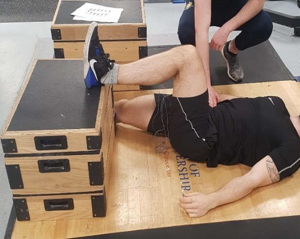
Sports 2019, 7, 13                                                                                   2 of 8

players [4,5]. The knee flexors are also key antagonist muscles that contribute to knee joint
stabilisation, reducing the magnitude of anterior shear force during high velocity actions [6]. Previous
data indicate that hamstring strength [7] and between-limb imbalances are risk factors for injury [8].
Strength assessment is thus warranted in order to identify soccer players who may be at a heightened
injury risk, allowing more individually targeted training interventions.
      Assessment of hamstring strength has traditionally been conducted using isokinetic
dynamometry [9,10,11]. These tests have limited utility as a screening tool to identify players who
subsequently sustain a hamstring injury [12] and are not practically viable for screening large
numbers of athletes because of time inefficiency and the need for expensive laboratory equipment.
An alternative approach is to examine hamstring strength using isometric protocols that involve
minimal structural muscle damage [13] and can thus be used regularly in-season. Additionally, rate
of force production can more easily be quantified [14], which has connotations for soccer players
where constraints exist for the time availability of force production.
      A simple and practically viable test has recently been developed in professional soccer players
to measure isometric hamstring strength and asymmetry using force plate diagnostics [14]. This
assessment is reliable (intraclass correlation coefficient (ICC) = 0.93–0.95; coefficient of variation (CV)
= 4.3%–6.3%) and has shown sensitivity to identify changes in performance following soccer match
play [14]. Field-based measurements that examine hamstring strength and monitor both acute and
chronic changes following soccer training and competitions can be readily used in applied settings.
However, before this approach can be recommended to coaches, it is necessary to examine the level
of muscle activation during the execution of the task. This information is required to ensure time
efficiency in testing and that the correct muscles are targeted, which has implications for injury risk
screening, longitudinal monitoring, and training prescription [15].
      The research by McCall et al. [14] included two different test positions; specifically, lying supine,
with the foot placed on top of a box and the knee positioned at both 30° and 90° of flexion. In the
development of this assessment, it was suggested that altering the knee position changes the
recruitment of the hamstring muscles [14] based on previous data [16]. The lateral and medial
hamstrings have been shown to be maximally activated at 15° to 30° and 90° to 105° of knee flexion
respectively [16]; however, these values were obtained during an open chain isokinetic test. Onishi
et al. [16] also included isometric tests that showed greater activation of the biceps femoris at lower
knee flexion angles, but it should be acknowledged that this test was performed in a prone position
with the knee placed at both 60° and 90° of flexion. In addition, several other muscles will contribute
to the development of peak force force during this test, such as the gluteals and plantar flexors; their
level of activation is currently unknown.
      The aim of this study was to examine the level of muscle activation in the biceps femoris, medial
hamstrings, gluteus maximus, and gastrocnemius during a previously used isometric hamstring
strength test [14] with the knee at two designated positions, as previously proposed (either 30° and
90° of flexion). It was hypothesised that hamstring recruitment and, in particular, biceps femoris
activation would be greater at the shallower knee angle (30°) as a result of magnified muscle activity
when the bi-articular hamstrings are at peak stretch [17,18].

2. Materials and Methods

2.1. Participants
     Ten recreational male soccer players (age: 28 ± 2.4 year; height: 177 ± 25 cm; body mass: 80.5 ±
6.4 kg) volunteered for this study. Inclusion criteria were as follows: (1) to have undertaken a
minimum of six months of resistance training encompassing lower limb and posterior chain exercises;
(2) participating regularly in soccer training and competitions; (3) free from injury within the last
three months; and (4) no previous history of hamstring strain injury. Physical activity readiness
questionnaires were collected prior to the commencement of testing. Ethical approval (JHUGHES17-
18) was granted by the institutional ethics committee in accordance with the declaration of Helsinki.
Sports 2019, 7, 13 3 of 8
Participants provided informed consent and were reserved the right to withdraw from the study at
any time without consequence.
2.2. Experimental Design
A cross-sectional design was used to examine muscle activation during a recently developed
field-based isometric hamstring strength test. Participants were required to attend the laboratory on
two separate occasions separated by a period of seven days. The first session was used to familiarise
participants with the assessment protocols and equipment, and the second session was used for data
collection. Standardised procedures were replicated at each test session including the warm up, test
set-up, and participant instructions. Participants were asked to refrain from strenuous exercise for at
least 48 h prior to testing and to eat according to their normal diet, and drink only water one hour
prior to each test session.
2.3. Procedures
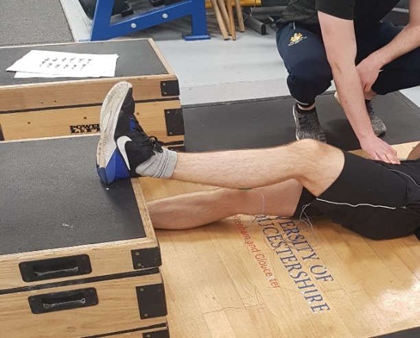
The test was performed on the dominant leg only with the knee placed in flexion at two pre-
selected angles (30° and 90°; Figure 1a,b where the 30° position corresponds to an internal joint angle
of 150°) using a manual goniometer, as previously suggested [14]. Participants were positioned
supine and asked to lie flat on an exercise mat. The heel of the dominant limb was then placed on top
of a 30 cm box, while the non-working limb lay flat on the floor facing forwards. Standardised
encouragement was provided, with instructions to push their heel as hard as possible into the box
and hold the contraction for three seconds while simultaneously keeping their head, upper body,
hands, and buttocks flat on the floor [14]. To restrict vertical elevation of the contralateral hip, a
member of the research team provided manual resistance to the participant throughout the test,
ensuring maintenance of the correct position [14]. The order of testing for each respective knee angle
was counterbalanced to avoid an order effect. Three repetitions were performed on the dominant leg
only with two minutes of recovery between trials based on previous recommendations [14].
(a)Sports 2019, 7, 13 4 of 8
(b)
Figure 1. Test position of the isometric posterior chain lower limb muscle test measured at 90° (a) and
30° (b) of knee flexion.
2.4. Surface Electromyography (sEMG)
The subjects’ body hair was shaved at the site of electrode placement, and the skin was cleaned
with an alcohol wipe and lightly abrased to remove dead skin cells before affixing the surface EMG
electrode. Bipolar active disposable dual Ag/AgCl snap electrodes spanning 1 cm in diameter for each
circular conductive area with 2 cm center-to-center spacing were used for all trials. Electrodes were
placed on the dominant limb along the axes of the muscle fibers for the gluteus maximus (GM),
medial hamstrings (MH), biceps femoris (BF), and medial gastrocnemius (MG) according to the
Surface Electromyography for the Non-Invasive Assessment of Muscles (SENIAM) protocol [19]. A
ground electrode was placed on the right medial malleolus. The sEMG signals were recorded by an
EMG acquisition system (Biometrics MWX8 DataLog; Biometrics Ltd., Newport, UK) with a sampling
rate of 1000 Hz using a commercially designed software program (Biometrics DATALOG Software
v8.51: Biometrics Ltd., Newport, UK). EMG activity was amplified (bipolar differential amplifier,
input impedance = 2 MΩ, common mode rejection ratio 100 dB minute [60 Hz], gain x 20, noise > 5
µV) and converted from an analog to digital signal (12 bit). The digitised surface EMG data were
band-pass filtered at 20–400 Hz using a fourth-order Butterworth filter with a zero lag. For muscle
activation time domain analysis, root mean square (RMS) (30 ms moving window) was calculated
during the maximum voluntary isometric contraction (MVIC). The sEMG data were then normalised
to the RMS average of the peak MVICs for each amplitude and muscle. The RMS analysis was defined
from the average of the first three repetitions for each condition and muscle.
EMG signals collected during all conditions were normalised to a maximum voluntary isometric
contraction (MVIC) against a fixed resistance. Two trials of 5 s MVICs were performed for each
muscle with a 1 min rest interval between actions for the dominant leg. The first MVIC was performed
to familiarise the participant with the procedure. For GM, BF, and MH MVIC, subjects were in the
prone position with their knee fully extended and locked into a leg curl bench (Power Lift, Jefferson,
IA, USA) with the restraint placed on the distal region of the Achilles tendon as the subject attempted
to maximally and simultaneously extend the hip and flex the knee. For MG MVICs, subjects stood
with their knee fully extended and with vertical resistance through the shoulders via fixed squat rack
while they attempted to maximally plantar flex. Standardised, verbal encouragement was given
during all MVICs. The order of MVICs was counterbalanced to reduce the potential for
neuromuscular fatigue.
2.5. Statistical Analysis
Descriptive statistics (mean ± SD) for both mean and peak EMG were calculated for each test
position. A Shapiro–Wilks test was used to check the normality distribution of the data. To examine
potential differences in the level of muscle activation between test positions, a two-way repeated
measure analysis of variance (ANOVA) with two factors (knee angle (two levels); and muscle (four
levels)) was utilised with statistical significance set at p < 0.05. Cohen’s d effect sizes were also
calculated to interpret the magnitude of change in the RMS EMG signal for each muscle measured
between test positions using the following classifications: standardised mean differences of 0.2, 0.5,
and 0.8 for small, medium, and large effect sizes, respectively [20]. Within-session reliability was
assessed for each muscle measured in both the 30° and 90° test positions via intraclass correlation
coefficient (ICC) using a two-way random model with absolute agreement and the coefficient of
variation (CV = (SD/mean × 100). The latter analysis was included to allow further interpretation of
the magnitude of change in muscle activation between test positions.
3. ResultsSports 2019, 7, 13 5 of 8
Within-session reliability statistics are shown in Table 1. Absolute mean EMG activity indicated
different magnitudes of hamstring, gluteal, and plantar flexor activation between the two test
positions; however, significant mean differences were present only for the biceps femoris, where
greater absolute values were shown at the 30° knee angle F(1–9) = 19.207 (p = 0.002; d = 1.19). EMG
percentage MVIC and percentage differences between the two knee positions (Table 2) showed a
trend of greater muscle activation with the knee positioned at 30°. Significant differences were shown
between test positions for the biceps femoris only corresponding to a very large effect size (p < 0.05;
d = 1.19). Medial hamstring, gastrocnemius, and gluteus maximus activation was higher with the
knee positioned at 30°, but these differences were not meaningful and effect sizes were small (d =
0.07–0.40).
Table 1. Reliability statistics for each muscle measured in both test positions. ICC—intraclass
correlation coefficient; CV—coefficient of variation.
30° Knee Angle 90° Knee Angle
ICC CV% ICC CV%
Medial gastrocnemius 0.93 31% 0.97 22.6%
Medial hamstrings 0.96 13.1% 0.88 8.8%
Biceps femoris 0.95 14.3% 0.95 13.1%
Gluteus maximum 0.96 36.1% 0.99 19.8%
Table 2. Percentage maximum voluntary isometric contraction (MVIC) for each muscle at the two
different knee flexion angles.
Knee Flexion Angle (Degrees)
% MVIC 30° 90° % Difference Effect Size d
Medial gastrocnemius 15 ± 11 14 ± 10 5 0.07
Medial hamstrings 34 ± 14 30 ± 12 15 0.40
Biceps femoris 31 ± 9 22 ± 7 30 1.19
Gluteus maximus 27 ± 10 24 ± 11 7 0.18
4. Discussion
The aim of this study was to examine the level of posterior chain muscle activation during a
hamstring strength test developed for soccer players. It has previously been suggested that a change
in knee angle is required to preferentially alter muscle recruitment; specifically, activation of the
medial hamstrings and biceps femoris [14]. The results showed that while changes in muscle
activation of the medial hamstrings and biceps femoris were present in the two test positions (30°
and 90° of knee flexion) with heightened activation observed with the knee positioned at 30°,
significant differences were observed for the biceps femoris only, which confirms the test hypothesis.
Greater biceps femoris, medial hamstring, and gluteal activation were seen in the current study
with the knee positioned at 30° of flexion. This is likely the result of magnified muscle activation
when the hamstrings are in an elongated position [17,18] and increases in the distal moment arm [21].
Also, peak torque of the knee flexors has been shown to occur with knee angles between 21–49° of
knee flexion, with reductions in torque concomitant with greater flexion angles [16]. Thus, greater
recruitment of key posterior chain musculature can be expected with the knee positioned at 30° of
flexion during this supine isometric closed chain strength assessment.
It has previously been suggested [14] that biceps femoris and medial hamstring activation peaks
between 15–30° and 90–105° of knee flexion, respectively; however, these results were obtained
during an open chain, isokinetic test [16]. Previous research has shown differences in the magnitude
of muscle activity and preferential muscle recruitment between open and closed chain exercises [22].
The original recommendations in the development of the novel isometric posterior chain lower limb
muscle test examined in the current study were to alter the knee flexion position to preferentially
recruit either the biceps femoris (30°) or medial hamstrings (90°) [14]. The results of the current studySports 2019, 7, 13 6 of 8
do not support this notion as greater recruitment of both the biceps femoris and medial hamstrings
were observed with the knee positioned at 30° of flexion. Previous literature has shown no changes
in EMG activity with different flexion angles, albeit at the elbow joint [23]. Conversely, Lunnen et al.
[21] observed changes in biceps femoris activation by altering the angle of hip flexion, which was
consistent with the current study; however, it should be noted that the test set up used by Lunnen et
al. [21] was different to the present study because in their work, the knee position was maintained at
60°. The assessment proposed by McCall et al. [14] is performed in supine and requires a closed
kinetic chain test position, allowing greater hip extension force production in comparison with open
chain tasks and higher biceps femoris long head activation [24].
When interpreting the results of this study, it should be considered that the ICC values were
excellent, indicating good between-participant variance [25] and maintenance of rank order between
trials. However, greater variations in muscle activation were present, as indicated by large standard
deviations for the medial hamstrings, gastrocnemius, and gluteus maximum. In addition, the CV%
shown in the medial gastrocnemius and gluteus maximus would be considered as being large [26],
with lower values seen in the medial hamstrings and biceps femoris (Table 1). These data indicate
that pronounced variation in performance was shown within individual participants. This was
consistent across both test positions and may be in part because of the participants’ interpretation of
the task. In spite of this, the CV values reported were lower than the percentage differences between
test positions for the medial hamstrings and biceps femoris (Table 2), suggesting that the observed
differences were ‘real’ and indicative of a meaningful change.
The observed within-subject variability in the supine test position could be in part because of
the instructions to ‘push’ with their heel into the box [14], which may create confusion in how the
participant should produce their maximum force. The term ‘push’ may be associated with an
attentional focus of moving an object away from their body, encouraging a hip extension dominant
production of force in some participants. This interpretation is supported by the need of the
researcher to anchor the hips of the participant to the floor [14]. Conversely, some participants may
have produced force by driving the heel into the box and towards their hip via a greater contribution
of knee flexion. Cumulatively, this may have contributed to the greater variation in either gluteal and
gastrocnemius contributions during both test positions. It should, therefore, be considered that the
instruction to ‘push’ may not be optimal in order to standardise muscle recruitment strategies, and
alternate cues warrant further investigation. The level of medial gastrocnemius activation could also
be affected by the position of the ankle where the athlete may have plantar flexed or dorsiflexed to a
greater amount. Thus, practitioners are advised to provide adequate standardisation and
familiarisation prior to testing and ensure that participants are clear on the task requirements to
obtain reliable measures that can be used for test/re-test comparison following targeted training
interventions.
5. Conclusions
The current study indicates that altering the knee position to 90° of flexion does not
preferentially recruit the medial hamstrings, as has been previously suggested. Significantly greater
biceps femoris activation was present with a shallower knee flexion angle, which was also combined
with heightened recruitment of other key posterior chain musculature, including the medial
hamstrings and gluteus maximus. However, caution should be applied as these differences were not
statistically significant and pronounced within-subject variability was present. On the basis of these
results, it could be recommended that following a period of extensive familiarisation, practitioners
wishing to use this test as part of a regular screening and monitoring programme with soccer players
should prioritise the 30° knee flexion position because of the observed increases in posterior chain
muscle recruitment. Deficiencies measured during this test could be used to identify baseline injury
risk factors for hamstring strains and knee ligamentous injury, in addition to monitoring the
effectiveness of targeted strength and conditioning programs.Sports 2019, 7, 13 7 of 8
Author Contributions: All authors contributed to this research and subsequent manuscript. The research
question was conceptualized by P.J.R., J.H., A.N.T., and R.C. Data collection was completed by S.A. Analysis
was completed by J.H. and all authors contributed to preparation of the manuscript.
Funding: The authors confirm that this research was conducted without any funding or research contracts.
Conflicts of Interest: The authors certify that there is no conflict of interest with any financial organization
regarding the material discussed in the manuscript.
References
1. Buchheit, M.; Mendez-Villanueva, A.; Simpson, B.M.; Bourdon, P.C. Match Running performance and
fitness in youth soccer. Int. J. Sports Med. 2010, 31, 818–825.
2. Ekstrand, J.; Hagglund, M.; Walden, M. Injury incidence and injury patterns in professional football: The
UEFA injury study. Br. J. Sports Med. 2011, 45, 553–558.
3. Ekstrand, J.; Hagglund, M.; Kristenson, K.; Magnusson, H.; Walden, M. Fewer ligament injuries but no
preventive effect on muscle injuries and severe injuries: An 11-year follow- up of the UEFA Champions
League injury study. Br. J. Sports Med. 2013, 47, 732–737.
4. Price, R.J.; Hawkins, R.D.; Hulse, M.A.; Hodson, A. The Football Association medical research programme:
An audit of injuries in academy youth football. Br J Sports Med 2004, 38, 466–471.
5. Read, P.J.; Oliver, J.L.; De Ste Croix, M.; Myer, G.D.; Lloyd, R.S. An audit of injuries in six professional
soccer academies. J. Sports Sci. 2017, 36, 1542–1548.
6. Draganich, L.F.; Vahey, J.W. An in-vitro study of ACL strain induced by quadriceps and hamstring forces.
J. Orthop. Res. 1990, 8, 57–63, 1990.
7. Opar, D.A.; Williams, M.; Timmins, R.; Hickey, J.; Duhig, S.; Shield, A. Eccentric hamstring strength and
hamstring injury risk in Australian footballers. Med. Sci. Sports Exerc. 2015, 47, 857–865.
8. Crosier, J.L.; Ganteaume, S.; Binet, J.; Gent, M.; Ferret, J.M. Strength imbalances and prevention of
hamstring injury in professional soccer players: A prospective study. Am. J. Sport Med. 2008, 36, 1469–1475.
9. Rahnaman, N.; Reilly, T.; Lees, A.; Graham-Smith, P. Muscle fatigue induced by exercise simulating the
work rate of competitive soccer. J. Sports Sci. 2003, 21, 933–942.
10. Robineau, J.; Jouaux, T.; Lacroix, M.; Babault, N. Neuromuscular fatigue induced by a 90-minute soccer
game modelling. J. Strength Cond. Res. 2012, 26, 555–562.
11. Small, K.; McNaughton, L.; Greig, M.; Lovell, R. The effects of multidirectional football-specific fatigue on
markers of hamstring injury risk. J. Sci. Med. Sport 2010, 13, 120–125.
12. Van Dyk, N.; Bahr, R.; Whiteley, R.; Tol, J.L.; Kumar, B.D.; Hamilton, B.; Farooq, A.; Witvrouw, E.
Hamstring and quadriceps isokinetic strength deficits are weak risk factors for hamstring strain injuries: A
4-year cohort study. Am. J. Sports Med. 2016, 44, 1789–1795.
13. Faulkner, J.A.; Brooks, S.V.; Opiteck, J.A. Injury to skeletal muscle fibers during contractions: Conditions
of occurrence and prevention. Phys. Ther. 1993, 73, 911–921.
14. McCall, A.; Nedelec, M.; Carling, C.; Le Gall, F.; Berthoin, S.; Dupont, G. Reliability and sensitivity of a
simple isometric posterior lower limb muscle test in professional football players. J. Sports Sci. 2015, 33,
1298–1304.
15. Maffiuletti, N.A.; Aagaard, P.; Blazevich, A.J.; Folland, J.; Tillin, N. Rate of force development: Physiological
and methodological considerations. Eur. J. Appl. Phys. 2016, 116, 1091–1116.
16. Onishi, H.; Yagi, R.; Oyama, M.; Akasaka, K.; Ihashi, K.; Handa, Y. EMG-angle relationship of the hamstring
muscles during maximum knee flexion. J. Electromyogr. Kinesiol. 2002, 12, 399–406.
17. Chumanov, E.S.; Schache, A.G.; Heiderscheit, B.C.; Thelen, D.G. Hamstrings are most susceptible to injury
during the late swing phase of sprinting. Br. J. Sports Med. 2012, 46, 90.
18. Thelen, D.G.; Chumanov, E.S.; Best, T.M.; Swanson, S.C.; Heiderscheit, B.C. Simulation of biceps femoris
musculotendon mechanics during the swing phase of sprinting. Med. Sci. Sports Exerc. 2005, 37, 1931–1938.
19. Hermens, H.J.; Freriks, B.; Disselhorst-Klug, C.; Rau, G. Development of recommendations for SEMG
sensors and sensor placement procedures. J. Electromyogr. Kinesiol. 2000, 10, 361–374.
20. Cohen, J. Statistical Power Analysis for the Behavioural Sciences, 2nd ed.; L. Erlbaum Associates: Hillsdale, NJ,
USA, 1988; pp. 284–287.
21. Lunnen, J.D.; Yack, J.; LeVeau, B.F. Relationship between muscle length, muscle activity, and torque of the
hamstring muscles. Phys. Ther. 1981, 61, 190–195.
22. Escamilla, R.F.; Fleisig, G.S.; Zheng, N.; Barrentine, S.W.; Wilk, K.E.; Andrews, J.R. Biomechanics of theSports 2019, 7, 13 8 of 8
knee during closed kinetic chain and open kinetic chain exercises. Med. Sci. Sport Exerc. 1998, 30, 556–569.
23. Leedham, J.S.; Dowling, J.J. Force-length, torque-angle and EMG-joint angle relationships of the human in
vivo biceps brachii. Eur. J. Appl. Physiol. 1995, 70, 421–426.
24. Bourne, M.N.; Wiliams, M.D.; Opar, D.A.; Najjar, A.A.; Kerr, G.K.; Shield, A.J. Impact of exercise selection
on hamstring muscle activation. Br. J. Sports Med. 2016, 51, 1021–1028.
25. Koo, T.K.; Li, M.Y. A Guideline of Selecting and Reporting Intraclass Correlation Coefficients for Reliability
Research. J. Chiropr. Med. 2016, 15, 155–163.
26. Cormack, S.; Newton, R.; McGuigan, M.; Doyle, T. Reliability of Measures Obtained During Single and
Repeated Countermovement Jumps. Int. J. Sports Physiol. Perform. 2018, 3, 131–144.
© 2019 by the authors. Licensee MDPI, Basel, Switzerland. This article is an open
access article distributed under the terms and conditions of the Creative Commons
Attribution (CC BY) license (http://creativecommons.org/licenses/by/4.0/).You can also read